. 2011 Mar 15;108 Suppl 1(Suppl 1):4659-65.
doi: 10.1073/pnas.1006451107. Epub 2010 Jun 25.

The two mucus layers of colon are organized by the MUC2 mucin, whereas the outer layer is a legislator of host-microbial interactions

Affiliations

The two mucus layers of colon are organized by the MUC2 mucin, whereas the outer layer is a legislator of host-microbial interactions

Malin E V Johansson et al. Proc Natl Acad Sci U S A. .

Abstract

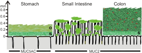
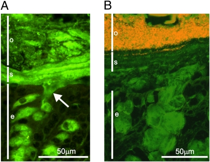
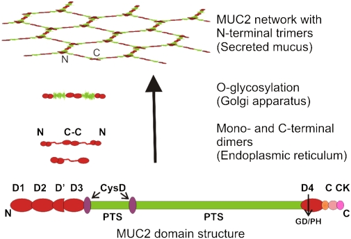
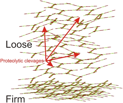
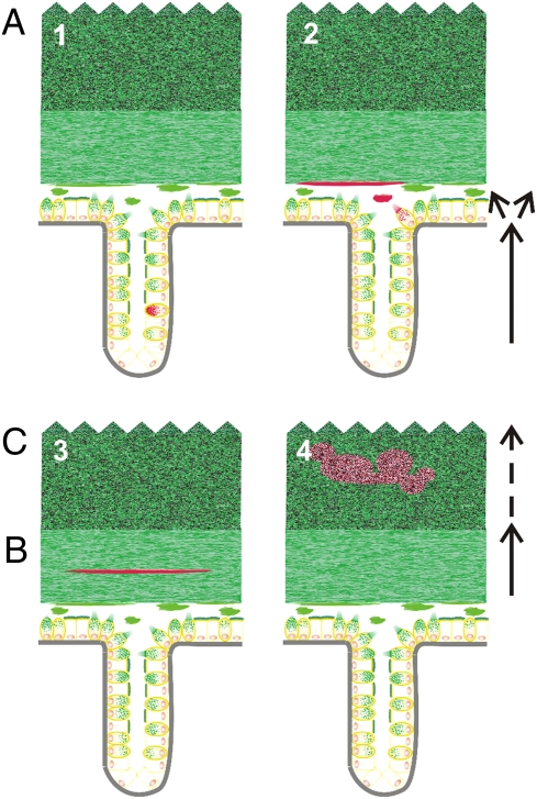
The normal intestinal microbiota inhabits the colon mucus without triggering an inflammatory response. The reason for this and how the intestinal mucus of the colon is organized have begun to be unraveled. The mucus is organized in two layers: an inner, stratified mucus layer that is firmly adherent to the epithelial cells and approximately 50 μm thick; and an outer, nonattached layer that is usually approximately 100 μm thick as measured in mouse. These mucus layers are organized around the highly glycosylated MUC2 mucin, forming a large, net-like polymer that is secreted by the goblet cells. The inner mucus layer is dense and does not allow bacteria to penetrate, thus keeping the epithelial cell surface free from bacteria. The inner mucus layer is converted into the outer layer, which is the habitat of the commensal flora. The outer mucus layer has an expanded volume due to proteolytic activities provided by the host but probably also caused by commensal bacterial proteases and glycosidases. The numerous O-glycans on the MUC2 mucin not only serve as nutrients for the bacteria but also as attachment sites and, as such, probably contribute to the selection of the species-specific colon flora. This observation that normal human individuals carry a uniform MUC2 mucin glycan array in colon may indicate such a specific selection.


Conflict of interest statement

The authors declare no conflict of interest.

Figures

Fig. 1.
Fig. 1.
Schematic proposal of how mucus is organized in the gut. The thicknesses given are from rat and adapted from the work of Holm and colleagues (13). The red dots in the outer mucus layer of colon illustrate bacteria. The genes encoding the gel-forming mucins (green) expressed by the surface goblet cells in the different parts of the intestine are marked by name. o, outer loose mucus layer; s, inner stratified, firmly attached mucus layer. That the mucus thickness and length of the villi vary along the length of the gut is not illustrated.
Fig. 2.
Fig. 2.
Muc2 mucin builds up a stratified mucus layer devoid of bacteria in the distal mouse colon. (A) Immunostaining of Muc2 (green) in the distal mouse colon reveals mucus-filled goblet cells in the epithelium (e) and secreted mucus. The secreted mucus forms two layers: a stratified inner, firmly attached mucus layer (s) and an outer, nonattached mucus (o). Arrow indicates release of Muc2 mucin from a goblet cell. (B) FISH using a general bacterial probe visualizes the bacteria (red) in combination with staining for Muc2 (green). The inner mucus layer (s) forms a barrier impervious to bacteria and thus protects the colonic epithelium.
Fig. 3.
Fig. 3.
Domain structure and biosynthesis of MUC2 mucin. MUC2 has cysteine-rich N- and C-terminal parts with four complete von Willebrand D domains in total. The central PTS domains are rich in serine, threonine, and proline, and these domains become heavily O-glycosylated to generate mucin domains. MUC2 forms dimers in the endoplasmic reticulum by disulfide bonds between the C termini. The dimer is translocated into the Golgi apparatus, where it is O-glycosylated, resulting in a size of ≈5 MDa. The MUC2 network is formed by disulfide-linked trimers connecting the N termini. The large polymers are stored in mucin granulae in the goblet cells before being secreted.
Fig. 4.
Fig. 4.
The firmly adherent mucus is converted to the loose mucus and expanded in volume. The secreted mucus is organized into the firmly adherent mucus that shows a stratified appearance. This mucus layer contains densely packed Muc2 that is insoluble in the chaotropic salt guanidinium chloride. At a certain distance from the epithelium it is converted into a nonattached, soluble mucus layer (loose). This is expanded in volume four times owing to proteolysis within the cysteine-rich parts of MUC2, in such a way that the polymeric network is maintained. The net-like structure will also allow limited proteolysis, disrupting the polymeric network without dissolving the gel. Arrows indicate both type of cleavages.
Fig. 5.
Fig. 5.
MUC2 mucin from the human sigmoid colon of different individuals has, in contrast to mucins from other organs, a relatively uniform O-glycan composition. (A) Schematic composite structure describing a majority of the O-linked MUC2 oligosaccharides found in human sigmoid colon. This general composite structure is never observed with all substituents present at the same time. The structure is based on a core 3 structure and a repeated polylactosamine chain with up to three repeats on the C-3 position of the innermost N-acetylgalactosamine (GalNAc), as previously described (35). The most used positions observed for different substituents are indicated by large letters, and the ones used less frequently are indicated by small letters. The residue marked HexNAc (N-acetylhexosamine) is most often a GlcNAc, but it can also be a terminal GalNAc. (B) Four examples showing the relative amounts of O-glycans present on MUC2 mucin from sigmoid colon in 25 individual control patients (each individual is represented by one bar). The relative amounts of glycans are in the same range for all patients. Overall, the MUC2 O-glycan repertoire and their relative amounts were uniform among healthy individuals.
Fig. 6.
Fig. 6.
Schematic view of the turnover of goblet cells and mucus in the colon. The subsequent steps in this process are marked by red MUC2. Schematic explanation of how (i) goblet cells are formed in the crypt and MUC2 mucin is formed in the crypt goblet cells and during transportation to the surface (Upper Left); (ii) MUC2 is released and expanded in volume to form a sheet that forms the inner mucus layer from below (A) (Upper Right); (iii) MUC2 are transported with the inner mucus layer (B) (Lower Left); and (iv) MUC2 is converted to the outer loose mucus layer at a sharp border and then expanded in volume in the outer loose mucus layer (C) (Lower Right). Finally, MUC2 mucin is dissolved by bacterial enzymes and transported away with the fecal stream (not illustrated).

References

    1. Xu J, Gordon JI. Inaugural article: Honor thy symbionts. Proc Natl Acad Sci USA. 2003;100:10452–10459. - PMC - PubMed
    1. Macpherson AJ, McCoy KD, Johansen FE, Brandtzaeg P. The immune geography of IgA induction and function. Mucosal Immunol. 2008;1:11–22. - PubMed
    1. Hooper LV. Do symbiotic bacteria subvert host immunity? Nat Rev Microbiol. 2009;7:367–374. - PubMed
    1. Boman HG. Innate immunity and the normal microflora. Immunol Rev. 2000;173:5–16. - PubMed
    1. Dann SM, Eckmann L. Innate immune defenses in the intestinal tract. Curr Opin Gastroenterol. 2007;23:115–120. - PubMed

Publication types