. 2010 Jul 13;107(28):12611-6.
doi: 10.1073/pnas.1006542107. Epub 2010 Jun 28.

Programmed necrosis induced by asbestos in human mesothelial cells causes high-mobility group box 1 protein release and resultant inflammation

Affiliations

Programmed necrosis induced by asbestos in human mesothelial cells causes high-mobility group box 1 protein release and resultant inflammation

Haining Yang et al. Proc Natl Acad Sci U S A. .

Abstract

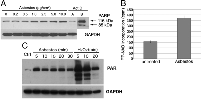
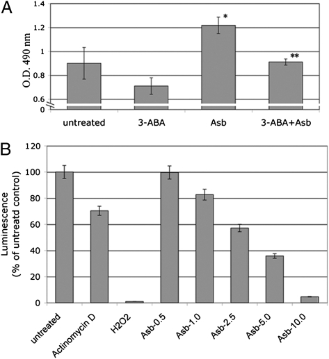
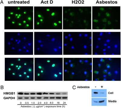
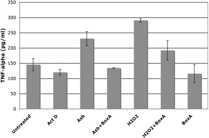
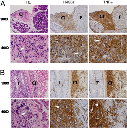
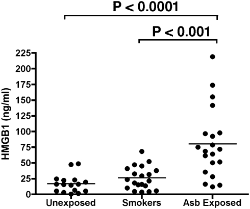
Asbestos carcinogenesis has been linked to the release of cytokines and mutagenic reactive oxygen species (ROS) from inflammatory cells. Asbestos is cytotoxic to human mesothelial cells (HM), which appears counterintuitive for a carcinogen. We show that asbestos-induced HM cell death is a regulated form of necrosis that links to carcinogenesis. Asbestos-exposed HM activate poly(ADP-ribose) polymerase, secrete H(2)O(2), deplete ATP, and translocate high-mobility group box 1 protein (HMGB1) from the nucleus to the cytoplasm, and into the extracellular space. The release of HMGB1 induces macrophages to secrete TNF-alpha, which protects HM from asbestos-induced cell death and triggers a chronic inflammatory response; both favor HM transformation. In both mice and hamsters injected with asbestos, HMGB1 was specifically detected in the nuclei, cytoplasm, and extracellular space of mesothelial and inflammatory cells around asbestos deposits. TNF-alpha was coexpressed in the same areas. HMGB1 levels in asbestos-exposed individuals were significantly higher than in nonexposed controls (P < 0.0001). Our findings identify the release of HMGB1 as a critical initial step in the pathogenesis of asbestos-related disease, and provide mechanistic links between asbestos-induced cell death, chronic inflammation, and carcinogenesis. Chemopreventive approaches aimed at inhibiting the chronic inflammatory response, and especially blocking HMGB1, may decrease the risk of malignant mesothelioma among asbestos-exposed cohorts.


Conflict of interest statement

Conflict of interest statement: M.E.B. is founder and part owner of HMGBiotech, a company that provides goods and services related to HMGB proteins.

Figures

Fig. 1.
Fig. 1.
Asbestos induces PARP activation in HM. (A) Increasing amounts of asbestos induced a parallel increase in PARP levels, without inducing PARP cleavage. HM were exposed to asbestos at 0–10 μg/cm2 or to actinomycin D (0.1 μM) for 24 h. Whole-cell extracts (30 μg) were analyzed by Western blot with anti-PARP monoclonal antibody. GAPDH was used as loading control. Actinomycin D (Act D, control for apoptosis) induces PARP cleavage. Act D, lane B: Film was overexposed to show cleavage of PARP more clearly. No PARP cleavage was seen after overexposure of the film in any of the asbestos lanes. (B) PARP activity is induced by asbestos. Representative results of three separate experiments. HM were exposed to 5.0 μg/cm2 crocidolite for 24 h. PARP activity was measured by incorporation of radiolabeled NAD using a PARP assay kit. PARP activity was induced ∼2.4-fold following asbestos exposure. (C) Amounts of PAR were increased following asbestos exposure, confirming that PARP activity is induced by asbestos. PARP activity was detected measuring PAR by Western blot. HM exposed to crocidolite (5 μg/cm2) or to 1 mM H2O2 (positive control for necrosis and PARP activation) for the indicated times.
Fig. 2.
Fig. 2.
Asbestos-induced HM death is PARP dependent and causes ATP depletion. (A) PARP inhibitor 3-ABA decreases asbestos cytotoxicity. HM were incubated with or without 3-ABA (0.5 mM) for 1 h before asbestos exposure (5 μg/cm2). Representative results of three separate experiments are shown. Cytotoxicity was detected using the LDH assay. 3-ABA decreased asbestos cytotoxicity and protected HM from asbestos-induced cell death. *Significantly different compared with HM without asbestos exposure. **Significantly different compared with HM exposed to asbestos without 3-ABA pretreatment (P < 0.05). (B) Asbestos induces ATP depletion in HM. HM were treated with actinomycin D (0.1 μM, positive control for apoptosis), H2O2 (200 μM, positive control for necrosis) or with asbestos (0.5–10 μg/cm2) for 24 h. Cellular ATP was measured by a bioluminescence assay. Increasing amounts of asbestos significantly induced ATP depletion. Each column represents the average of three separate experiments.
Fig. 3.
Fig. 3.
HMGB1 is released from HM into the extracellular space after asbestos exposure. (A) HMGB1 translocates from the nucleus to the cytosol upon asbestos exposure. Top row: HMGB1 staining (FITC conjugated antibody, green). Middle row: DAPI staining (blue). Bottom row: Overlay of HMGB1 (green) and DAPI staining (blue). HM were exposed to crocidolite asbestos (5 μg/cm2), H2O2 (100 μM) or actinomycin D (0.1 μM) for 6 h before immunofluoresence staining. Nuclei were visualized by DAPI staining. HMGB1 is localized in the nuclei in controls and actinomycin D–treated cells. In HM exposed to crocidolite or treated with H2O2, HMGB1 translocates to the cytosol and is seen mainly outside the nuclei (“overlay” panels, bottom row, where HMGB1 and DAPI stains are overlayed to identify location of HMGB1). (B) HMGB1 is released from HM after asbestos exposure. HM were exposed to 5 μg/cm2 asbestos for 0.5–24 h. Cell extracts were analyzed by Western blot. The amount of HMGB1 decreases 16 h after asbestos exposure when cell death becomes detectable. (C) HMGB1 is released from HM into the extracellular medium during asbestos exposure. HM were exposed to asbestos (5 μg/cm2) for 24 h. (Upper) cell extract. (Lower) Cell culture medium was collected, concentrated, and tested by Western blot with a HMGB1-specific antibody.
Fig. 4.
Fig. 4.
Asbestos-induced cell death and the release of HMGB1 promote TNF-α secretion by macrophages. HM were exposed to asbestos (5 μg/cm2), treated with H2O2 (1 mM) or actinomycin D (0.5 μM) for 1 h. Drugs were washed away and cells were fed with fresh media. After 24 h, cell culture media were collected, concentrated by ultrafiltration, and added to 105 macrophages in tissue culture with or without preincubation with Box A. The amount of TNF-α released by macrophages was measured 24 h later. TNF-α secretion is induced by culture medium from asbestos-exposed HM and from H2O2-treated HM but not by the medium from actinomycin D–treated HM. TNF-α secretion is significantly inhibited by preincubation of medium with HMGB1 antagonist Box A (100 ng/mL) (P < 0.05).
Fig. 5.
Fig. 5.
Immunohistochemical analyses show strong HMGB1 and TNF-α staining around areas of asbestos deposits in two representative murine specimens. (A) (Left) H&E staining shows nodular area of chronic inflammation (CI) around asbestos deposits under peritoneum, top left quadrant. P, pancreas. HMGB1 (Center) and TNF-α (Right) are present in area with chronic inflammation and not in pancreas (some staining is seen also along connective tissue that divides pancreatic lobes that contains lymphatic vessels). (Original magnification: top row, 100×; lower row, 400×.) (B) Top row. (Left) H&E staining. Testis (T) is shown on left half of figure. Tunical albuginea separate testis from tunical vaginalis (i.e., peritoneum), where chronic inflammation (CI) has formed around areas containing asbestos fibers. HMGB1 (Center) and TNF-α (Right) stain area with chronic inflammation (some staining can be seen also along the connective tissue, right portion of the panel, which contains lymphatic vessels with inflammatory cells). (Original magnification: top row, 100×; lower row, 400×.). H&E staining: Arrows point to asbestos fibers surrounded by inflammatory cells, macrophages, giant cells, and lymphocytes. HMGB1 and TNF-α are detected in cytoplasm and extracellular space.
Fig. 6.
Fig. 6.
HMGB1 levels in serum from individuals exposed to asbestos, in heavy smokers, and in nonsmoker,non‐asbestos-exposed controls. Bars show mean of HMGB1 levels. Mean serum HMGB1 level in asbestos-exposed individuals was significantly higher than in unexposed controls (P < 0.0001) and also significantly higher than in heavy smokers with lung inflammation (P < 0.001). Twenty individuals were studied in each group. ELISAs shown were performed in parallel and blindly. Results were reproduced in an additional 15 asbestos-exposed individuals from the same cohorts (Fig. S6).

References

    1. Carbone M, Bedrossian CW. The pathogenesis of mesothelioma. Semin Diagn Pathol. 2006;23:56–60. - PubMed
    1. Carbone M, et al. A mesothelioma epidemic in Cappadocia: scientific developments and unexpected social outcomes. Nat Rev Cancer. 2007;7:147–154. - PubMed
    1. Philip M, Rowley DA, Schreiber H. Inflammation as a tumor promoter in cancer induction. Semin Cancer Biol. 2004;14:433–439. - PubMed
    1. Vakkila J, Lotze MT. Inflammation and necrosis promote tumour growth. Nat Rev Immunol. 2004;4:641–648. - PubMed
    1. Quinlan TR, Marsh JP, Janssen YM, Borm PA, Mossman BT. Oxygen radicals and asbestos-mediated disease. Environ Health Perspect. 1994;102(Suppl 10):107–110. - PMC - PubMed

Publication types

MeSH terms