. 2010 Jul 13;107(28):12499-504.
doi: 10.1073/pnas.0914987107. Epub 2010 Jun 28.

LIM-domain proteins, LIMD1, Ajuba, and WTIP are required for microRNA-mediated gene silencing

Affiliations

LIM-domain proteins, LIMD1, Ajuba, and WTIP are required for microRNA-mediated gene silencing

Victoria James et al. Proc Natl Acad Sci U S A. .

Abstract

In recent years there have been major advances with respect to the identification of the protein components and mechanisms of microRNA (miRNA) mediated silencing. However, the complete and precise repertoire of components and mechanism(s) of action remain to be fully elucidated. Herein we reveal the identification of a family of three LIM domain-containing proteins, LIMD1, Ajuba and WTIP (Ajuba LIM proteins) as novel mammalian processing body (P-body) components, which highlight a novel mechanism of miRNA-mediated gene silencing. Furthermore, we reveal that LIMD1, Ajuba, and WTIP bind to Ago1/2, RCK, Dcp2, and eIF4E in vivo, that they are required for miRNA-mediated, but not siRNA-mediated gene silencing and that all three proteins bind to the mRNA 5' m(7)GTP cap-protein complex. Mechanistically, we propose the Ajuba LIM proteins interact with the m(7)GTP cap structure via a specific interaction with eIF4E that prevents 4EBP1 and eIF4G interaction. In addition, these LIM-domain proteins facilitate miRNA-mediated gene silencing by acting as an essential molecular link between the translationally inhibited eIF4E-m(7)GTP-5(')cap and Ago1/2 within the miRISC complex attached to the 3'-UTR of mRNA, creating an inhibitory closed-loop complex.


Conflict of interest statement

The authors declare no conflict of interest.

Figures

Fig. 1.
Fig. 1.
In vivo colocalization and binding of LIMD1 to eIF4E, Dcp2, Ago2, and RCK within P-bodies. (A) U2OS cells transiently cotransfected to express mTAN-LIMD1 and the indicated YFP-tagged P-body proteins, were fixed 48 h posttransfection and proteins visualized by confocal microscopy. (B) Digital zoom showing greater detail of the degree of colocalization for the indicated merged panels. (C) Endogenous LIMD1 and GW182 colocalize in U2OS cells. U2OS cells were cotransfected with mTan-LIMD1 and the indicated EYFP-P-body marker proteins. At 48 h posttransfection, cells were fixed and then immunocytochemistry performed using anti-GW182 (human serum #18033). Triple colocalization is indicated by white coloration in merged panel. Scale bar = 10 μm. (D) U2OS cells were fixed and then immunocytochemistry performed using anti-LIMD1 (monoclonal 3F2/C6) and anti-GW182 (human serum #18033 ) to detect endogenous proteins. Scale bar = 10 μm. (E) Higher magnification of boxed region in (D), Scale bar = 5 μm and (F) digital zoom of boxed region in (E) to highlight colocalized (yellow) LIMD1/GW182 positive foci/P-bodies (white arrows). (G) Extracts derived from U2OS cells expressing Xpress®-tagged LIMD1 together with the indicated YFP-tagged proteins, were incubated in the absence or presence of RNAse A and proteins recovered by immunoprecipitation with anti-GFP antibodies. (H) Endogenous coprecipitation of Ago2 with LIMD1. 2.5 μg of anti-LIMD1 (monoclonal 3F2/C6) or isotype specific controls were used for immunoprecipitation. Proteins detected by Western blotting with anti-Ago2 (C34C6) antibody and antimouse IgG shown as input control.
Fig. 2.
Fig. 2.
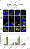
Depletion of endogenous LIMD1 decreases P-body number. U2OS cells were transduced with lentivirus to stably express scrambled shRNA (a negative control), LIMD1-5′-UTR targeted shRNA (sh-LIMD1 (5′) shRNA) or a LIMD1 targeted shRNA, with concurrent expression of an RNAi resistant Flag-His-tagged LIMD1 protein (LIMD1-FL). (A) Stable knockdown of endogenous LIMD1 and expression of the RNAi resistant protein, LIMD1-FL confirmed by Western blotting. The band at approximately 75 kDa in the rrΔ472–676 lane represents residual endogenous LIMD1. (B) YFP fused P-body protein markers were overexpressed in these cell lines and visualized by confocal microscopy. (C) P-body numbers were quantified for all cells in the field of view (× 63 magnification) and data from at least 3 random fields were collected and analyzed using Prism 4 software.
Fig. 3.
Fig. 3.
LIMD1 is specifically involved in miRNA-mediated gene silencing. Release of gene repression by siRNA or miRNA-targeted events are presented as fold induction of RL over FFL (see Methods for details). (A) Depletion of endogenous LIMD1 derepresses miRNA-mediated gene silencing. The stable cell lines described in Fig. 2 expressing scrambled, sh-LIMD1-5′, rrLIMD1-FL or rrΔ472–676-FL were transfected with the si-Let7 and mi-Let7 RL/FFL reporters. After 24 h, total cell extracts were analyzed for dual-luciferase activity. and changes in relative luciferase activities presented as fold induction of RL over FFL (see Methods for details). (B) miRNA-mediated gene silencing in Limd1-/- mouse embryonic fibroblasts (MEFs). si-Let7 and mi-Let7a-pSiCheck reporters were transfected into wild-type and Limd1-/- derived MEFs. After 24 h luciferase activities determined, as described. (C) Rates of protein synthesis for wild-type MEF and LIMD1 null-/- MEFs were determined via 35S-Met incorporation assay. Values shown as % of wild type control set to 100%. (D) U2OS cells were transfected with the indicated siRNAs. At 48 h posttransfection, cells were further transfected with si-Let7 and mi-Let7 RL/FFL luciferase reporters; following a further 24 h cell extracts were assayed for luciferase activity as described above.
Fig. 4.
Fig. 4.
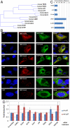
LIMD1, Ajuba, and WTIP are a new family of positive regulators of miRNA-mediated gene silencing. (A) Phylogenetic analysis based on amino acid homologies between the six established members of the Zyxin family of LIM domain containing proteins. This figure shows a clear division into two subfamilies containing LIMD1, Ajuba, and WTIP (LAW) or Zyxin, LPP, and TRIP6 (ZLiP). gi_13517496 mouse TRIP6, gi_2558592 human TRIP6, gi_1537030 human LPP, gi_55154563, mouse LPP, gi_58530845, human zyxin; gi_6756085 , mouse zyxin; gi_7305237 , mouse LIMD1; gi_7657307 , human LIMD1; gi_29470380, human Ajuba; gi_31981662 , mouse Ajuba; gi_46402183 , mouse WTIP; gi_89057404 , human WTIP (B) Ajuba and WTIP colocalize with LIMD1 to form P-bodies. Vector encoding a LIMD1-Asred fusion protein was cotransfected into U2OS cells with EGFP fusion proteins of all six family members. At 48 h posttransfection cells were fixed and visualized via confocal microscopy. (C) Percentage colocalization efficiency of the indicated EGFP-fusion proteins with LIMD1-Asred fluorescent fusion protein. (D) Comparative analysis of siRNA-targeted depletion of all six LIM domain family members together with Ago2 and GW182 and affects on siRNA and miRNA-mediated repression. U2OS cells were transiently transfected with the indicated siRNA. At 48 h posttransfection, cells were further transfected with si-Let7 and mi-Let7 RL/FFL reporter constructs and following a further 24 h, luciferase activity assayed as described Fig. 3. * indicates p-value ≤ 0.01
Fig. 5.
Fig. 5.
LIMD1, Ajuba, and WTIP are recovered specifically with the m7GTP cap structure in association with eIF4E. (A) Extracts derived from cells transfected with Xpress-tagged LIM-domain proteins were incubated with m7GTP-Sepharose or GTP-Sepharose beads, in the absence (-) and presence (+) of the m7GpppG cap analog. Inputs (5%) and bound fractions (40%) were analyzed by Western blotting using anti-Xpress antibody (LIM family member proteins) or a polyclonal anti-eIF4E antiserum. (B) m7GTP-Sepharose binding assays were carried out as in panel (A) but with the inclusion of incubation with RNase A where indicated. (C) GST-VO, GST-Δ472–676, GST-LIMD1, or GST-eIF4E proteins were incubated with U2OS and HEK 293 cell extracts. The GST-tagged proteins were recovered on Glutathione-Sepharose and the recovery of the indicated endogenous proteins determined by Western blotting. (D) LIMD1 influences the level of Ago2 that immunoprecipitates with eIF4E. Equal amounts Xpress-tagged LIMD1, Xpress-VO, HA-AGO2, EYFP-eIF4E, or EYFP-VO were transfected into U2OS cells in the combinations shown. EYFP-tagged proteins were recovered by IP and the association with the indicated proteins determined by Western blotting; the Xpress vector served as a negative control.

References

    1. Kato M, Slack FJ. microRNAs: Small molecules with big roles—C. elegans to human cancer. Biol Cell. 2008;100:71–81. - PubMed
    1. Valencia-Sanchez MA, Liu J, Hannon GJ, Parker R. Control of translation and mRNA degradation by miRNAs and siRNAs. Genes Dev. 2006;20:515–524. - PubMed
    1. Eulalio A, Behm-Ansmant I, Schweizer D, Izaurralde E. P-body formation is a consequence, not the cause, of RNA-mediated gene silencing. Mol Cell Biol. 2007;27:3970–3981. - PMC - PubMed
    1. Kadrmas JL, Beckerle MC. The LIM domain: From the cytoskeleton to the nucleus. Nat Rev Mol Cell Bio. 2004;5:920–931. - PubMed
    1. Kumar MS, Lu J, Mercer KL, Golub TR, Jacks T. Impaired microRNA processing enhances cellular transformation and tumorigenesis. Nat Genet. 2007;39:673–677. - PubMed

Publication types