. 2010 Jul 20;107(29):12901-6.
doi: 10.1073/pnas.1001499107. Epub 2010 Jul 2.

Important roles of PI3Kgamma in osteoclastogenesis and bone homeostasis

Affiliations

Important roles of PI3Kgamma in osteoclastogenesis and bone homeostasis

Heeseog Kang et al. Proc Natl Acad Sci U S A. .

Abstract

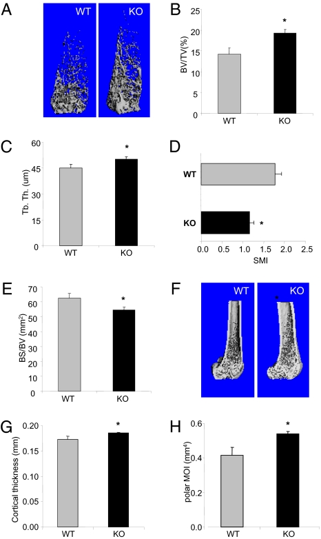
G protein-coupled receptor-regulated PI3Kgamma is abundantly expressed in myeloid cells and has been implicated as a promising drug target to treat various inflammatory diseases. However, its role in bone homeostasis has not been investigated, despite the fact that osteoclasts are derived from myeloid lineage. We therefore carried out thorough bone phenotypic characterization of a PI3Kgamma-deficient mouse line and found that PI3Kgamma-deficient mice had high bone mass. Our analyses further revealed that PI3Kgamma deficiency did not affect bone formation because no significant changes in osteoblast number and bone formation rate were observed. Instead, the lack of PI3Kgamma was associated with decreased bone resorption, as evidenced by decreased osteoclast number in vivo and impaired osteoclast formation in vitro. The decreased osteoclast formation was accompanied by down-regulated expression of osteoclastogenic genes, compromised chemokine receptor signaling, and an increase in apoptosis during osteoclast differentiation. Together, these data suggest that PI3Kgamma regulates bone homeostasis by modulating osteoclastogenesis. Our study also suggests that inhibition of PI3Kgamma, which is being considered as a potential therapeutic strategy for treating chronic inflammatory disorders, may result in an increase in bone mass.


Conflict of interest statement

The authors declare no conflict of interest.

Figures

Fig. 1.
Fig. 1.
PI3Kγ KO mice show increased bone mass and rigidity. (A–E) μCT analysis of cancellous bone in the distal metaphysic of femurs. (A) 3D μCT reconstruction of representative femurs from PI3Kγ-null and WT mice (4 mo old, male). Trabecular bone parameters shown are (B) bone volume fraction (BV/TV, %), (C) trabecular thickness (μm), (D) structural model index (SMI, quantifies the characteristic form of the cancellous bone in terms of plate-like to rod-like; for an ideal plate and rod structure, the SMI value is 0 and 3, respectively), and (E) bone surface/bone volume (BS/BV). (F–H) μCT analyses of cortical bone in the middiaphysis of the femurs. (F) Representative images of 3-D μCT, (G) cortical thickness, and (H) polar moment of inertia (pMOI). Data are presented as means ± SEM (n = 12). *P < 0.05 vs. WT.
Fig. 2.
Fig. 2.
Deletion of PI3Kγ has no impact on bone formation. (A–C) Static histomorphometry analysis of cancellous bone in metaphysis of tibiae from PI3Kγ WT and KO mice (4 mo old, male). (A) Representative images of toluidine blue–stained sections of tibiae from PI3Kγ WT and KO mice. (B) Bone volume fraction (BV/TV, %). (C) Number of osteoblasts per bone perimeter (N.Ob/B.Pm.). (D–G) Dynamic histomorphometry of tibiae from PI3Kγ WT and KO mice. (D) Representative epifluorescence images of endosteal surface of middiaphysis of tibiae. (E) Mineral apposition rate (MAR). (F) Mineralizing surface (MS/BS). (G) Bone formation rate (BFR/BS). (H) Osteocalcin level in serum was measured by RIA. Data are plotted as mean ± SEM (n = 12). *P < 0.05 vs. WT. NS, not statistically significant.
Fig. 3.
Fig. 3.
Impaired osteoclast development in PI3Kγ-deficient bones. Histomorphometric analysis of TRAP-stained tibiae from PI3Kγ-null and WT mice (4 mo old, male). (A) Representative images of TRAP-stained tibia sections. (B) Osteoclast surface per bone surface (Oc.S/BS). (C) Number of osteoclasts per bone perimeter (N.Oc/B.Pm.). *P < 0.05 vs. WT. Data are shown as mean ± SEM, n = 12.
Fig. 4.
Fig. 4.
Loss of PI3Kγ impairs osteoclastogenesis in vitro. (A) TRAP-stained multinucleated osteoclasts generated from BMMs of PI3Kγ WT and KO mice (4 mo old, male). BMMs were cultured in the presence of M-CSF (25 ng/mL) and RANKL (100 ng/mL) for 6 d, with refreshing media every other day. Quantification of (B) total surface area occupied by multinucleated TRAP-positive osteoclasts, (C) cell number per well, and (D) mean osteoclast surface. The experiment was performed in triplicate, and representative data from three experiments are shown. Data are presented as mean ± SEM. *P < 0.05 vs. WT, n = 12.
Fig. 5.
Fig. 5.
Down-regulated expression of osteoclast-associated genes in PI3Kγ-null cells. Real-time quantitative RT-PCR analysis for relative expression of (A) nuclear factor of activated T cells c1 (NFATc1), (B) TRAP (Acp5), (C) d2 isoform of vacuolar [H(+)] ATPase (v-ATPase) V(0) domain (Atp6v0d2), (D) DC-STAMP, (E) cathepsin K, (F) OSCAR, (G) calcitonin receptor, (H) fos-related antigen-2 (Fra-2, fosl2), (I) c-fos, and (J) microphthalmia-associated transcription factor (MitF). Expression level of WT at day 0 was set to 1. Graphs are plotted with mean values of triplicates in a representative experiment. Error bars present SEM. *P < 0.02 vs. WT at each time point.
Fig. 6.
Fig. 6.
Effect of PI3Kγ deficiency on Akt phosphorylation and apoptosis in BMM. (A) PI3Kγ deficiency does not affect Akt phosphorylation by M-CSF. BMM cells from PI3Kγ WT and KO mice were serum-starved for 3 h and treated with M-CSF (25 ng/mL), RANKL (100 ng/mL), or both for durations as indicated. (B) PI3Kγ deficiency abrogated Akt phosphorylation by SDF-1α. BMM cells were serum-starved for 3 h and treated with SDF-1α (200 ng/mL) for durations as indicated. (C) PI3Kγ deficiency increases caspase activity. BMM cells were cultured in M-CSF (25 ng/mL), RANKL (100 ng/mL) or M-CSF plus RANKL for 2 d. Caspase activity was determined by a homogeneous caspase assay kit. (D) PI3Kγ deficiency does not influence cell proliferation. BMMs were labeled with 10 μM BrdU for 4 h, and the incorporated BrdU was detected by chemiluminescent ELISA.

References

    1. Cantley LC. The phosphoinositide 3-kinase pathway. Science. 2002;296:1655–1657. - PubMed
    1. Hirsch E, et al. Central role for G protein-coupled phosphoinositide 3-kinase gamma in inflammation. Science. 2000;287:1049–1053. - PubMed
    1. Laffargue M, et al. Phosphoinositide 3-kinase gamma is an essential amplifier of mast cell function. Immunity. 2002;16:441–451. - PubMed
    1. Barber DF, et al. PI3Kgamma inhibition blocks glomerulonephritis and extends lifespan in a mouse model of systemic lupus. Nat Med. 2005;11:933–935. - PubMed
    1. Camps M, et al. Blockade of PI3Kgamma suppresses joint inflammation and damage in mouse models of rheumatoid arthritis. Nat Med. 2005;11:936–943. - PubMed

Publication types

MeSH terms

LinkOut - more resources