. 2010 Jul 27;107(30):13426-31.
doi: 10.1073/pnas.1007884107. Epub 2010 Jul 7.

Reversal of hyperglycemia in diabetic mouse models using induced-pluripotent stem (iPS)-derived pancreatic beta-like cells

Affiliations

Reversal of hyperglycemia in diabetic mouse models using induced-pluripotent stem (iPS)-derived pancreatic beta-like cells

Zaida Alipio et al. Proc Natl Acad Sci U S A. .

Abstract

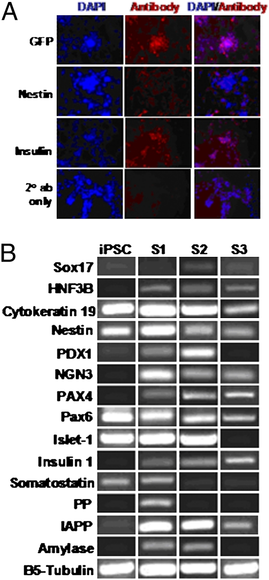
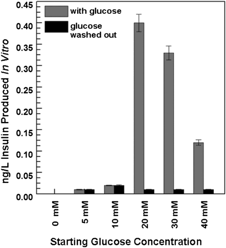
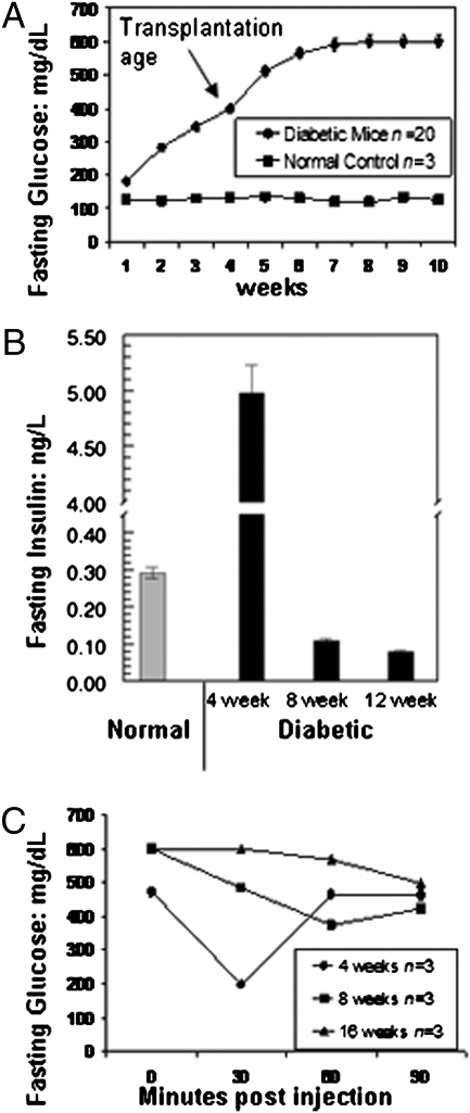
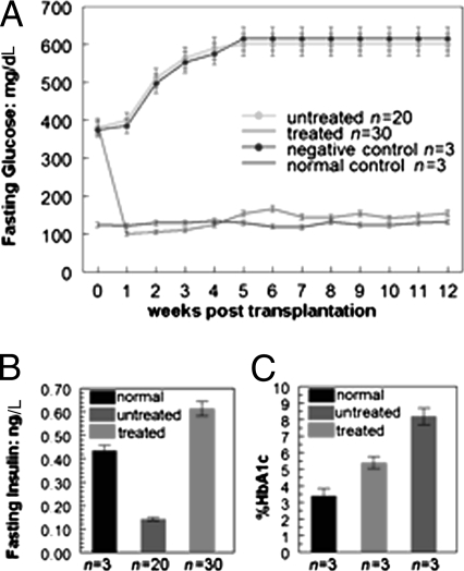
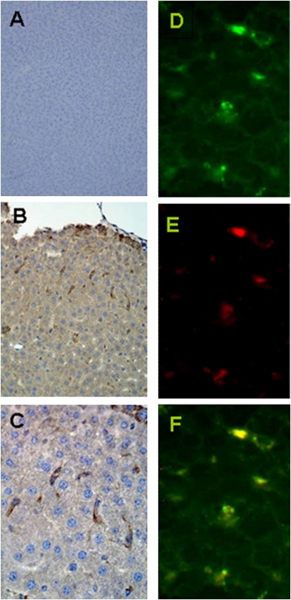
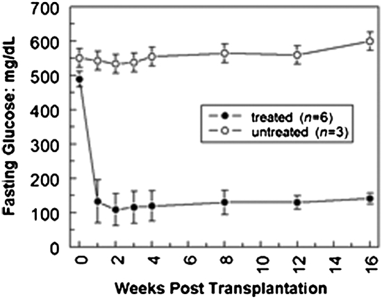
Diabetes mellitus is characterized by either the inability to produce insulin (type 1 diabetes) or as insensitivity to insulin secreted by the body (type 2 diabetes). In either case, the body is unable to move blood glucose efficiently across cell membranes to be used. This leads to a variety of local and systemic detrimental effects. Current treatments for diabetes focus on exogenous insulin administration and dietary control. Here, we describe a potential cure for diabetes using a cellular therapy to ameliorate symptoms associated with both reduced insulin secretion and insulin sensitivity. Using induced pluripotent stem (iPS) cells, we were able to derive beta-like cells similar to the endogenous insulin-secreting cells in mice. These beta-like cells secreted insulin in response to glucose and corrected a hyperglycemic phenotype in two mouse models of type 1 and 2 diabetes via an iPS cell transplant. Long-term correction of hyperglycemia was achieved, as determined by blood glucose and hemoglobin A1c levels. These data provide an initial proof of principle for potential clinical applications of reprogrammed somatic cells in the treatment of diabetes type 1 or 2.


Conflict of interest statement

The authors declare no conflict of interest.

Figures

Fig. 1.
Fig. 1.
Characterization of iPS cell-derived insulin-producing β-like cells. (A) Immunofluorescent staining: stage 3 on day 20. In vitro-derived β-like cells were stained with antibodies to GFP, nestin, and insulin on day 20 of stage 3. Cells were counterstained with DAPI. Nestin staining was reduced to minimal, although insulin staining persisted. (B) Gene expression profile of in vitro-derived β-like cells. Gene expression analysis by RT-PCR with various pancreatic cell markers during iPS cell-derived β-like cell differentiation. Total RNA was isolated from cells at stage 1 (S1), stage 2 (S2), and stage 3 (S3) of β-like cell development. B5-tubulin was used as an endogenous control. IAPP, islet amyloid polypeptide; iPSC, cells before differentiation.
Fig. 2.
Fig. 2.
In vitro insulin induction. On glucose treatment, stage 3 on day 20, iPS cell-derived β-like cells secrete insulin in vitro. Cells were first exposed to five glucose concentrations (5, 10, 20, 30, and 40 mM), and the insulin levels in their supernatants were determined. Glucose was then removed from the medium, and insulin levels were again measured in supernatants from the same cells.
Fig. 3.
Fig. 3.
In vivo characterization of diabetes mellitus type 2 model. (A) Glucose concentration of the untransplanted type 2 diabetes mellitus mouse model. Basal glucose concentrations were obtained from untransplanted type 2 diabetes mellitus mouse models (n = 20). As shown, this model began to exhibit diabetic features (glucose >300 mg/dL) at 3 wk of age. Glucose levels continued to increase until week 6 and then remained elevated at 600 mg/dL. Normal control animals retained lower glucose levels throughout the entire period. (B) Insulin levels of the untransplanted type 2 diabetes mellitus mouse model. Basal insulin concentrations were obtained from untransplanted type 2 diabetes mellitus mouse models at 4 wk old (n = 20), 8 wk old (n = 3), and 12 wk old (n = 3). Insulin was initially high at 4 wk but dropped off and remained reduced from 8 wk on. Insulin concentrations from normal mice (n = 3) are shown for comparison. (C) Insulin tolerance test. Untransplanted type 2 diabetes mellitus mouse models at 4 wk old (n = 3), 8 wk old (n = 3), and 16 wk old (n = 3) were i.p. injected with 0.75 U/kg human insulin. Fasting glucose concentration was measured before injection and at 30, 60, and 90 min postinjection via tail vein bleed and a handheld glucometer. At 4 wk, the mice showed sensitivity to insulin therapy, but they were resistant by 8 and 16 wk of age.
Fig. 4.
Fig. 4.
Transplanted diabetes mellitus type 2 model. (A) Glucose concentration of engrafted type 2 diabetes mellitus mouse model. Before transplantation, all mice presented a hyperglycemic phenotype. Stage 3 day 7 iPS cell-derived pancreatic β cells engrafted into type 2 diabetes mellitus mice (n = 30) were able to regulate glucose levels and ameliorate hyperglycemia for more than 3 mo. Glucose levels were obtained from the tail vein and measured by a handheld glucometer every 2–3 d. For untreated and control mice, n = as marked. Weekly tracking of the decrease in treated mice attributable to both postsurgical loss and harvest of animals for histology is illustrated in Fig. S6. (B) Insulin production in the engrafted type 2 diabetes mellitus mouse model. Serum insulin levels were measured 3 wk posttransplantation in type 2 diabetes mellitus mice (n = 30) engrafted with stage 3 day 7 iPS cell-derived β-like cells. Data indicate that treated mice had markedly increased insulin levels as compared with untreated mice. (C) Hemoglobin (Hb) A1c levels of the engrafted type 2 diabetes mellitus mouse model. At 4 wk posttransplantation, blood samples from engrafted type 2 diabetes mellitus mice (n = 3) were tested by an independent company (DTI) to measure the level of Hb A1c. The Hb A1c level of unengrafted mice was ∼2.5 times higher than normal. After engraftment, the Hb A1c level in the treated mice (n = 3), although still higher than that in normal control mice, had decreased to a level ∼40% lower than that of the untreated cohort (n = 3).
Fig. 5.
Fig. 5.
Immunohistochemistry and immunofluorescent staining of liver tissue from the engrafted type 2 diabetes mellitus mouse model. Mice were killed at 7 d (n = 3 for each) for immunostaining of liver tissues to analyze the distribution of engrafted cells. (A) Liver of C57BL6 control as a negative control for GFP staining. (B) At low magnification, the engraftment of spindle-shaped GFP-positive cells (brown) scattered throughout the liver parenchyma is shown. (C) Higher magnification of the GFP-positive cells. (D) At high magnification, GFP-positive cells (green) are scattered throughout the tissue. (E) Same section with insulin-positive cells (red). (F) Merged images demonstrate colocalization of GFP and insulin.
Fig. 6.
Fig. 6.
Type 1 diabetes mellitus mouse model. The type 1 diabetes mellitus mouse model was derived from a single i.p. injection of 180 mg/kg STZ and stabilized for 10 d before transplantation. The mice used in this study (n = 9) all demonstrated hyperglycemia of greater than 400 mg/dL glucose in at least three levels before transplantation. Six mice were then injected with iPS cell-derived β-like cells via hepatic portal vein injection. Fasting glucose levels obtained every 2–3 d, starting at 2 d posttransplantation and continuing for ∼4 mo, show amelioration of hyperglycemia in this type 1 diabetic mouse model. The data shown here are for weeks 0 through 16.

References

    1. Jones PM, Courtney ML, Burns CJ, Persaud SJ. Cell-based treatments for diabetes. Drug Discov Today. 2008;13:888–893. - PubMed
    1. Urbán VS, et al. Mesenchymal stem cells cooperate with bone marrow cells in therapy of diabetes. Stem Cells. 2008;26:244–253. - PubMed
    1. Karnieli O, Izhar-Prato Y, Bulvik S, Efrat S. Generation of insulin-producing cells from human bone marrow mesenchymal stem cells by genetic manipulation. Stem Cells. 2007;25:2837–2844. - PubMed
    1. Sun B, Roh KH, Lee SR, Lee YS, Kang KS. Induction of human umbilical cord blood-derived stem cells with embryonic stem cell phenotypes into insulin producing islet-like structure. Biochem Biophys Res Commun. 2007;354:919–923. - PubMed
    1. Lumelsky N, et al. Differentiation of embryonic stem cells to insulin-secreting structures similar to pancreatic islets. Science. 2001;292:1389–1394. - PubMed

Publication types

MeSH terms