. 2010 Sep;31(8):1408-12.
doi: 10.3174/ajnr.A2212. Epub 2010 Jul 8.

Imaging characteristics of schwannoma of the cervical sympathetic chain: a review of 12 cases

Affiliations

Imaging characteristics of schwannoma of the cervical sympathetic chain: a review of 12 cases

G Anil et al. AJNR Am J Neuroradiol. 2010 Sep.

Abstract

Background and purpose: SCSCs are rare. This study reviews our experience with CT and MR imaging of SCSCs.

Materials and methods: We retrospectively reviewed the CT and MR imaging studies as well as clinical data of 12 patients (6 men, 6 women; mean age, 41 years; range, 27-55 years) with surgicopathologic evidence of SCSC, referred to our institution between January 1999 to October 2008. Images were evaluated with respect to the location, number, morphology, attenuation/signal intensity, enhancement characteristics, and patterns of mass effect of the schwannomas.

Results: The schwannomas were solitary, well-circumscribed, and medial to the carotid sheath. Seven were hypoattenuated to skeletal muscle on CT with poor postcontrast enhancement, 4 were isoattenuated, and a single lesion showed intense heterogeneous enhancement. At MR imaging, they were heterogeneously bright on T2WI with intense inhomogeneous postgadolinium enhancement. The ICA was displaced anteriorly in 9 patients with a component of lateral displacement in 8 of these patients. The ICA was in a neutral position in 2 patients and posterolaterally displaced in 1 patient. A single patient demonstrated separation of the ICA and IJV. There was splaying of the carotid bifurcation in 4 patients.

Conclusions: We present the patterns of mass effect and the spectrum of CT and MR imaging characteristics of SCSC, including certain observations that are infrequently described in the published literature.


Figures

Fig 1.
Fig 1.
These postcontrast axial CT images of 3 different patients demonstrate the variable appearances of the SCSC with respect to their texture and attenuation. All of them are well-defined and round to oval with anterior (A) or anterolateral (B and C) displacement of the vessels in the carotid sheath. There is no separation of the IJV and ICA. A, The schwannoma is homogeneously hypoattenuated to the skeletal muscle, with almost no enhancement. B, The schwannoma is predominantly isoattenuated with enhancement similar to that of the skeletal muscle. Internal heterogeneity is noted as an irregular poorly enhancing hypoattenuated area in its anteromedial quadrant. C, The schwannoma shows marked internal heterogeneity. Central areas are intensely enhancing, while the rest of the tumor is moderately or poorly enhancing.
Fig 2.
Fig 2.
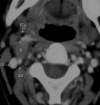
The schwannoma shows intense heterogeneous enhancement on the postgadolinium fat-saturated T1WI (A) and appears heterogeneously bright on the fat-saturated T2WI (B). The nonenhancing areas in the center of the mass correspond to the areas of fluid signal intensity seen in the T2WI. The entire carotid sheath (arrows) is displaced in an anterolateral direction.
Fig 3.
Fig 3.
The postcontrast axial CT image shows the SCSC as a hypoattenuated mass with poor enhancement. There is posterolateral displacement of the right ICA and IJV. This is unlike the anterior displacement of the ICA that is normally seen in carotid space masses (compare with the earlier figures). However, true to its carotid space location, there is anterior displacement of the fat in the prestyloid parapharyngeal space, and it displaces the visceral space medially.
Fig 4.
Fig 4.
This postcontrast axial CT image demonstrates the separation of the right IJV and ICA by the SCSC. Tumors of the sympathetic chain usually displace the ICA and IJV together, without separating them. On imaging, it is almost impossible to distinguish the tumor in this image from a vagal schwannoma. Such a lesion may be considered as a caveat to the paradigm of Furukawa et al.
Fig 5.
Fig 5.
In this postcontrast axial CT image, the schwannoma separates the ICA and ECA along a sagittal plane. The arteries are being splayed but not encased by the tumor, and there is <180° of contact between the tumor and the vessel. The ICA is posterior to the tumor; still, it is in a neutral position compared with the contralateral side. In this case, the tumor is probably too small to displace the arteries or adjoining fat planes.

References

    1. Malone JP, Lee WJ, Levin RJ. Clinical characteristics and treatment outcome for nonvestibular schwannomas of the head and neck. Am J Otolaryngol 2005;26:108–12 - PubMed
    1. Som PM, Curtin HD. Parapharyngeal space. In: Som PM, Curtin HD, eds. Head and Neck Imaging. Vol 2. 3rd ed. St Louis: Mosby; 1996:915–51
    1. Civelek E, Karasu A, Cansever T, et al. . Surgical anatomy of the cervical sympathetic trunk during anterolateral approach to cervical spine. Eur Spine J 2008;17:991–95. Epub 2008 Jun 12 - PMC - PubMed
    1. Lyons AJ, Mills CC. Anatomical variants of the cervical sympathetic chain to be considered during neck dissection. Br J Oral Maxillofac Surg 1998;36:180–82 - PubMed
    1. Harnsberger HR, Osborn AG. Differential diagnosis of head and neck lesions based on their space of origin. 1. The suprahyoid part of the neck. AJR Am J Roentgenol 1991;157:147–54 - PubMed

MeSH terms