Review
. 2011 Jun-Jul;32(6):984-92.
doi: 10.3174/ajnr.A2171. Epub 2010 Jul 8.

Central nervous system lymphoma: characteristic findings on traditional and advanced imaging

Affiliations
Review

Central nervous system lymphoma: characteristic findings on traditional and advanced imaging

I S Haldorsen et al. AJNR Am J Neuroradiol. 2011 Jun-Jul.

Abstract

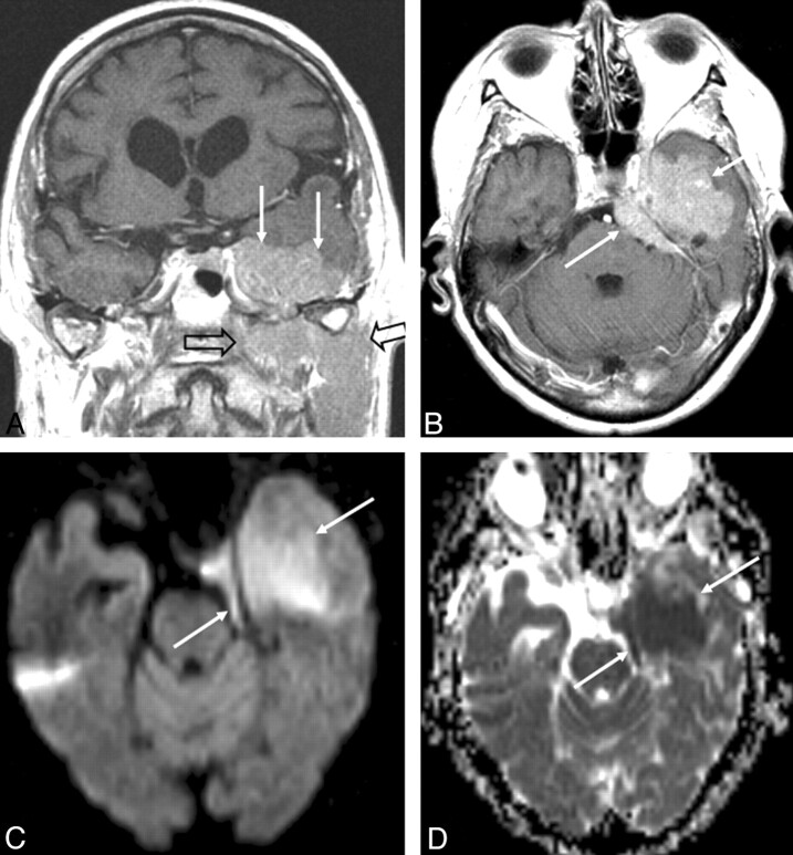
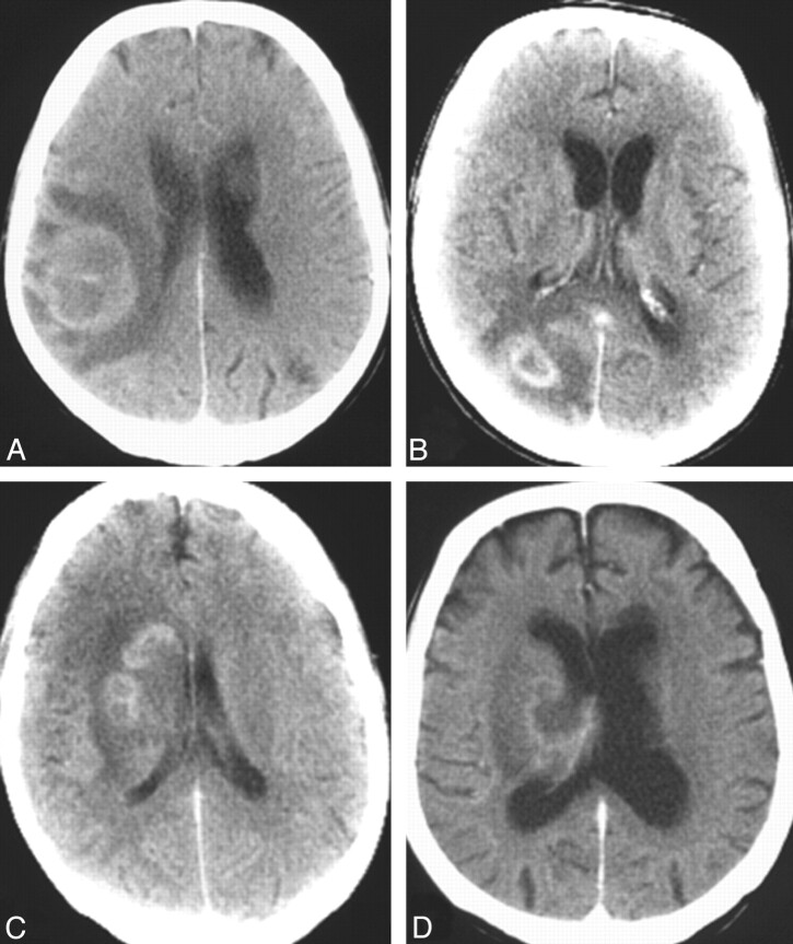
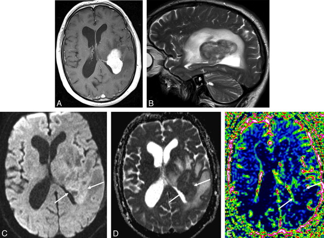
CNS lymphoma consists of 2 major subtypes: secondary CNS involvement by systemic lymphoma and PCNSL. Contrast-enhanced MR imaging is the method of choice for detecting CNS lymphoma. In leptomeningeal CNS lymphoma, representing two-thirds of secondary CNS lymphomas, imaging typically shows leptomeningeal, subependymal, dural, or cranial nerve enhancement. Single or multiple periventricular and/or superficial contrast-enhancing lesions are characteristic of parenchymal CNS lymphoma, representing one-third of secondary CNS lymphomas and almost 100% of PCNSLs. New CT and MR imaging techniques and metabolic imaging have demonstrated characteristic findings in CNS lymphoma, aiding in its differentiation from other CNS lesions. Advanced imaging techniques may, in the future, substantially improve the diagnostic accuracy of imaging, ultimately facilitating a noninvasive method of diagnosis. Furthermore, these imaging techniques may play a pivotal role in planning targeted therapies, prognostication, and monitoring treatment response.


Figures

Fig 1.
Fig 1.
Axial (A) and sagittal (B) T1-weighed contrast-enhanced MR images in a patient with CNS metastases from NHL show diffuse subependymal contrast enhancement (arrows) and 2 parenchymal lesions (open arrows) in the right basal ganglia (A) and left cerebellum (B).
Fig 2.
Fig 2.
Solitary lesions on a noncontrast CT scan (A), contrast-enhanced CT scans (B and C), and an MR image (D) in 3 patients with non-AIDS PCNSL. Note a hyperattenuated lesion in the frontal lobe on the noncontrast CT scan (A) with marked enhancement on the contrast series (B) and focal contrast-enhancing lesions in the left basal ganglia (C) and temporal lobe (D). One lesion has ring enhancement (D).
Fig 3.
Fig 3.
Multiple lesions on contrast-enhanced axial (B and D) and coronal (A and C) MR images in 4 patients with non-AIDS PCNSL. Note lesions in the basal ganglia (A), ventricles (B), frontal lobes (C), and cerebellar lobes (D).
Fig 4.
Fig 4.
Coronal (A) and axial (B) contrast-enhanced T1-weighted MR images and an axial DWI (C) and ADC map (D) in a patient with primary dural B-cell lymphoma (arrows) with tumor spread below the skull base (open arrow). The contrast-enhancing tumor at the caudal surface of the left temporal lobe (A and B) displaces the temporal lobe cranially and resembles a meningioma. DWI reveals restricted diffusion within the tumor with high signal intensity on DWI (C) and corresponding low signal intensity on the ADC map (D).
Fig 5.
Fig 5.
Contrast-enhancing lesions on CT scans (A–D) in 4 patients with AIDS-related PCNSL. Note irregularly enhancing lesions in the right parietal lobe (A), right occipital lobe (B), and right periventricular white matter (C and D); most of the lesions show ring enhancement (A, B, and C).
Fig 6.
Fig 6.
Axial contrast-enhanced T1-weighted MR image (A), sagittal T2-weighted MR image (B), axial DWI (C), an ADC map (D), and an rCBV map (E) in a patient with PCNSL. The periventricular contrast-enhancing tumor (A) in the left parietal lobe has restricted diffusion with high signal intensity on DWI (C) with corresponding low signal intensity on the ADC map (arrows, D). Perfusion MR imaging shows low perfusion within the contrast-enhancing tumor on the rCBV map (arrows, E).
Fig 7.
Fig 7.
Axial contrast-enhanced CT scan (A), coronal (B) and axial (C) contrast-enhanced T1-weighted MR images, and methionine PET (coronal, D, and axial, E) images in a patient with PCNSL. The contrast-enhancing tumor in the right temporal lobe (A–C) shows high uptake of methionine on PET (D and E).

References

    1. Wong ET. Management of central nervous system lymphomas using monoclonal antibodies: challenges and opportunities. Clin Cancer Res 2005; 11: 7151s– 57s - PubMed
    1. Mohile NA, Abrey LE. Primary central nervous system lymphoma. Semin Radiat Oncol 2007; 17: 223– 29 - PubMed
    1. Hill QA, Owen RG. CNS prophylaxis in lymphoma: who to target and what therapy to use. Blood Rev 2006; 20: 319– 32 - PubMed
    1. Montoto S, Lister TA. Secondary central nervous system lymphoma: risk factors and prophylaxis. Hematol Oncol Clin North Am 2005; 19: 751– 63 - PubMed
    1. Haldorsen IS, Krossnes BK, Aarseth JH, et al. . Increasing incidence and continued dismal outcome of primary central nervous system lymphoma in Norway 1989–2003: time trends in a 15-year national survey. Cancer 2007; 110: 1803– 14 - PubMed

MeSH terms