. 2011 May;44(5):682-91.
doi: 10.1165/rcmb.2009-0309OC. Epub 2010 Jul 8.

Lung self-assembly is modulated by tissue surface tensions

Affiliations

Lung self-assembly is modulated by tissue surface tensions

Margaret A Schwarz et al. Am J Respir Cell Mol Biol. 2011 May.

Abstract

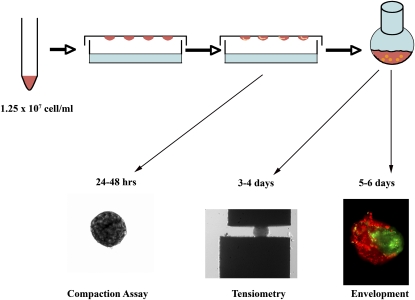
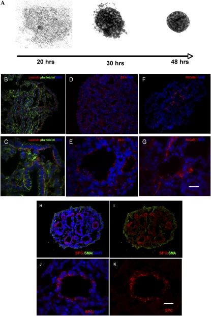
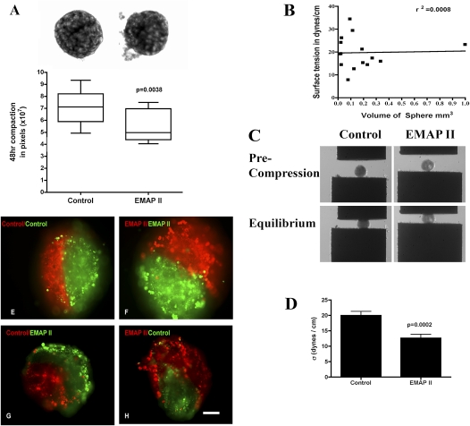
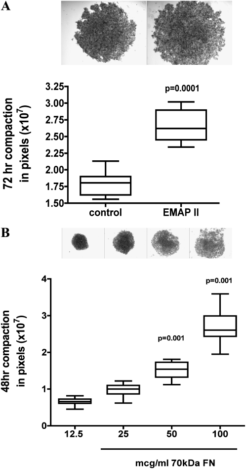
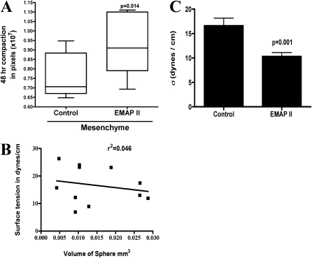
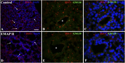
To identify cell-intrinsic properties that facilitate interaction between epithelial endodermal and mesenchymal mesodermal cells during lung morphogenesis, we developed a model of lung self-assembly that mimics fetal lung formation in structure, polarity, vasculature, and extracellular matrix expression. Three-dimensional pulmonary bodies (PBs) spontaneously self-assemble from single-cell suspensions and exhibit liquid-like properties that allow measurements of compaction rate and cohesion, and that may help to specify cellular self-organization. We hypothesized that changes in one or more of these parameters could potentially explain the lung hypoplasia associated with abnormal lung development. We examined the impact of endothelial/monocyte-activating polypeptide (EMAP) II in PBs, because EMAPII is highly expressed in lung hypoplasia. EMAPII significantly increased compaction rate and decreased overall cohesion of PBs composed of both epithelial and mesenchymal cells. Moreover, the effects of EMAPII on compaction and cohesion act exclusively through the mesenchymal cell population by interfering with fibronectin matrix assembly. We also show that EMAPII alters epithelial cell polarity and surfactant protein C expression. Our findings demonstrate, for the first time, that PBs possess liquid-like properties that can help to guide the self-assembly of fetal lungs, and that EMAPII expression can influence both mesenchymal and epithelial cells but through different molecular mechanisms.


Figures

Figure 1.
Figure 1.
Fetal pulmonary cells in three-dimensional (3D) suspension self-assemble to form pulmonary bodies (PBs). Fetal lungs isolated at Embryologic Day 14.5 were enzymatically dissociated and resuspended in 3D hanging drops (HDs). Pulmonary cells (1.25 × 10⁷ cell/ml) self-assembled or compacted over 48 hours to form pulmonary sheets (compaction assay). Pulmonary sheets placed in a shaker flask for 24–48 hours formed spherical PBs. These were subjected to tissue surface tensiometry (TST) to measure aggregate cohesivity (tensiometry), or to envelopment assays in which pairs of differentially stained PBs were apposed in 3D HDs and examined by fluorescence microscopy after 24–48 hours (envelopment).
Figure 2.
Figure 2.
PBs form blood vessels, polarize epithelial cells, and express surfactant protein C (SPC). Dissociated fetal lung cells aggregate over 48 hours to form sheets (A). After orbital shaking for 24–48 hours, immunofluorescent analysis of PBs indicate that laminin α (B and C) (cy3, FITC-phalloidin) localized to the basilar surface of the epithelium at the epithelial–mesenchymal interface, zona occludens (ZO)–1 expression was confined to the apical region of the epithelial cyst (D and E), as demonstrated by their SPC expression (HK) (cy3, FITC-SMA), and platelet endothelial cell adhesion molecule-1 (PECAM-1) distribution was confined to the mesenchyme (F and G). DAPI, 4′,6-diamidino-2-phenyindole (denotes nuclear staining) (BH and J). Scale bar, 60 μm (B, D, F, H, I) and 20 μm (C, E, G, J, K).
Figure 3.
Figure 3.
Pulmonary sheet compaction is accelerated by endothelial/monocyte–activating polypeptide (EMAP) II, whereas cohesivity is decreased. The assembly of dissociated fetal lung cells into an aggregate within 48 hours into pulmonary sheets was assessed. HDs treated with EMAPII exhibited a more rapid rate (A) of compaction as compared with controls (P = 0.0038, Student's t test, representative experiment performed at least 10 times). Magnification, 100×. PBs subjected to TST were noted to have liquid properties and surface tension that, by linear regression, was shown to be independent of aggregate size (B) (r2=0.0008). (C) PB aggregates at precompression (top panels) and after PBs have reached force and shape equilibria (bottom panels). PB cohesivity was markedly decreased in aggregates treated with EMAPII as compared with controls (D) (P = 0.0002; n = 14). Dissociated fetal lung cells were stained with membrane intercalating green or red fluorescent dyes. After formation, PBs were fused in 3D HDs and envelopment assessed. The less cohesive EMAPII-treated PBs enveloped the more cohesive untreated PBs (G and H). Combinations in which like-aggregates were used fused along their midline and formed a planar interface (E and F). Scale bar, 40 μm.
Figure 4.
Figure 4.
Disruption of fibronectin (FN) matrix assembly alters compaction in α5-integrin expressing Chinese hamster ovary (CHO) cells and in PBs. CHO-α5 cells in 3D HDs treated with EMAPII had a marked suppression in compaction as compared with controls (P = 0.0001, representative experiment n = 10, performed 3 times). PBs treated with the 70 kD fragment of FN had a dose-dependent near-linear relationship in compaction with higher doses (50–100 μg/ml; P = 0.05 and 0.001, respectively) inhibiting compaction (representative experiment n = 10, performed three times).
Figure 5.
Figure 5.
EMAPII inhibits PBs fibrillogenesis and collagen I deposition in PBs. Fibrillogenesis was assessed in PBs treated with EMAPII. Immunofluorescence analysis indicates that EMAPII inhibits insoluble FN matrix deposition, as noted by the lack of insoluble FN strands at the epithelial/mesenchymal interface (B) (FN–FITC, arrows) as compared with the insoluble strands of FN noted in controls (A) (arrows). Isolation of insoluble deoxycholate (DOC) FN supports this observation, as EMAPII (C) (lane E) suppressed the insoluble form of FN (P = 0.009) (C) (n = 6; lane marked with dash). Collagen I deposition was also altered by EMAPII (E) (arrows, Cy3) as compared with controls (D) (arrows). DAPI denotes nuclear staining (A, B, D, E). Scale bar, 20 μm.
Figure 6.
Figure 6.
Mesoderm compaction and cohesivity is inhibited by EMAPII, whereas endoderm compaction is not. Aggregate formation was examined in isolated epithelial and mesenchymal cells. Mesenchymal sheet compaction was significantly inhibited by EMAPII (A) (P = 0.014, n = 10, representative experiment, performed 10 times) as compared with controls. Mesenchymal bodies (MBs) subjected to TST were noted to have liquid properties and surface tension that was independent of aggregate size (B) (r2 = 0.046). MB cohesivity was markedly decreased in aggregates treated with EMAPII as compared with controls (C) (P = 0.001, n = 10).
Figure 7.
Figure 7.
EMAPII alters epithelial apical alignment. PBs treated with EMAPII showed disrupted epithelial apical distribution of ZO-1 (DF) (arrow, Cy3) and GM130 (E and F) (asterisk indicates epithelial cluster, FITC) as compared with controls (AC) (arrow and asterisk). DAPI denotes nuclear staining (A and B). Scale bar, 50 μm (A and D) 16 μm (B, C, E, F).

References

    1. Zeng X, Wert SE, Federici R, Peters KG, Whitsett JA. VEGF enhances pulmonary vasculogenesis and disrupts lung morphogenesis in vivo. Dev Dyn 1998;211:215–227. - PubMed
    1. Schwarz MA, Zhang F, Gebb S, Starnes V, Warburton D. EMAP II inhibits lung neovascularization and airway epithelial morphogenesis. Mech Dev 2000;95:123–132. - PubMed
    1. Dickson MC, Martin JS, Cousins FM, Kulkarni AB, Karlsson S, Akhurst RJ. Defective haematopoiesis and vasculogenesis in transforming growth factor–beta 1 knock out mice. Development 1995;121:1845–1854. - PubMed
    1. Zhou L, Dey CR, Wert SE, Whitsett JA. Arrested lung morphogenesis in transgenic mice bearing an SP-C–TGF–beta 1 chimeric gene. Dev Biol 1996;175:227–238. - PubMed
    1. Schuger L, O'Shea KS, Nelson BB, Varani J. Organotypic arrangement of mouse embryonic lung cells on a basement membrane extract: involvement of laminin. Development 1990;110:1091–1099. - PubMed

Publication types

MeSH terms