Review
. 2010 Aug;10(8):554-67.
doi: 10.1038/nri2808. Epub 2010 Jul 9.

The polarization of immune cells in the tumour environment by TGFbeta

Affiliations
Review

The polarization of immune cells in the tumour environment by TGFbeta

Richard A Flavell et al. Nat Rev Immunol. 2010 Aug.

Abstract

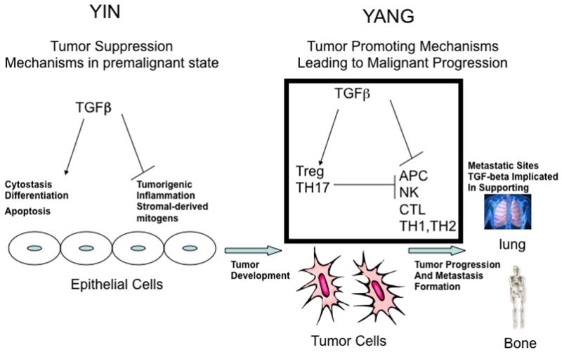
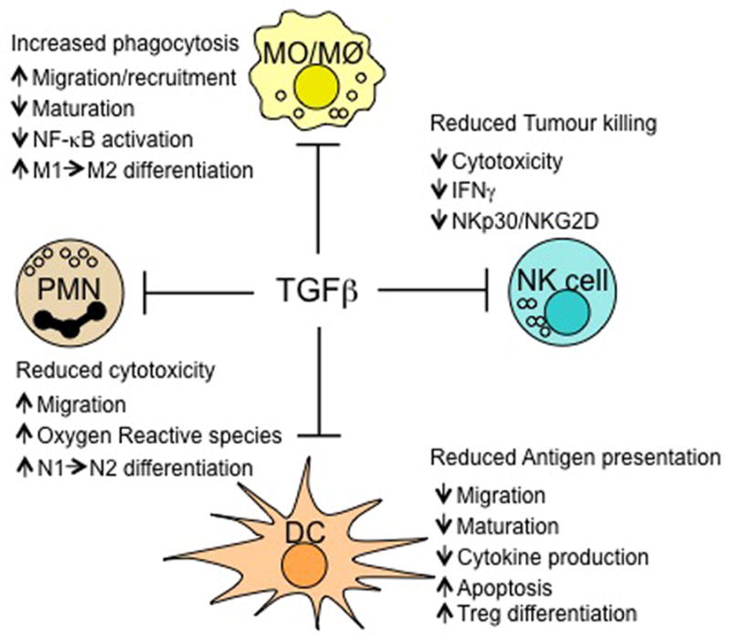
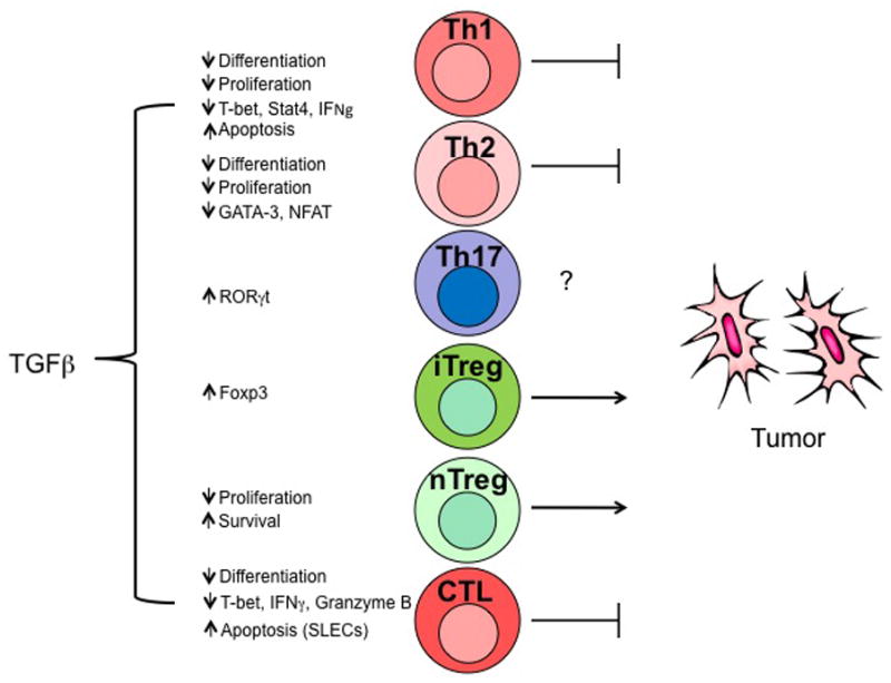
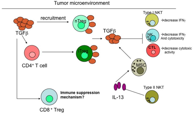
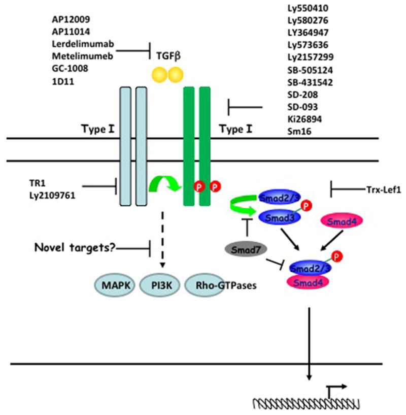
Transforming growth factor-beta (TGFbeta) is an immunosuppressive cytokine produced by tumour cells and immune cells that can polarize many components of the immune system. This Review covers the effects of TGFbeta on natural killer (NK) cells, dendritic cells, macrophages, neutrophils, CD8(+) and CD4(+) effector and regulatory T cells, and NKT cells in animal tumour models and in patients with cancer. Collectively, many recent studies favour the hypothesis that blocking TGFbeta-induced signalling in the tumour microenvironment enhances antitumour immunity and may be beneficial for cancer therapy. An overview of the current drugs and reagents available for inhibiting TGFbeta-induced signalling and their phase in clinical development is also provided.


Conflict of interest statement

The authors declare that they have no competing financial interests.

Figures

Figure 1
Figure 1. The yin and yang of TGFβ in tumour development maintenance, and metastasis formation
Before epithelial cells transform into a malignant tumour, transforming growth factor-β (TGFβ) functions as a tumour-suppressor, by blocking expression of stromal-derived mitogens and suppressing pro-tumourogenic inflammation. Furthermore, TGFβ supports the cytostasis, terminal differentiation and apoptosis of premalignant cells which harbor either an overexpressed oncogene or suppressed tumour suppressor gene. Once the epithelial cells become fully malignant, TGFβ has the opposite effect by blocking the antitumour immune response through support for the activity of regulatory cells and through direct inhibition of effector cell mechanisms from clearing the established tumour, as described in the main text and summarised in Figure 2. Once tumours are established, TGFβ further supports the formation of metastases to several sites, including bone and lung tissues. Additional nonimmune mechanisms outside the scope of this Review that support tumorigenesis and metastasis formation are addressed in References and .
Figure 2
Figure 2. Effects of TGFβ on innate immune cells
Transforming growth factor-β (TGFβ) has an inhibitory effect on innate immunity in the tumour microenvironment through several pathways. It inhibits natural killer (NK) cell function by downregulating interferon-γ (IFNγ) production and expression of the activating receptors NKp30 and NKG2D, thereby decreasing NK cell killing activity. In the presence of TGFβ, dendritic cells (DCs) acquire a tolerogenic phenotype involving decreased migration, maturation and cytokine production and increased apoptosis; they gain the ability to induce regulatory T (TReg) cell differentiation. TGFβ can also convert N1 neutrophils to a N2 phenotype, which is less cytotoxic. Similarly, TGFβ can promote the recruitment of M2 over M1 macrophages and of tumour-associated macrophages (TAMs), and can decrease cytokine production by these macrophages by inhibiting nuclear factor-κB (NF-κB) activity.
Figure 3
Figure 3. Effects of TGFβ on effector T cells
Transforming growth factor-β (TGFβ) differentially regulates the survival, differentiation, proliferation and apoptosis of T cell subsets. Among TH subpopulations, both TH1 and TH2 can provide antitumour responses, however TH1 seem to be more efficient. Both nTreg and iTreg populations inhibit antitumour immune responses. Within the tumour microenvironment, TGFβ can promote tumour growth by the maintenance of TReg cell and differentiation of iTreg subpopulations. TGFβ can also inhibit TH1 cell and CTL functions by downregulating T-bet and IFNγ expression and probably promoting a shift towards TH2 differentiation. CTLs are potent antitumour effector cells. TGFβ could also inhibit tumour immune surveillance by the induction of apoptosis in short-lived effector CTLs. The role of TH17 cells in tumour biology is still controversial and requires further characterization.
Figure 4
Figure 4. Effects of TGFβ on regulatory cells
Within the tumour microenvironment, transforming growth factor-β (TGFβ) has been implicated in recruiting natural regulatory T (nTReg) cells as well as converting CD4+ effector T cells to induced TReg (iTReg) cells. These TReg cells can express cell surface-bound TGFβ and can inhibit effector cells, including natural killer (NK) cells and CD8+ T cells, in the tumour microenvironment by cell–cell contact to dampen the antitumour response. Type I NKT cells, which are responsible for recruiting effector immune cells to the tumour through the production of large amounts of IFNγ, can be suppressed by intratumoural TGFβ, whereas Type II NKT cells support increased TGFβ production by myeloid-derived suppressor cells (MDSCs) through the generation of interleukin-13 (IL-13). CD8+ regulatory T cells have been observed in lung tumours and these might result from the production of IL-10 by antigen-presenting cells, leading to increased TGFβ production in the tumour microenvironment. The precise immunosuppressive mechanisms of CD8+ regulatory T cells in regulating the antitumour immune response have yet to be identified.
Figure 5
Figure 5. Targets for inhibiting TGFβ and downstream signalling events
The transforming growth factor-β (TGFβ)-dependent signalling pathway depends on Type I and Type II serine-threonine kinase receptors and transcription factors known as SMADs. The dimeric bioactive ligand binds to a Type II receptor, which in turn phosphorylates and activates a Type I receptor. Once the Type I receptor is activated, it phosphorylates the receptor SMADs (R-SMADs: SMAD2 and SMAD3), which promotes their interaction with the common mediator SMAD (SMAD4) and translocation to the nucleus. The inhibitory SMAD7 negatively regulates TGFβ signalling by competing with R-SMADs for interaction with the Type I receptor or SMAD4. Current TGFβ signalling inhibitors (listed in Table I ans shown in scheme) include ligand, receptor and SMAD antagonists. However, additional SMAD-independent pathways have been reported to be induced in response to TGFβ, including the activation of MAPK, Rho-like GTPase and phosphatidylinositol 3-Kinase (PI3K) signalling pathways; a complete understanding of these alternative pathways could potentially offer additional downstream molecules that could be targeted in future therapeutic approaches. (Figure adapted from )

Similar articles

Cited by

References

    1. Massague J. TGFbeta in Cancer. Cell. 2008;134:215–30. - PMC - PubMed
    1. Wrzesinski SH, Wan YY, Flavell RA. Transforming Growth Factor-{beta} and the Immune Response: Implications for Anticancer Therapy. Clin Cancer Res. 2007;13:5262–5270. - PubMed
    1. Padua D, Massague J. Roles of TGFbeta in metastasis. Cell Res. 2009;19:89–102. - PubMed
    1. Tian M, Schiemann WP. The TGF-beta paradox in human cancer: an update. Future Oncol. 2009;5:259–71. - PMC - PubMed
    1. Bierie B, Moses HL. Transforming growth factor beta (TGF-beta) and inflammation in cancer. Cytokine Growth Factor Rev. 2009 - PMC - PubMed

Publication types

MeSH terms