. 2010 Jul 1;6(7):e1000975.
doi: 10.1371/journal.ppat.1000975.

HIV gp41 engages gC1qR on CD4+ T cells to induce the expression of an NK ligand through the PIP3/H2O2 pathway

Affiliations

HIV gp41 engages gC1qR on CD4+ T cells to induce the expression of an NK ligand through the PIP3/H2O2 pathway

Hugues Fausther-Bovendo et al. PLoS Pathog. .

Abstract

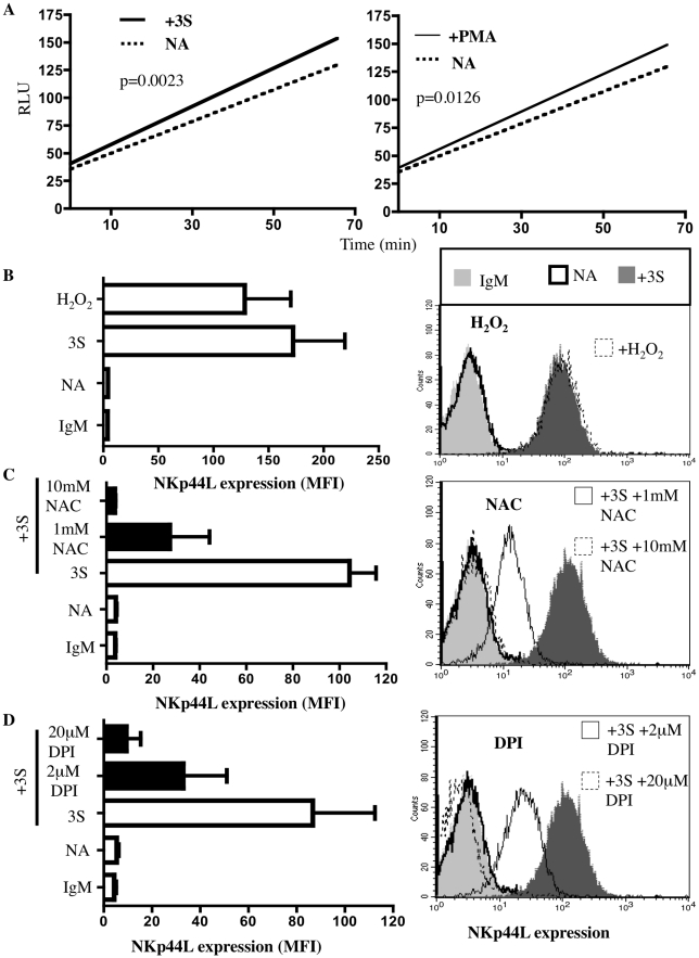
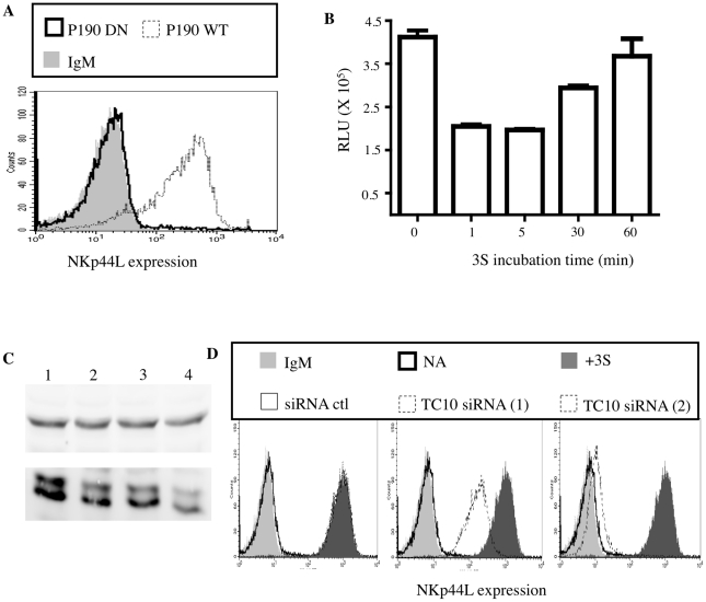
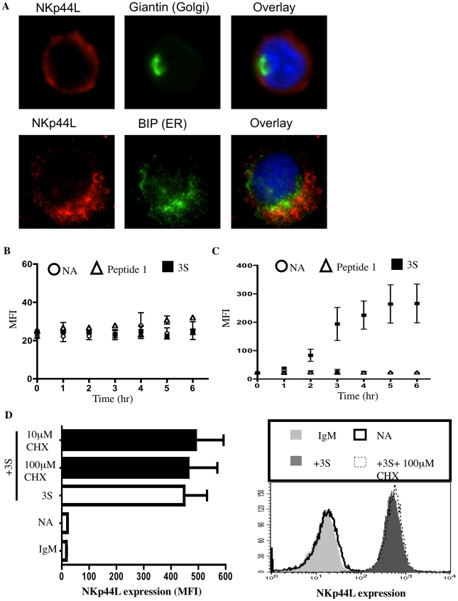
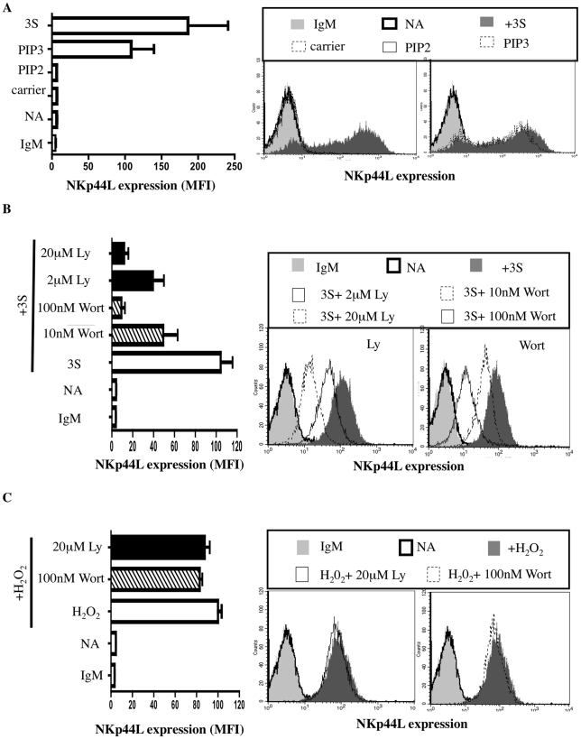
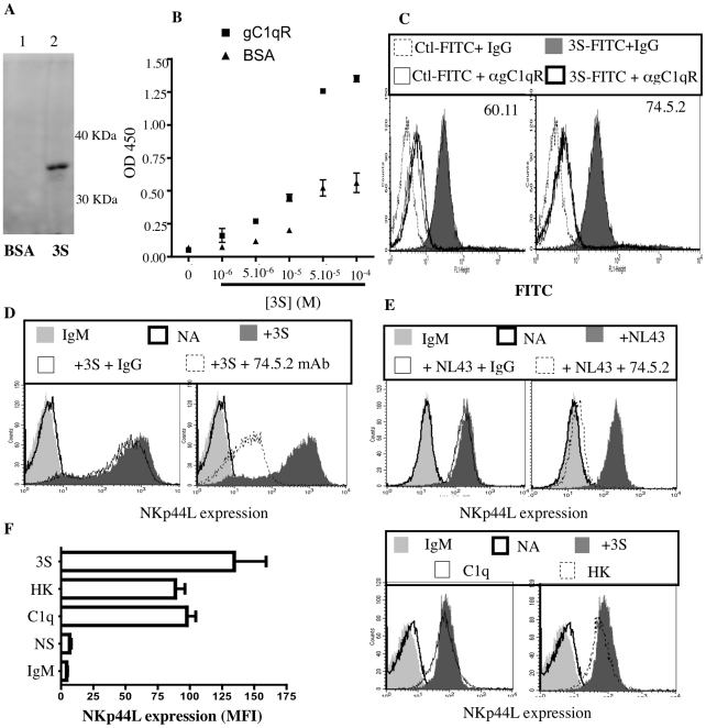
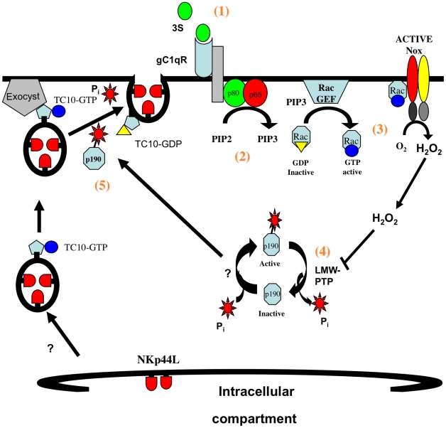
CD4(+) T cell loss is central to HIV pathogenesis. In the initial weeks post-infection, the great majority of dying cells are uninfected CD4(+) T cells. We previously showed that the 3S motif of HIV-1 gp41 induces surface expression of NKp44L, a cellular ligand for an activating NK receptor, on uninfected bystander CD4(+) T cells, rendering them susceptible to autologous NK killing. However, the mechanism of the 3S mediated NKp44L surface expression on CD4(+) T cells remains unknown. Here, using immunoprecipitation, ELISA and blocking antibodies, we demonstrate that the 3S motif of HIV-1 gp41 binds to gC1qR on CD4(+) T cells. We also show that the 3S peptide and two endogenous gC1qR ligands, C1q and HK, each trigger the translocation of pre-existing NKp44L molecules through a signaling cascade that involves sequential activation of PI3K, NADPH oxidase and p190 RhoGAP, and TC10 inactivation. The involvement of PI3K and NADPH oxidase derives from 2D PAGE experiments and the use of PIP3 and H2O2 as well as small molecule inhibitors to respectively induce and inhibit NKp44L surface expression. Using plasmid encoding wild type or mutated form of p190 RhoGAP, we show that 3S mediated NKp44L surface expression on CD4(+) T cells is dependent on p190 RhoGAP. Finally, the role of TC10 in NKp44L surface induction was demonstrated by measuring Rho protein activity following 3S stimulation and using RNA interference. Thus, our results identify gC1qR as a new receptor of HIV-gp41 and demonstrate the signaling cascade it triggers. These findings identify potential mechanisms that new therapeutic strategies could use to prevent the CD4(+) T cell depletion during HIV infection and provide further evidence of a detrimental role played by NK cells in CD4(+) T cell depletion during HIV-1 infection.


Conflict of interest statement

The authors have declared that no competing interests exist.

Figures

Figure 1
Figure 1. 3S peptide stimulation induces ROS production by NADPH oxidase.
(A) Intracellular production of reactive oxygen species (ROS) was measured with ROS-sensitive DCFH-DA. CD4+ T cells were pre-incubated with 10 µM DCFH-DA. Samples were then incubated in the absence of peptide (dotted line), in the presence of 20 µg/ml 3S peptide (thick solid line) or in the presence of 0.2 µg/ml PMA (thin solid line). DCFH-DA fluorescence was then measured by luminometer every 3 min for 66 min. The curves shown are representative of experiments performed on purified CD4+ T cells from 5 individuals. (B) Induction of NKp44L expression by H2O2. CD4+ T cells were stained with IgM isotype control (IgM), or CD4+ T cells that were unstimulated (NA), or that were stimulated with 5 µg/ml 3S peptide (3S) or with 100 µM H2O2 were stained with anti-NKp44L mAb. (C) NAC, an antioxidant, blocks NKp44L surface expression. CD4+ T cells were pre-incubated without inhibitor (open bar) and with 1 mM NAC or with 10 mM NAC (black bars), and then stimulated with the 3S peptide. They were then stained with anti-NKp44L mAb. Unstimulated CD4+ T cells stained with anti-NKp44L mAb (NA) were used as negative controls. (D) DPI, a NADPH oxidase inhibitor, also blocks NKp44L surface expression. CD4+ T cells were pre-incubated without inhibitor (open bar) and with 2 µM DPI, or with 20 µM DPI (black bars), and then stimulated with the 3S peptide, before staining with anti-NKp44L mAb. Unstimulated CD4+ T cells stained with anti-NKp44L mAb (NA) were used as negative controls. For (B), (C) and (D) (Left) The mean +/− SD of NKp44L Mean fluorescence intensity (MFI) from experiments using CD4+ T cells from 3 individuals are indicated. (Right) One representative experiment is illustrated as FACS histograms.
Figure 2
Figure 2. 3S peptide stimulation downregulates Rho activity via p190 RhoGAP A.
(A) Purified CD4+ T cells transfected with deficient p190 RhoGAP A (bold line) or wild-type p190 RhoGAP A (dotted line) were stained with anti-NKp44L antibodies. CD4+ T cells stained with IgM isotype control (light gray) were used as the negative control. (B) Rho activity was measured in lysate of CD4+ T cells incubated with 5 µg/ml 3S peptide for various lengths of time ranging from 1 to 60 min. (C) Western blot using lysate of CD4+ T cells pre-incubated in absence of siRNA (Lane 1) or with control siRNA (Lane 2) or with siRNA against TC10 (Lane 3 & 4). Both Tubulin (top panel) and TC10 (bottom panel) were monitored. (D) TC10 siRNA inhibits NKp44L cell-surface expression. Unstimulated CD4+ T cells stained with IgM isotype control (light gray) or with anti-NKp44L mAb (bold line) were used as negative controls. CD4+ T cells were pre-incubated without siRNA (dark gray), with 1 mM control siRNA (thin solid line), 1 mM of either siRNA against TC10 (dotted lines) before stimulation with the 5 µg/ml 3S peptide. Samples were then stained with anti-NKp44L mAb.
Figure 3
Figure 3. The 3S peptide induces translocation of NKp44L to the cell surface.
(A) NKp44L intracellular localization. CD4+ T cells were permeabilized and stained for NKp44L (red) and the golgi marker, giantin (green) or the ER marker, GRP78 BiP (green). Nuclei were stained using DAPI (blue). (B) & (C) Kinetic study of total and surface NKp44L expression. CD4+ T cells were incubated in absence of peptide (circle), in presence of the 3S peptide (square) or scramble peptide (peptide 1) (triangle) for various length of time (ranging from 0 to 6hrs). Samples were either (B) permeabilized and stained for total NKp44L or (C) stained for surface NKp44L. (D) Cycloheximide does not impede NKp44L surface expression. (Left) CD4+ T cells were pre-incubated without inhibitor (open bar) and with 100 µM Cycloheximide (black bars), and then stimulated with the 3S peptide. They were then stained with anti-NKp44L mAb. Unstimulated CD4+ T cells stained IgM isotype (IgM) or with anti-NKp44L mAb (NA) were used as negative controls. Means +/− SD of NKp44L MFI from 3 experiments using CD4+ T cells from 3 individuals are illustrated. (Right) One representative experiment is illustrated as FACS histograms.
Figure 4
Figure 4. The 3S peptide activates the PI3K.
(A) PIP3 induces cell-surface expression of NKp44L. CD4+ T cells were stained with IgM isotype control (IgM) or CD4+ T cells without stimulation (NA), stimulated with the 5 µg/ml 3S peptide (3S), or 7 µM carrier alone; 7 µM PIP2, or 7 µM PIP3 were then stained with anti-NKp44L mAb. (B) Inhibitors of PI3K block cell-surface expression of NKp44L. CD4+ T cells pre-incubated in the absence of inhibitor (Open bar), with 2 or 20 µM Ly294,002 (Black bars), or with 10 or 100 nM wortmannin (Hatched bars) before stimulation with 5 µg/mL 3S peptide were stained with anti-NKp44L mAb. Unstimulated CD4+ T cells stained with anti-NKp44L mAb (NA) were used as negative controls. (C) Inhibitors of PI3K do not inhibit cell-surface expression of NKp44L following H2O2 treatment. CD4+ T cells pre-incubated in the absence of inhibitor (Open bar), with 20 µM Ly294,002 (Black bars), or with 100 nM wortmannin (Hatched bars), before stimulation with 100 µM H2O2 were stained with anti-NKp44L mAb. Unstimulated CD4+ T cells stained with anti-NKp44L mAb (NA) were used as negative controls. (Left) The mean +/− SD of NKp44L MFI from 3 experiments using CD4+ T cells from 3 individuals is illustrated. (Right) One representative experiment is illustrated as FACS histograms.
Figure 5
Figure 5. gC1qR is the receptor for the 3S motif of HIV-1 gp41.
(A) Immunoprecipitation of gC1qR. Magnetic beads were coated with either BSA or 3S peptide. Coated beads were used to immunoprecipitate the receptor of the 3S motif. Purified proteins were analyzed by Western blot with anti-gC1qR mAbs (74.5.2 and 60.11). Lane 1: Eluate using BSA coated beads; Lane 2: Eluate using 3S coated beads. (B) Interaction of gC1qR with 3S peptide by Elisa. Microplate wells were coated with gC1qR or BSA and incubated with increasing concentrations of biotin-conjugated 3S peptide. Biotin was detected with HRP-conjugated streptavidin. (C) Inhibition of 3S interaction by anti-gC1qR mAbs. CD4+ T cells were pretreated with 10 µg/ml anti-gC1qR mAb (74.5.2 or 60.11) or IgG1 isotype control, before incubation with 16µg FITC-conjugated 3S peptide. CD4+ T cells incubated with 16µg FITC-conjugated scramble peptide (peptide 1) was used as negative control. (D, E) Inhibition of 3S-dependent and HIV-mediated NKp44L stimulation by anti-gC1qR mAbs. CD4+ T cells pre-incubated in the absence of antibodies (dark gray), with 10 ug/ml mouse IgG1 (thin solid line), or 10 µg/ml anti-gC1qR 74.5.2 clone (dotted line) before stimulation during 4 hr with (D) 5 µg/mL 3S peptide or (E) wild type HIV virus (NL4,3), were stained with anti-NKp44L mAb. As controls, unstimulated CD4+ T cells were stained with IgM isotype control (light gray) or anti-NKp44L antibodies (bold line). (F) Induction of NKp44L surface expression by the natural ligand of gC1qR. CD4+ T cells, either unstimulated (NA), or stimulated with 5 µg/ml 3S peptide (3S), 10 µg/ml C1q (C1q) or 10 µg/ml HK (HK), were stained with anti-NKp44L mAb. As control, CD4+ T cells were stained with IgM isotype control (IgM). (Left) The mean +/− SD of NKp44L MFI from 3 experiments using CD4+ T cells from 3 individuals is illustrated. (Right) One representative experiment is illustrated as FACS histograms.
Figure 6
Figure 6. Model of 3S mediated NKp44L translocation on CD4+ T cells.
1) The 3S motif of HIV-gp41 binds to its receptor, gC1qR; 2) upon binding gC1qR activates PI3K; 3) activated PI3K indirectly induces H2O2 production by NADPH-oxidase; 4) H2O2 produced promotes p190 RhoGAP-A activity; and 5) activated p190 RhoGAP-A then induces GTP hydrolysis by Rho-A and TC10, which are implicated in exocytosis of NKp44L bearing vesicles.

References

    1. Alimonti JB, Ball TB, Fowke KR. Mechanisms of CD4+ T lymphocyte cell death in human immunodeficiency virus infection and AIDS. J Gen Virol. 2003;84:1649–1661. - PubMed
    1. Stevenson M. HIV-1 pathogenesis. Nat Med. 2003;9:853–860. - PubMed
    1. Finkel TH, Tudor-Williams G, Banda NK, Cotton MF, Curiel T, et al. Apoptosis occurs predominantly in bystander cells and not in productively infected cells of HIV- and SIV-infected lymph nodes. Nat Med. 1995;1:129–134. - PubMed
    1. Varbanov M, Espert L, Biard-Piechaczyk M. Mechanisms of CD4 T-cell depletion triggered by HIV-1 viral proteins. AIDS Rev. 2006;8:221–236. - PubMed
    1. Kaplan D, Sieg S. Role of the Fas/Fas ligand apoptotic pathway in human immunodeficiency virus type 1 disease. J Virol. 1998;72:6279–6282. - PMC - PubMed

Publication types

MeSH terms