. 2010 Jul;13(4):211-8.
doi: 10.1111/j.1463-5224.2010.00782.x.

Efficacy and safety of mitomycin C as an agent to treat corneal scarring in horses using an in vitro model

Affiliations

Efficacy and safety of mitomycin C as an agent to treat corneal scarring in horses using an in vitro model

Dylan G Buss et al. Vet Ophthalmol. 2010 Jul.

Abstract

Objective: Mitomycin C (MMC) is used clinically to treat corneal scarring in human patients. We investigated the safety and efficacy of MMC to treat corneal scarring in horses by examining its effects at the early and late stages of disease using an in vitro model.

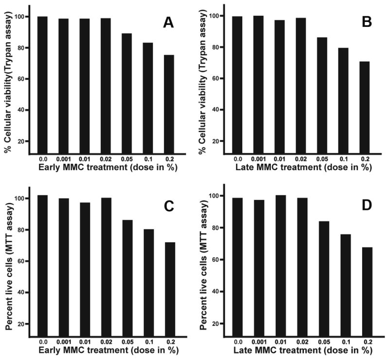
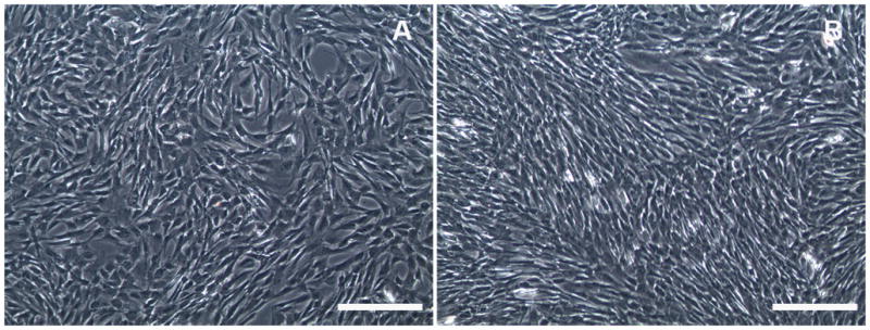
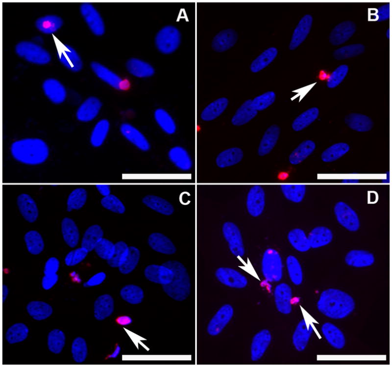
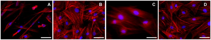
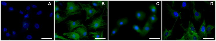
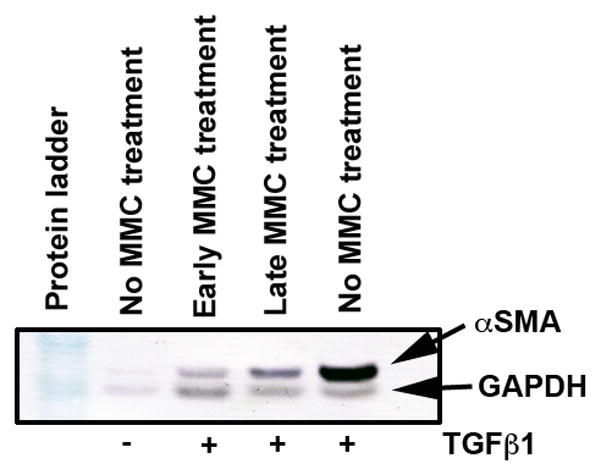
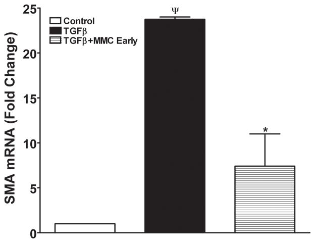
Procedure: An in vitro model of equine corneal fibroblast (ECF) developed was used. The ECF or myofibroblast cultures were produced by growing primary ECF in the presence or absence of transforming growth factor beta-1 (TGFbeta1) under serum-free conditions. The MMC dose for the equine cornea was defined with dose-dependent trypan blue exclusion and (3-4,5-dimethylthiazol-2-yl)-2,5-diphenyltetrazolium bromide (MTT) assays after applying MMC to the cultures once for 2 min. The efficacy of MMC to control corneal scarring in horses was determined by measuring mRNA and protein expression of corneal scarring markers (alpha-smooth muscle actin and F-actin) with western blotting, immunocytochemistry and/or quantitative real-time polymerase chain reactions.

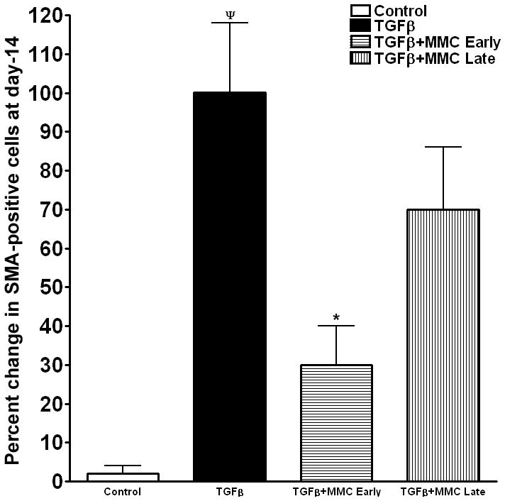
Results: A single 2-min treatment of 0.02% or less MMC did not alter ECF phenotype, viability, or cellular proliferation whereas 0.05% or higher MMC doses showed mild-to-moderate cellular toxicity. The TGFbeta1 at 1 ng/mL showed significant myofibroblast formation in ECF under serum-free conditions. A single 2-min, 0.02% MMC treatment 24 h (early) after TGFbeta1 stimulation significantly reduced conversion of ECF to myofibroblasts, however, a single 0.02% MMC treatment 11 days after TGFbeta1 stimulation showed moderate myofibroblast inhibition.

Conclusions: That MMC safely and effectively reduced scarring in ECF by reducing the degree of transdifferentiation of corneal fibroblasts to myofibroblasts in vitro. Further clinical in vivo investigations are warranted using MMC in horses.


Figures

Figure 1
Figure 1
Dose-dependent effect of early and late MMC treatment to ECF. Panels A and B show data collected with trypan blue assay and panels C and D show MTT assay data. The doses of MMC (0.02% or less) did not alter ECF viability whereas MMC doses higher than 0.02% showed mild-to-moderate toxicity.
Figure 2
Figure 2
Phase-contrast light microscopy images of ECF. No morphological changes to ECF were observed when 0.02% of MMC early (A) or late (B) treatment was performed, thus establishing MMC safety to ECF in-vitro. Bar = 100μm
Figure 3
Figure 3
TUNEL assay detecting apoptosis induced by 0.02% MMC in ECF. Panel A demonstrates the early control of no MMC treatment, panel B shows effects of early MMC treatment, panel C is late control (no MMC) and panel D shows effects of late MMC treatment on ECF. TUNEL-positive cells are stained in red and nuclei in blue with DAPI. Arrows denote TUNEL-positive cells. Magnification 400x. Bar = 100μm
Figure 4
Figure 4
Immunocytochemical staining with phalloidin showing levels of F-actin in ECF control group (A; no MMC treatment), in the presence of TGFβ1 (B), early MMC treatment group (C) and late MMC treatment group (D). F-actin stained cells are red and nuclei are stained blue with DAPI. Magnification 400x. Bar = 100μm
Figure 5
Figure 5
Immunocytochemical staining of α-SMA (green) showing anti-fibrotic effects of MMC on ECF. The TGFβ1 untreated cultures did not show any α-SMA (A) while the TGFβ1 treated cultures showed significant α-SMA staining (B). Early MMC treatment (C) showed significant inhibition in TGFβ1-induced myofibroblast formation in ECF whereas late MMC treatment showed moderate inhibition (D). The DAPI stained nuclei are shown in blue. Magnification 400x. Bar = 100μm
Figure 6
Figure 6
Quantification of α-SMA-stained cells in ECF cultures treated with or without MMC. Myofibroblast formation was significantly induced in the presence of TGFβ1. Early MMC treatment significantly decreased TGFβ1-induced myofibroblast formation in ECF compared to untreated controls, whereas late MMC treatment showed moderate reduction. ¬denotes p <0.01 (Con vs TGFβ1) and * represents p<0.01 (TGFβ1 vs TGFβ1+MMC).
Figure 7
Figure 7
Western blot analysis showing quantitative measurement of α-SMA (myofibroblast marker) in ECF treated with or without TGFβ1 (1ng/ml) and 0.02% MMC. Equal quantity of protein (50 μg) was loaded in each lane. GAPDH was used as house keeping gene. A single early MMC treatment showed significant decrease in TGFβ1-induced myofibroblast formation in ECF compared to untreated controls. The late MMC treatment showed moderate reduction in α-SMA.
Figure 8
Figure 8
Quantitative real-time PCR reactions showing measurements of α-SMA in MMC treated or untreated ECF. TGFβ1 exposure induced significant α-SMA formation in ECF and early MMC treatment significantly decreased TGFβ1-induced myofibroblast formation in ECF. Late MMC treatment showed moderate decrease in α-SMA RNA (data not shown). ¬indicates p <0.01 (Con vs TGFβ1) and * represents p<0.01 (TGFβ1 vs TGFβ1+MMC early).

References

    1. Haber M, Cao Z, Panjwani N, et al. Effects of growth factors (EGF, PDGF-BB and TGF-beta 1) on cultured equine epithelial cells and keratocytes: implications for wound healing. Veterinary Ophthalmology. 2003;6:211–217. - PubMed
    1. Michau TM, Schwabenton B, Davidson MG, et al. Superficial, nonhealing corneal ulcers in horses: 23 cases (1989–2003) Veterinary Ophthalmology. 2003;6:291–297. - PubMed
    1. Nasisse MP, Nelms S. Equine ulcerative keratitis. Vet Clinics North America Equine Practice. 1992;8:537–555. - PubMed
    1. Buss DG, Giuliano EA, Sharma A, et al. Isolation and cultivation of equine corneal keratocytes, fibroblasts and myofibroblasts. Veterinary Ophthalmology. 2010;13:37–42. - PMC - PubMed
    1. Buss DG, Sharma A, Giuliano EA, et al. Gene delivery in the equine cornea: a novel therapeutic strategy. American Journal of Veterinary Research. 2009 In Press. - PMC - PubMed

Publication types

MeSH terms