. 2011 Jan;138(1):35-45.
doi: 10.1017/S0031182010000922. Epub 2010 Jul 12.

Low levels of transforming growth factor-beta (TGF-beta) and reduced suppression of Th2-mediated inflammation in hyperreactive human onchocerciasis

Affiliations
Free PMC article

Low levels of transforming growth factor-beta (TGF-beta) and reduced suppression of Th2-mediated inflammation in hyperreactive human onchocerciasis

S Korten et al. Parasitology. 2011 Jan.
Free PMC article

Abstract

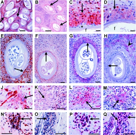
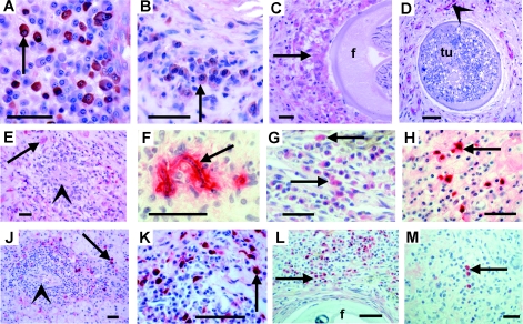
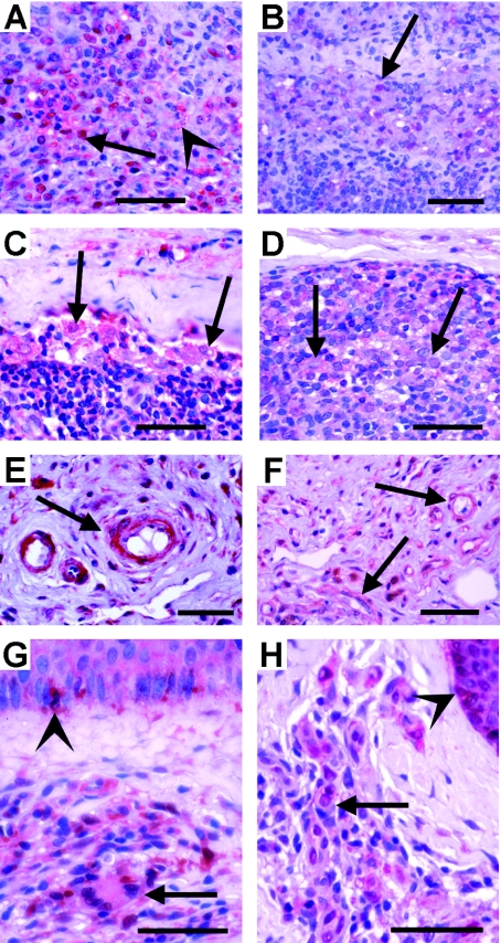
Th2-biased inflammation with eosinophilia and IgE production is a hallmark of helminth infections. It is pronounced in hyperreactive onchocerciasis patients ('sowda' or 'local form'), who efficiently kill microfilariae resulting in severe dermatitis and lymphadenitis. In contrast, hyporeactive patients ('generalised form') tolerate high microfilarial loads. This is thought to be mediated by regulatory CD4+ T cells and macrophages producing suppressive cytokines such as IL-10 and transforming growth factor-beta (TGF-β). We investigated whether hyperreactivity was reflected by lower local TGF-β production, analysing stable latent TGF-β1 expression in onchocercomas, lymph nodes and skin from hyperreactive and hyporeactive patients by immunohistochemistry. TGF-β expression was compared with that of IgE, IgG1, IgG4, and the antigen-presenting, CD4+ T cell-inducing MHC class II molecule HLA-DR. TGF-β was weakly and less frequently expressed by various cell types in onchocercomas, skin and lymph nodes from hyperreactive compared to hyporeactive patients. This applied to reactions around living and dead adult worms as well as dead microfilariae. Antigen-presenting cells strongly expressed HLA-DR in both forms, but their numbers were reduced in hyperreactive nodules. Plasma cells produced more IgE and IgG1, but less of the anti-inflammatory antibody IgG4 in hyperreactive onchocercomas. In conclusion, hyperreactivity is linked with reduced local expression of TGF-β, HLA-DR and IgG4, which might contribute to the insufficient down-regulation of inflammation via TGF-β- and HLA-DR-induced regulatory lymphocytes.


Figures

Fig. 1
Fig. 1
Immunohistological comparison of TGF-β and HLA-DR expression by macrophages and CD3+ T cells in onchocercomas from untreated hyporeactive and hyperreactive patients in Liberia and Ghana. (A-B) Nodule centre with strong (hyporeactive) and weak (hyperreactive) TGF-β expression (arrows) around older female worms. (C-D) Details of TGF-βhigh (hyporeactive) and TGF-βlow (hyperreactive) nuclei and cytoplasm of macrophages (arrow) adjacent to female worms (f), while a small giant cell, attached to the cuticle, is negative (*). (E-H) Serial sections with strong (hyporeactive) and weak or negative (hyperreactive) TGF-β (E-F) and HLA-DR (G-H) expression (arrows) mainly in macrophages adjacent to productive female worms and an attached HLA-DR+ giant cell (*). Immune cells further distant from the worm are HLA-DR+ (arrowhead in H), whereas they are TGF-β-positive (F) and macrophages adjacent to the worm are HLA-DR-negative (H). (J-M) Moribund or dead mf in a nodular microgranuloma being attacked by macrophages strongly (hyporeactive) or faintly (hyperreactive) expressing TGF-β or HLA-DR (arrows). (K+M) are serial sections. (N-Q) Mainly CD3+ T lymphocytes express TGF-β strongly in a nodule from a hyporeactive and weakly in a nodule from a hyperreactive patient. (N+P and O+Q) are serial sections. (P+Q) CD3-staining. Scale bars=50 µm.
Fig. 2
Fig. 2
Immunohistological comparison of TGF-β and immunoglobulin production by plasma cells and macrophages in onchocercomas from hypo- and hyperreactive patients in Liberia and Uganda. (A-B) Plasma cells (arrow) strongly (hyporeactive) and faintly (hyperreactive) expressing TGF-β. Untreated patients. (C) Weakly TGF-β+ macrophages (arrows) attached to a productive female worm (f) 2 months after IVM. Hyperreactive. (D) Only TGF-β+ endothelia (arrowhead) are seen but nearly no immune cells near to a moribund female worm harbouring a pleomorphic neoplasm (tu) filling the entire pseudocoeloma cavity (Duke et al. ; Brattig et al. 2010). Hyperreactive. (E) Weakly TGF-β+ macrophages (arrow) around a microgranuloma (arrowhead) with a dead mf in a nodule from a hyperreactive patient 2 months after IVM treatment. (F) A dead mf coated by IgG1 (arrow) in a nodule from a hyperreactive German child after IVM. (G-H) Weakly (hyporeactive, 48 h after IVM) and strongly (hyperreactive, untreated) IgG1 producing plasma cells (arrows). TGF-β (J-K) or HLA-DR (L-M) (arrows) faintly (hyporeactive) and strongly (hyperreactive) IgE producing plasma cells (arrows) in perivascular infiltrates. Untreated patients. (L-M) Many (hyporeactive) and few (hyperreactive) IgG4 producing plasma cells (arrows) in nodules with productive female worms (f); 2–3 days after IVM treatment. Scale bars=20 µm (A, B, E); 50 µm (C-D, F-M).
Fig. 3
Fig. 3
Immunohistological comparison of TGF-β expression in lymph nodes (A-F) and skin (G-H) from hypo- and hyperreactive patients in Liberia and Uganda. (A-B) Strong (hyporeactive) and faint TGF-β expression (hyperreactive) in lymphocytes (arrows) adjacent to remnants of nearly resorbed mf (arrowhead). Untreated patients. (C-D) Strong (hyporeactive) and faint TGF-β expression (hyperreactive) in macrophages (arrows); 1 day or 5 weeks after IVM treatment. (E-F) Strongly (hyporeactive) TGF-β+ and faintly (hyperreactive) TGF-β expressing endothelia of blood vessels; 1 day or 5 weeks after IVM treatment. (G-H) Skin from untreated patients. (G) Macrophages and lymphocytes with varying degrees of TGF-β expression (arrow) accumulating in a dermal infiltrate and strongly TGF-β+ Langerhans cells in the epidermis (arrowhead; hyporeactive). (H) A similar accumulation of less strongly TGF-β expressing cells in a dermal infiltrate and of Langerhans cells in the epidermis (arrowhead) of a hyperreactive patient. Scale bars=50 µm.

References

    1. Aalberse R. C., Stapel S. O., Schuurman J., Rispens T.. Immunoglobulin G4: an odd antibody. Clinical and Experimental Allergy. 2009;39:469–477. - PubMed
    1. Albiez E. J. Büttner D. W. Duke B. O. 1988aDiagnosis and extirpation of nodules in human onchocerciasis Tropical Medicine and Parasitology 39331–346. - PubMed
    1. Albiez E. J., Gallin M., Erttmann K. D., Racz P., Büttner D. W.. Characteristics of chronic severe onchodermatitis in Liberia. Journal of the Liberian Medical and Dental Association. 1985;15:120–125.
    1. Albiez E. J. Walter G. Kaiser A. Ranque P. Newland H. S. White A. T. Greene B. M. Taylor H. R. Büttner D. W. 1988bHistological examination of onchocercomata after therapy with ivermectin Tropical Medicine and Parasitology 3993–99. - PubMed
    1. Battaglia M., Roncarolo M. G.. The fate of human Treg cells. Immunity. 2009;30:763–765. - PubMed

Publication types