Review
. 2010 Jun;20(3):153-60.
doi: 10.1016/j.semcancer.2010.06.005. Epub 2010 Jul 7.

Tumorigenic and adhesive properties of heparanase

Affiliations
Review

Tumorigenic and adhesive properties of heparanase

Flonia Levy-Adam et al. Semin Cancer Biol. 2010 Jun.

Abstract

Heparanase is an endo-β-glucuronidase that cleaves heparan sulfate side chains presumably at sites of low sulfation, activity that is strongly implicated with cell invasion associated with cancer metastasis, a consequence of structural modification that loosens the extracellular matrix barrier. In addition, heparanase exerts pro-adhesive properties, mediated by clustering of membrane heparan sulfate proteoglycans (i.e., syndecans) and activation of signaling molecules such as Akt, Src, EGFR, and Rac in a heparan sulfate-dependent and -independent manner. Activation of signaling cascades by enzymatically inactive heparanase and by a peptide corresponding to its substrate binding domain not only increases cell adhesion but also facilitates cancer cell growth. This notion is supported by preclinical and clinical settings, encouraging the development of anti-heparanase therapeutics. Here, we summarize recent progress in heparanase research emphasizing the molecular mechanisms that govern its pro-tumorigenic and pro-adhesive properties. Pro-adhesive properties of the heparanase homolog, heparanase 2 (Hpa2), are also discussed. Enzymatic activity-independent function of proteases (i.e., matrix metalloproteinases) is discussed in the context of cell adhesion and tumor progression. Collectively, these examples suggest that enzyme function exceeds beyond the enzymatic aspect, thus significantly expanding the scope of the functional proteome. Cross-talk with matrix metalloproteinases and the role of heparanase in pathological settings other than cancer are also described.


Conflict of interest statement

Conflict of interest: The authors declare that there are no conflicts of interest

Figures

Figure 1
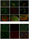
Figure 1. Heparanase stimulates syndecan clustering, Rac1 activation, and cell spreading
A. Syndecan-1 clustering and cell spreading. U87 glioma cells were serum starved for 24 h and were then incubated with control scrambled peptide (Scr; upper panel, left), KKDC peptide (50 μM, upper panel, right) or heparanase (Hepa; 1 μg/ml; upper panel, middle) for 1 h. Cells were then gently washed, fixed with cold methanol and subjected to fluorescent staining with anti-syndecan-4 antibody (green) merged with nuclear staining (red; upper panels). Cells were similarly stained with anti-syndecan-4 (green) and anti-heparanase (red) antibodies, merged with nuclear staining (blue; second panels). Co-localization of syndecan and heparanase in endocytic vesicles appears yellow. Cell spreading. Human fibroblasts were plated on the 110 kDa fibronectin-like protein and incubated with heparanase (Hepa; 1μg/ml) or the KKDC/Scr peptides (50 μM) for 1 h. Cells were then fixed with 4% paraformaldehyde and double stained with phalloidin-TRITC (red) and anti-vinculin (green) antibody (lower panels). Cells treated with heparanase or with the KKDC peptide appeared spread and formed more focal adhesions. B. Hpa2c clusters syndecan but fails to get internalized. Cal-27 tongue carcinoma cells were left untreated (Con, upper panels) or incubated with Hpa2c (second panels) protein for 30 min. Cells were then fixed and subjected to immunofluorescent staining applying anti-syndecan-1 (left panels, green) and anti-Hpa2 (Ab58, red). Merge images are shown in the right panels together with nuclear staining (blue). Note, that Hpa2 clusters syndecan-1 but fails to get internalized. C. Rac1 activation. U87 cells were kept in serum-free medium for 24 h and were then stimulated with the KKDC/Scr peptides (50 μM), wild type (Hepa; 1 μg/ml) or double mutated (DM; Glu225, Glu343) heparanase proteins for 30 min. Cell lysates were then subjected to pull-down with GST-PAK-agarose beads for detection of active Rac1 (upper panel) and subjected to immunoblotting with anti-Rac1 antibody (lower panel) (32). D. Hpa2c exerts high affinity to heparin/HS. HEK293 cells stably transfected with Myc-tagged heparanase or Hpa2c gene constructs were incubated without (0) or with the indicated concentration of heparin, heparan sulfate (HS), or hyaluronic acid (HA) and the conditioned medium was subjected to immunoblotting applying anti-Myc antibody.
Figure 2
Figure 2. HS-dependent and -independent function of heparanase
A. Clustering of syndecan family members, and possibly glypicans, by the KKDC peptide dimer or the two heparin binding domains of heparanase facilitates cell-adhesion and cell-spreading [32, 68]. Enhanced cell adhesion and spreading is mediated by the recruitment and activation of PKCα, Rac1, and Src. B. Heparanase is also thought to interact with heparanase-binding cell surface protein/receptor(s), leading to HS-independent Akt, p38, and Src activation. This results in enhanced transcription of genes such as vascular endothelial growth factor (VEGF-A, VEGF-C) [82, 105], tissue factor (TF) [81], and Cox2 [106], and further contributes to cell adhesion, spreading and motility. Src activation, in turn, phosphorylates a number of substrates including the EGFR, leading to increased cell proliferation and tumorigenesis [47, 83].

References

    1. Yilmaz M, Christofori G. EMT, the cytoskeleton, and cancer cell invasion. Cancer Metastasis Rev. 2009;28:15–33. - PubMed
    1. Pillay V, Dass CR, Choong PF. The urokinase plasminogen activator receptor as a gene therapy target for cancer. Trends Biotechnol. 2007;25:33–9. - PubMed
    1. Mohamed MM, Sloane BF. Cysteine cathepsins: multifunctional enzymes in cancer. Nat Rev Cancer. 2006;6:764–75. - PubMed
    1. Rowe RG, Weiss SJ. Breaching the basement membrane: who, when and how? Trends Cell Biol. 2008;18:560–74. - PubMed
    1. Bernfield M, Gotte M, Park PW, Reizes O, Fitzgerald ML, Lincecum J, et al. Functions of cell surface heparan sulfate proteoglycans. Annu Rev Biochem. 1999;68:729–77. - PubMed

Publication types