. 2010 Jul 23;142(2):256-69.
doi: 10.1016/j.cell.2010.06.006. Epub 2010 Jul 8.

A surveillance pathway monitors the fitness of the endoplasmic reticulum to control its inheritance

Affiliations

A surveillance pathway monitors the fitness of the endoplasmic reticulum to control its inheritance

Anna Babour et al. Cell. .

Abstract

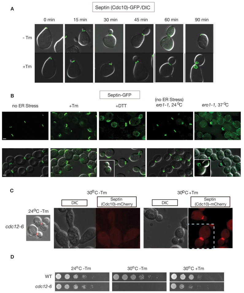
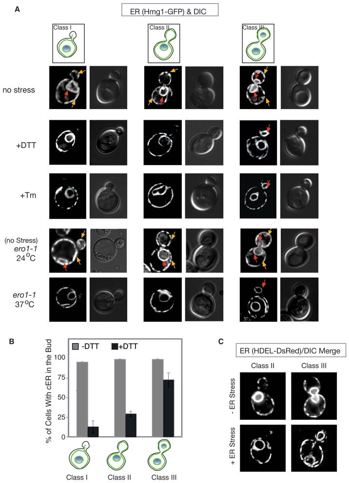
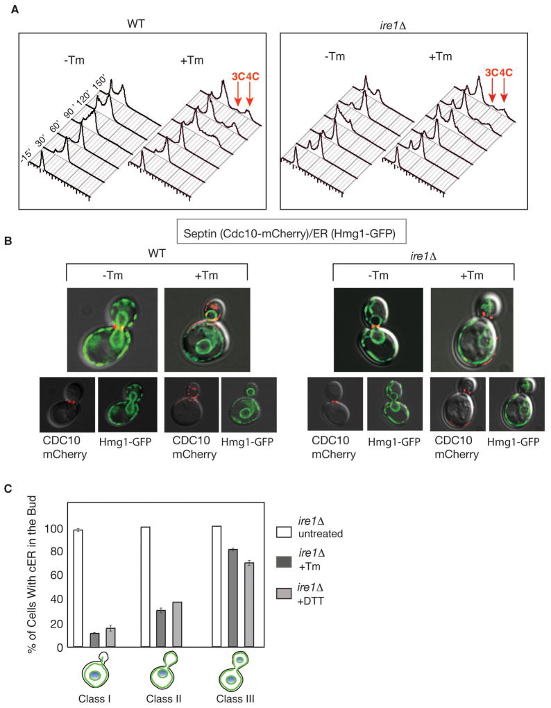
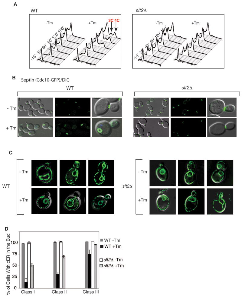
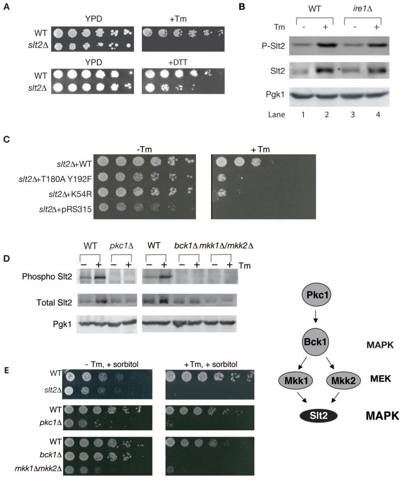
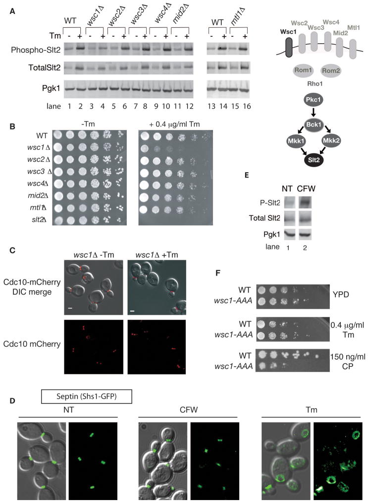
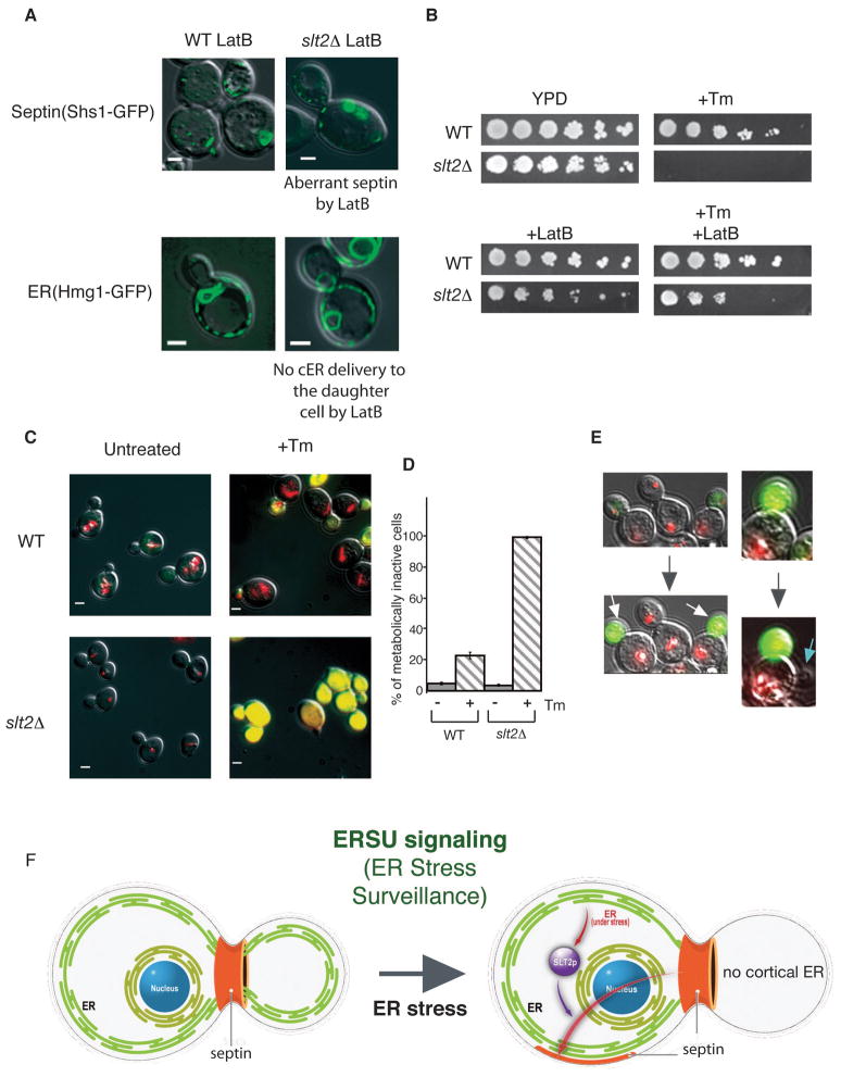
The endoplasmic reticulum (ER) plays an essential role in the production of lipids and secretory proteins. Because the ER cannot be generated de novo, it must be faithfully transmitted or divided at each cell division. Little is known of how cells monitor the functionality of the ER during the cell cycle or how this regulates inheritance. We report here that ER stress in S. cerevisiae activates the MAP kinase Slt2 in a new ER stress surveillance (ERSU) pathway, independent of the unfolded protein response. Upon ER stress, ERSU alters the septin complex to delay ER inheritance and cytokinesis. In the absence of Slt2 kinase, the stressed ER is transmitted to the daughter cell, causing the death of both mother and daughter cells. Furthermore, Slt2 is activated via the cell surface receptor Wsc1 by a previously undescribed mechanism. We conclude that the ERSU pathway ensures inheritance of a functional ER.


Figures

Figure 1
Figure 1. ER stress alters the localization and morphology of the septin ring
(A) Septin ring morphology during normal growth (−Tm) and under ER stress (+Tm). Upon release, α-factor synchronized cells expressing genomically GFP-tagged CDC10 (Jimenez et al., 1998)) were treated with or without tunicamycin (Tm) for the indicated amount of time (Bicknell et al., 2007). (B) Genomic loci encoding septin subunits, either SHS1 or CDC11, were C-terminally tagged with GFP. ER stress was induced in asynchronous populations of WT cells with 1 μg/ml Tm (3 hrs) or 2 mM DTT (3 hrs), or in ero1-1 cells by shifting from the permissive temperature (24°C) to the non-permissive temperature (37°C) for 3 hrs. Note that similar conditions were used for ER stress induction throughout this study unless otherwise stated. GFP alone (upper panels) and GFP merged with a DIC image (lower panel) are shown. Bars, 2 μm. (C) (D) Septin morphology in cdc12-6 cells carrying CDC10-mCherry in YPD or YPD+Tm at the indicated temperature. Upon shift to the non-permissive temperature without addition of Tm in the growth media (30°C, 3 hrs, −Tm), cells showed loss of normal septin structure (dispersed) and abnormal elongated morphology. However, addition of Tm (1 μg/ml, 3 hrs at 30°C) caused recovery of the septin ring and normal cell morphology to majorities of cdc12-6 cells, consistent with the partial recovery of cell growth with Tm at 30°C as shown in (D). cdc12-6 cells were not able to sustain their growth at 30°C without Tm. Cells were spotted on YPD or YPD+Tm (0.2 μg/ml) plates after five-fold serial dilutions and incubated at the indicated temperature for 3 days. See also Figure S1.
Figure 2
Figure 2. ER stress induces a delay in cortical ER (cER) inheritance
(A) ER stress was induced (by Tm, DTT, or ero1-1) in cells expressing the ER membrane marker, Hmg1-GFP, for examination by fluorescence microscopy. Three categories of cells are depicted for each condition and as described in Results section. Yellow arrow shows cER, while red arrow represents perinuclear ER. (B) For each of the three classes of cells, the number of cells containing elements of cER in the daughter cell was counted (n=300) during growth under normal conditions (grey bars) or in the presence of 2 mM DTT (3 hrs) (black bars). The average of 3 independent experiments is depicted; error bars represent standard deviation (SD). (C) ER visualized by a HDEL-DsRed ER reporter confirmed that cER inheritance was delayed when ER stress was induced by Tm. Images shown are HDEL-DsRed alone. See also Figure S2.
Figure 3
Figure 3. The UPR does not signal septin stabilization, cytokinesis block or ER inheritance delay during ER stress
(A) DNA content of α-factor synchronized wild-type and ire1Δ cells were monitored by FACS upon Tm treatment as described in experimental procedures. After 90 minutes, both cell types showed populations of cells with 3C or 4C DNA content (red arrows), indicating a cytokinesis delay (Bicknell et al., 2007). (B) Cortical cER inheritance (visualized by Hmg1-GFP) and septin morphology (by Cdc10-mCherry) were perturbed in both WT and ire1Δ cells when grown in Tm. DIC pictures merged with either Cdc10-mCherry or Hmg1-GFP are shown for both WT and ire1Δ cells. (C) Quantitation of ire1Δ cells with cER in the bud under ER stress inducing conditions (n=300 for each class, under normal conditions (white bars) or in the presence of 1 μg/ml Tm (dark gray bars) and 2 mM DTT (light gray bars). Error bars represent standard deviation (SD) of three independent experiments.
Figure 4
Figure 4. Slt2 MAP kinase mediates septin alterations, cytokinesis delay, and ER inheritance delay during ER stress
(A) ER stress did not induce a cytokinesis delay in slt2Δ cells, shown by no slt2Δ cells with 3C or 4C DNA content FACS peaks, in contrast to ER stressed wild-type cells (red arrows). Experimental conditions were as described in Figure 3A. (B) Unlike in ER-stressed WT cells, the septin ring remained at a normal position in ER-stressed slt2Δ cells. Septin was visualized by Cdc10-GFP in WT (left) and slt2Δ cells (right) grown with or without 1 μg/ml Tm, as indicated. Additional genes tested are shown in Supplemental Table 1. (C) cER entered the bud of slt2Δ cells treated with Tm (1 μg/ml) as visualized by Hmg1-GFP. (D) Quantitation of wild-type and slt2Δ cells with elements of cER in the daughter cell under normal (−Tm) or ER stress (+Tm) (1 μg/ml) conditions in each class (n=300). The average of three independent experiments is depicted; error bars represent SD. See also Figure S3.
Figure 5
Figure 5. Slt2p phosphorylation and kinase activity are essential for ER stress surveillance
(A) slt2Δ cells were not able to sustain growth on a plate containing Tm or DTT. Fivefold serial dilutions of WT and slt2Δ cells were spotted on plates containing no drug (YPD) or +Tm (0.1 μg/ml), or +DTT (4 mM). (B) Slt2 MAP kinase was phosphorylated upon ER stress in WT (lanes 1&2) and ire1Δ cells (lanes 3&4). Total cell lysate of either WT or ire1Δ cells with (+) or without (−) Tm treatment were blotted with anti-phospho specific Slt2, total Slt2, or Pgk1 (loading control) antibodies. In addition to increase in phosphorylation of Slt2, total protein level was also increased, although ER stress-induced phosphorylation does not require increase in Slt2 protein levels (see Figure S3B). (C) slt2Δ cells were transformed with empty plasmid, or plasmid containing WT SLT2, T180AY192F slt2, or K54R slt2. Cells were grown to log phase in synthetic complete (SC)-leu medium, diluted serially 5-fold, and spotted onto plates with or without 0.1 μg/ml Tm. (D) pkc1Δ and corresponding WT cells were grown in the presence of 1M sorbitol, while bck1Δ, mkk1Δ /mkk2Δ, and corresponding WT cells were grown in the absence of sorbitol. (E) Cells of the indicated genotype were grown to log phase in YPD + 1M sorbitol, diluted serially 5 fold, and spotted onto 0.2 μg/ml Tm or no Tm plates containing 1M sorbitol. Sorbitol had no effect on sensitivity of slt2Δ on Tm plate (compare Figures 5E vs 5A & 6B), revealing lack of the CWI pathway involvement. See also Figure S4.
Figure 6
Figure 6. The ERSU pathway is activated by Wsc1 and is independent of the cell wall integrity and arrest of secretion response pathways
(A) WSC1 is required for phosphorylation of Slt2 upon ER stress. Tm treated (+Tm) or untreated (−Tm) cells of indicated genotypes were collected and analyzed by western blot for Slt2 phosphorylation, total Slt2, and Pgk1. (B) wsc1Δ cells did not support growth on a YPD plate containing 0.4 μg/ml Tm. Fivefold serial dilutions of the indicated mutant cells were grown on medium with and without Tm. (C) In the absence of WSC1, septin rings (Cdc10-mCherry reporter) did not show altered morphology upon ER stress induced by Tm (1 μg/ml for 3 hrs), but were only observed at the bud neck (a similar septin phenotype as what was seen in slt2Δ cells). (D) Not all stresses that induce Slt2 activation affect septin ring dynamics. Wild-type cells expressing Shs1-GFP were visualized after 2 hours of treatment with 10μg/ml calcofluor white (CFW) or 1 μg/ml Tm for 3 hrs. NT, no treatment. GFP alone, and GFP merged with a DIC image are shown. (E) Samples from the experiment described in (D) were collected and analyzed by western blot for Slt2 phosphorylation. (F) Endocytosis of Wsc1 does not play a role in the Wsc1 function in ERSU signaling, although it is known to be involved in the cell wall integrity (CWI) pathway. Five-fold serial dilutions of WT cells, and cells with the genomic copy of WSC1 replaced by the endocytosis mutant wsc1-AAA (Piao et al., 2007) were spotted onto plates with and without Tm (0.4 μg/ml) and the CWI pathway activator, caspofungin (CP) (150 ng/ml). Growth was monitored after 2 days. See also Figure S5.
Figure 7
Figure 7. ERSU signaling protects mother cells during ER stress
(A) The effect of Latrunculin B (LatB) on ER and septin distribution was visualized with the septin reporter Shs1-GFP and the ER reporter Hmg1-GFP in wild-type (left) or slt2Δ cells (right) treated with 1 μg/ml Tm & 400 μM LatB for 2 hrs. In both cell types, septin morphology was altered and cER inheritance was inhibited by the presence of LatB. (B) Lat B rescues growth of slt2Δ cells during ER stress. Five-fold serial dilutions of WT and slt2Δ cells were spotted on plates containing no drug (YPD), +Tm (0.1 μg/ml) alone, +LatB alone (6 μM) or +Tm (0.1 μg/ml)/+LatB (6 μM). See also Figure S6. (C) & (D) Untreated and Tm treated WT and slt2Δ cells were stained with FUN-1. Upon ER stress induction by Tm treatment (+Tm) (1 μg/ml, 3 hrs), the daughter cells showed yellow staining, while the larger mother cells showed intravacuolar red rod-like structures (CIVSs), indicating that they were metabolically active. In contrast, slt2Δ cells following Tm treatment were stained yellow for both mother and daughter cells. Untreated WT and slt2Δ cells showed red CIVSs. Quantitation of yellow metabolically inactive cells is shown in (C). For WT and slt2Δ cells, the numbers of cells with yellow staining were counted. (E) Continuous observation of mother and daughter cell fate during ER stress. Following staining with Fun-1, Tm treated cells (1 μg/ml for 30 min) were spread onto a gelatine-YPD pad containing 0,1 μg/ml Tm for microscopic observation. Initially both mother and daughter cells were alive (stained red), although the daughter cell (white arrow) became metabolically inactive. Furthermore, a blue arrow indicates a new daughter cell started to emerge from the original mother cell, showing asymmetric protection of the mother cell. See also Figure S7. (F) During the cell cycle, the functional capacity of the ER is under surveillance and when ER stress is detected, ERSU activates Slt2, leading to septin re-organization and alteration so that the aberrant septin ring (shown in orange) is found in unusual places and is no longer at the bud neck. Concomitantly, ER inheritance, and cytokinesis are delayed. Thus, ERSU plays a critical role in correct transmission of a functional ER into the progeny cell.

References

    1. Bertin A, McMurray MA, Grob P, Park SS, Garcia G, III, Patanwala I, Ng H-l, Alber T, Thorner J, Nogales E. Saccharomyces cerevisiae septins: Supramolecular organization of heterooligomers and the mechanism of filament assembly. Proceedings of the National Academy of Sciences of the United States of America. 2008;105:8274–8279. - PMC - PubMed
    1. Bicknell AA, Babour A, Federovitch CM, Niwa M. A novel role in cytokinesis reveals a housekeeping function for the unfolded protein response. J Cell Biol. 2007;177:1017–1027. - PMC - PubMed
    1. Bicknell AA, Niwa M. Regulating Endoplasmic Reticulum Function through the Unfolded Protein Response. Handbook of Cell Signaling. 2009:2511–2525.
    1. Bonilla M, Cunningham KW. Mitogen-activated protein kinase stimulation of Ca(2+) signaling is required for survival of endoplasmic reticulum stress in yeast. Mol Biol Cell. 2003;14:4296–4305. - PMC - PubMed
    1. Bukau B, Weissman J, Horwich A. Molecular Chaperones and Protein Quality Control. Cell. 2006;123:443–551. - PubMed

Publication types

MeSH terms

Substances