. 2010 Nov;1803(11):1287-97.
doi: 10.1016/j.bbamcr.2010.06.011. Epub 2010 Jul 8.

Posttranslational regulation of membrane type 1-matrix metalloproteinase (MT1-MMP) in mouse PTEN null prostate cancer cells: Enhanced surface expression and differential O-glycosylation of MT1-MMP

Affiliations

Posttranslational regulation of membrane type 1-matrix metalloproteinase (MT1-MMP) in mouse PTEN null prostate cancer cells: Enhanced surface expression and differential O-glycosylation of MT1-MMP

Seaho Kim et al. Biochim Biophys Acta. 2010 Nov.

Abstract

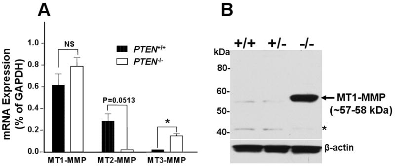
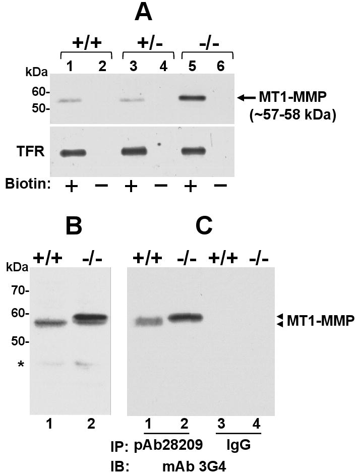
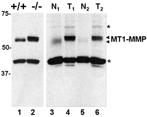
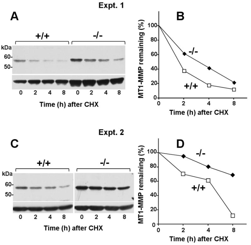
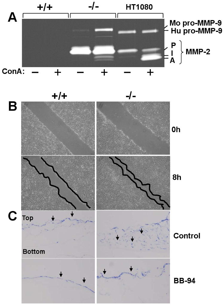
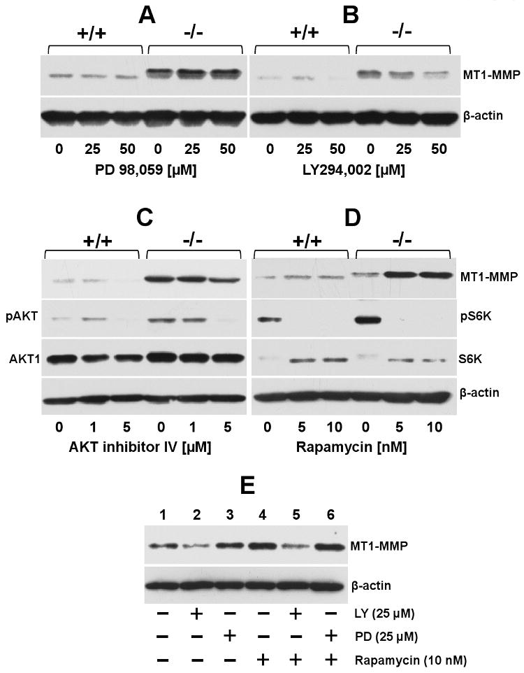
Membrane type 1 (MT1)-matrix metalloproteinase (MT1-MMP) is a membrane-tethered MMP that has been shown to play a key role in promoting cancer cell invasion. MT1-MMP is highly expressed in bone metastasis of prostate cancer (PC) patients and promotes intraosseous tumor growth of PC cells in mice. The majority of metastatic prostate cancers harbor loss-of-function mutations or deletions of the tumor suppressor PTEN (phosphatase and tensin homologue deleted on chromosome ten). However, the role of PTEN inactivation in MT1-MMP expression in PC cells has not been examined. In this study, prostate epithelial cell lines derived from mice that are either heterozygous (PTEN(+/-)) or homozygous (PTEN(-/-)) for PTEN deletion or harboring a wild-type PTEN (PTEN(+/+)) were used to investigate the expression of MT1-MMP. We found that biallelic loss of PTEN is associated with posttranslational regulation of MT1-MMP protein in mouse PC cells. PTEN(-/-) PC cells display higher levels of MT1-MMP at the cell surface when compared to PTEN(+/+) and PTEN(+/-) cells and consequently exhibited enhanced migratory and collagen-invasive activities. MT1-MMP displayed by PTEN(-/-) cells is differentially O-glycosylated and exhibits a slow rate of turnover. MT1-MMP expression in PTEN(-/-) cells is under control of the PI3K/AKT signaling pathway, as determined using pharmacological inhibitors. Interestingly, rapamycin, an mTOR inhibitor, upregulates MT1-MMP expression in PTEN(+/+) cells via PI3K activity. Collectively, these data in a mouse prostate cell system uncover for the first time a novel and complex relationship between PTEN loss-mediated PI3K/AKT activation and posttranslational regulation of MT1-MMP, which may play a role in PC progression.


Figures

Fig 1
Fig 1
Differential expression of MT-MMP mRNA and MT1-MMP protein in mouse prostate epithelial cells with different PTEN status. (A) Total RNA from PTEN+/+ (black bar) and PTEN-/- (white bar) cells cultured in complete media were used for cDNA synthesis and subsequently real time PCR to quantify MT1-, MT2-, and MT3-MMP mRNA. Ct value of each gene was normalized by the GAPDH Ct value. Results shown are representative of two independent experiments. *, p <0.05; Student's t test. NS, not statistically significant. (B) Cell lysates from PTEN+/+, PTEN+/-, and PTEN-/- cells were resolved by reducing 10% SDS-PAGE followed by immunoblotting analysis using mAb 3G4 against the catalytic domain of MT1-MMP (Mr ∼57-59 kDa). β-actin was used as a loading control. Asterisk shows non-specific bands.
Fig. 2
Fig. 2
Elevated surface expression of MT1-MMP in PTEN-/- cells. (A) PTEN+/+ (lanes 1 and 2), PTEN+/- (lane 3 and 4), and PTEN-/- cells (lane 5 and 6) were seeded on 6-well plates and incubated with (+) or without (-) cell-impermeable biotin for 30 min on ice. Cell lysates from each cell were then incubated with avidin-conjugated beads and biotinylated proteins were resolved by reducing 10% SDS-PAGE followed by immunoblot analysis using mAb 3G4. The same blot was reprobed with an antibody against transferrin receptor (TFR), as a control. (B) Lysates from PTEN+/+ and PTEN-/- cells were resolved by reducing 7.5% SDS-PAGE followed immunoblotting analysis using mAb 3G4. (C) Lysates from PTEN+/+ and PTEN-/- cells were immunoprecipitated with pAb28209 to the cytosolic tail of MT1-MMP or non-immune rabbit IgG and protein-A conjugated agarose beads. The immunoprecipitates were resolved by reducing 7.5% SDS-PAGE followed immunoblotting analysis using mAb 3G4. Arrowheads on the right in panes B and C indicate the different species of MT1-MMP detected. Asterisk shows non-specific bands.
Fig. 3
Fig. 3
Enhanced expression of MT1-MMP in lysates of wild type and PTEN-/- mouse prostate tissues. Mouse PTEN-/- prostate tumor (T1 and T2, lanes 4 and 6) and normal (N1 and N2, lanes 3 and 5) prostate lysates from different mice and lysates from cultured PTEN+/+ (lane 1) and PTEN-/- (lane 2) cells were resolved by reducing 8% SDS-PAGE followed by immunoblot analyses using mAb 3G4 to the catalytic domain. The antigen was detected using the TrueBlot™ ULTRA antibody against mouse IgG followed by chemiluminescence. Asterisks show non-specific bands.
Fig. 4
Fig. 4
MT1-MMP is differentially O-glycosylated in PTEN+/+, PTEN+/- and PTEN-/- cells. PTEN+/+ (lanes 1 and 4), PTEN+/- (lanes 2 and 5), and PTEN-/- cells (lanes 3 and 6) were incubated with (+) or without (-) the O-glycosylation inhibitor BGN (2 mM) for 48 h. The cells were then lysed, and the lysates were resolved by reducing 7.5% SDS-PAGE followed immunoblot analysis using the mAb 3G4. Arrowheads on the right indicate the different species of MT1-MMP detected.
Fig. 5
Fig. 5
Reduced turnover of MT1-MMP in PTEN-/- cells. (A-D) PTEN+/+ and PTEN-/- cells were incubated with 50 μg/ml of cycloheximide (CHX) for 8 h. At each time point (0, 2, 4, and 8 h), the cells were lysed and the lysates were resolved by reducing 10% SDS-PAGE followed by immunoblotting analysis using mAb 3G4. A and B show blots derived from two independent experiments. C and D show the results of the densitometry analyses, as described in Materials and Methods. Values represent the relative amount of MT1-MMP at each time point relative to time zero (100%).
Fig. 6
Fig. 6
Pro-MMP-2 activation, migration and invasiveness of PTEN-/- cells. (A) Cultures of PTEN+/+, PTEN-/-, and HT1080 cells in 6-well plates were untreated (-) or treated (+) with ConA and the lysates were subjected to gelatin zymography. P, pro-MMP-2 (∼72 kDa), I, intermediate form of MMP-2 (∼66 kDa), A, active MMP-2 (∼62 kDa). Mo, mouse, Hu, human. (B) Confluent cultures of PTEN+/+ and PTEN-/- cells in 6-well plates were scratched, and incubated in complete media supplemented with 5 μg/ml mitomycin C for 8 h. The cell monolayers were then photographed. (C) PTEN+/+ and PTEN-/- cells were seeded on top of a type I collagen gel in the presence or absence of BB-94. After 14 days, 5 μm-thick frozen sections were stained with toluidine blue to identify the cells, indicated as black arrows. The experiments presented in panels A, B, and C were repeated at least three times with similar results.
Fig. 7
Fig. 7
Effect of pharmacological inhibitors to PI3K, AKT, MAPK and mTOR on MT1-MMP expression. Serum starved PTEN+/+ and PTEN-/- cells were incubated (18 h) with (+) or without (-) the indicated doses of either PD 98,059 (A), LY 294,002 (B), Akt inhibitor IV (C) or rapamycin (D) in serum-free media. (E) Serum starved PTEN-/- cells were untreated (-) or treated (+) with either LY 294,002 or PD 98,059 for 18 h. Then, the cultures received rapamycin (+) or vehicle (-) and were incubated for 18 h incubation. At the end of the incubation period, the cells were lysed, and the lysates were resolved by reducing 10% SDS-PAGE followed by immunoblot analyses with the appropriate antibodies. The data presented in panels A-E were repeated at least three times with similar results.

Similar articles

Cited by

References

    1. Massova I, Kotra LP, Fridman R, Mobashery S. Matrix metalloproteinases: structures, evolution, and diversification. FASEB J. 1998;12:1075–1095. - PubMed
    1. Zucker S, Pei D, Cao J, Lopez-Otin C. Membrane type-matrix metalloproteinases (MT-MMP) Curr Top Dev Biol. 2003;54:1–74. - PubMed
    1. Osenkowski P, Toth M, Fridman R. Processing, shedding, and endocytosis of membrane type 1-matrix metalloproteinase (MT1-MMP) J Cell Physiol. 2004;200:2–10. - PubMed
    1. Itoh Y. MT1-MMP: a key regulator of cell migration in tissue. IUBMB Life. 2006;58:589–596. - PubMed
    1. Itoh Y, Ito N, Nagase H, Evans RD, Bird SA, Seiki M. Cell surface collagenolysis requires homodimerization of the membrane-bound collagenase MT1-MMP. Mol Biol Cell. 2006;17:5390–5399. - PMC - PubMed

Publication types

MeSH terms