

Identification, epidemiology, and chronicity of pediatric esophageal eosinophilia, 1982-1999
- PMID: 20620567
- PMCID: PMC4115587
- DOI: 10.1016/j.jaci.2010.05.027
Identification, epidemiology, and chronicity of pediatric esophageal eosinophilia, 1982-1999
Abstract
Background: Eosinophilic esophagitis (EE) is now a commonly encountered disorder that was rarely diagnosed a decade ago.
Objective: We aimed to determine the epidemiologic and histologic features of retrospective pediatric esophageal eosinophilia before the first case of EE at our institution was recognized.
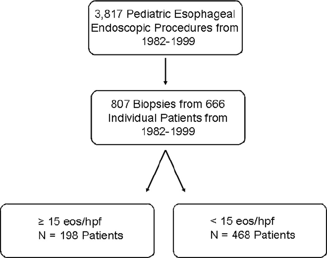
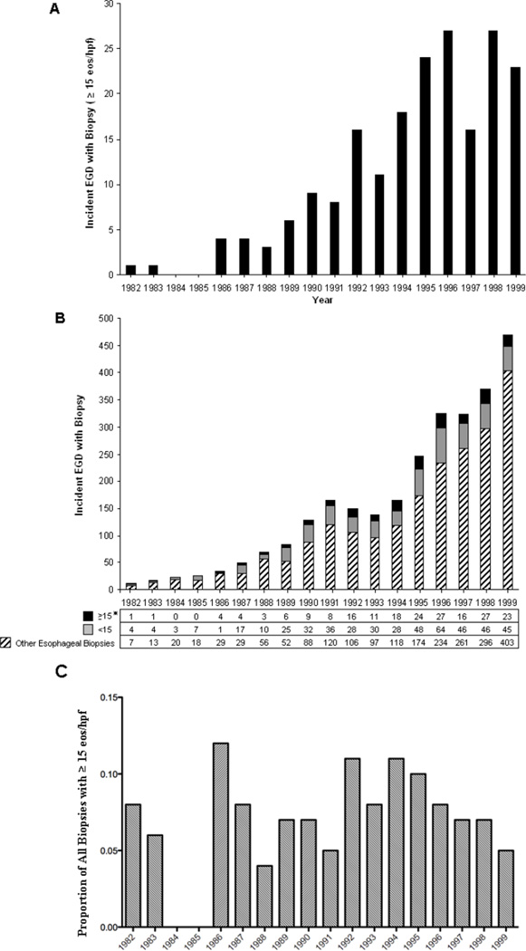
Methods: Esophageal biopsy specimens obtained between 1982 and 1999 with reflux esophagitis were re-examined and reorganized into 2 groups based on peak esophageal eosinophil number (<15 eosinophils per high-powered field [hpf] and > or =15 eosinophils/hpf). The epidemiology and histology of the entire cohort and a population-based cohort were evaluated.
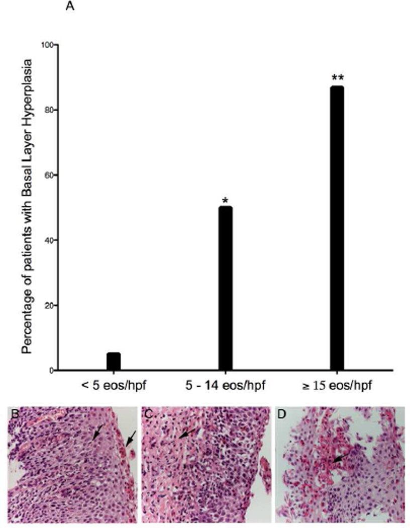
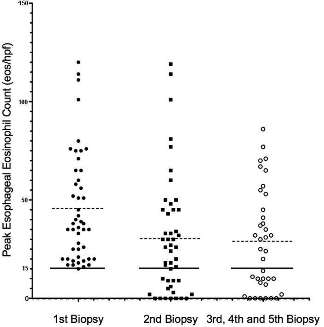
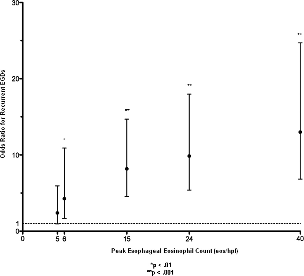
Results: Eight hundred seven biopsy specimens from 666 patients were re-examined; 198 patients had 15 eosinophils/hpf or greater. Among a population-based cohort of patients with 15 eosinophils/hpf or greater, there was a modest increase in incidence (P < .001; incidence rate ratio, 1.18; 95% CI, 1.09-1.28). After correcting for a 40-fold increase in the number of endoscopies during this time period, the proportion of biopsy specimens with 15 eosinophils/hpf or greater did not change (0.08 in 1982 vs 0.08 in 1996 [peak]; P = .9; incidence rate ratio, 1.02; 95% CI, 0.73-1.44). Patients who had as few as 5 eosinophils/hpf were more likely to have persistent esophageal eosinophilia on repeat esophagogastroduodenoscopy, evidence of basal layer hyperplasia, and lamina propria fibrosis compared with patients with less than 5 eosinophils/hpf (P < .001).
Conclusions: Esophageal eosinophilia at levels consistent with EE was present among 30% of patients given diagnoses of reflux esophagitis, and the incidence of esophageal eosinophilia did not change over time. Patients with 5 eosinophils/hpf or greater had evidence of other histologic abnormalities and were likely to have persistent esophageal eosinophilia.
Copyright 2010 American Academy of Allergy, Asthma & Immunology. Published by Mosby, Inc. All rights reserved.
Figures
Comment in
-
Escalating incidence of eosinophilic esophagitis: a 20-year prospective, population-based study in Olten County, Switzerland.J Allergy Clin Immunol. 2011 Dec;128(6):1349-1350.e5. doi: 10.1016/j.jaci.2011.09.013. Epub 2011 Oct 21. J Allergy Clin Immunol. 2011. PMID: 22019091 No abstract available.
References
-
- Atkins D, Kramer R, Capocelli K, Lovell M, Furuta GT. Eosinophilic esophagitis: the newest esophageal inflammatory disease. Nat Rev Gastroenterol Hepatol. 2009;6:267–278. - PubMed
-
- Franciosi JP, Liacouras CA. Eosinophilic esophagitis. Immunol Allergy Clin North Am. 2009;29:19–27. viii. - PubMed
-
- Liacouras CA, Bonis P, Putnam PE, Straumann A, Ruchelli E, Gupta SK, et al. Summary of the First International Gastrointestinal Eosinophil Research Symposium. J Pediatr Gastroenterol Nutr. 2007;45:370–391. - PubMed
Publication types
MeSH terms
Grants and funding
LinkOut - more resources
Full Text Sources
Other Literature Sources
Medical
