. 2010 Jun 24;66(6):848-58.
doi: 10.1016/j.neuron.2010.05.007.

Nova2 regulates neuronal migration through an RNA switch in disabled-1 signaling

Affiliations

Nova2 regulates neuronal migration through an RNA switch in disabled-1 signaling

Masato Yano et al. Neuron. .

Abstract

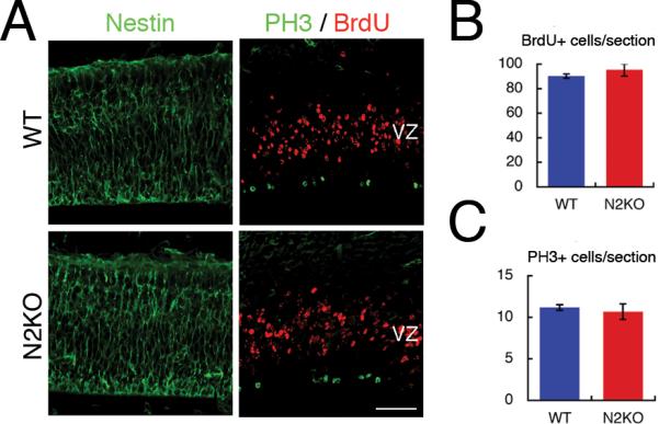
Neuronal migration leads to a highly organized laminar structure in the mammalian brain, and its misregulation causes lissencephaly and behavioral and cognitive defects. Reelin signaling, which is mediated in part by a key adaptor, disabled-1 (Dab1), plays a critical but incompletely understood role in this process. We found that the neuron-specific RNA-binding protein Nova2 regulates neuronal migration in late-generated cortical and Purkinje neurons. An unbiased HITS-CLIP and exon junction array search for Nova-dependent reelin-pathway RNAs at E14.5 revealed only one candidate-an alternatively spliced isoform of Dab1 (Dab1.7bc). In utero electroporation demonstrated that Dab1.7bc was sufficient to induce neuronal migration defects in wild-type mice and exacerbate defects when Dab1 levels were reduced, whereas Dab1 overexpression mitigates defects in Nova2 null mice. Thus, Nova2 regulates an RNA switch controlling the ability of Dab1 to mediate neuronal responsiveness to reelin signaling and neuronal migration, suggesting new links between splicing regulation, brain disease, and development.


Figures

Figure 1
Figure 1. Loss of Nova2 results in mislocation of late-generated cortical neurons, and, to a lesser degree, disorganization of early generated cortical neurons
(A) Sagittal sections of postnatal P10 WT and Nova2 KO (N2KO) neocortex at the level of the anterior hippocampus were immnnostained with layer-specific marker proteins, Brn-2 (layer II–III, V in part; green), Er81 (layer V; red) and DAPI (blue). (B) FoxP2 (layerV–VI; green) or Cux1 (layerII–IV; Red). High magnification of deep layers in Nova2 KO (box region in B). These two marker proteins are not completely merged. Scale bar; 100 μm. WT.
Figure 2
Figure 2. Nova2 controls laminar formation of late generated neurons
(A,C) BrdU-labeling was performed at E14.0 or E16.0 and cortical sections of WT or Nova2 KO animals were immunostained with BrdU antibody at P0 as indicated. (B,D) Graphs show a quantification of BrdU positive cells percentage in each of 10 bins (along the dorsal/ventral axis) relative to the total number of BrdU positive cells in WT or Nova2 KO. Data represents mean +/– standard deviation (SD) from experiments using three independent animals for each genotype. Results showed statistically significant differences for Nova2 KO relative to WT in both E14 and E16 BrdU injections (F(9,35) = 6.56, P<0.0001, F(9.35) = 2.9348, p=0.0106, respectively by repeated measures ANOVA). Scale bar; 100 μm.
Figure 3
Figure 3. Neural progenitor cells in WT and Nova2 KO cortex
(A) BrdU was injected 1 hr before fixation. Coronal sections of embryonic day 14.5 cortex in wild type and Nova2 KO brains were immunostained with antibodies against Nestin (left panel; green), which is neural progenitor marker protein, BrdU (right panel; red) and PH3 (right panel; green). (B) This graph represents the number of BrdU positive cells per section. (C) This graph represents the number of PH3 positive cells per sections. Error bar indicates SD of three biological replicates. Scale bar; 50 μm. PH3; phospho-Histone3.
Figure 4
Figure 4. Nova2 regulates Purkinje neurons migration
(A) Calbindin staining (green) and counter staining with DAPI (blue) in P10 wild type and Nova2 KO sagittal sections at the level of vermis in cerebellum. High magnification orthogonal picture was enlarged from the boxed region in Nova2 KO cerebellum and double stained with RORα (red). Quantitative result of ectopic calbindin positive neurons in cerebellum white matter in wild type and Nova2 KO using three biological replicates (each ~6 sections in cerebellar vermis). Error bar represents SD using three biological replicates. CaBP; calbindin, Scale bar; 100 μm (B,C) BrdU-labeling at E11.5 and immunostained with BrdU (red) and calbindin (green) antibodies at P4 cerebellum. (B) Whole cerebellum in WT and Nova2 KO at the level of vermis in cerebellum (C) High magnifications of Purkinje cell layers in WT (box region in B in wild type) and Nova2 KO (box region in B in N2KO-1) and white matter in Nova2 KO (box region in B in N2KO-2). Arrow indicates BrdU/calbindin double positive cells. Bar graph represents the percent of BrdU/calbindin double positive cells in white matter per total double positive cells in whole cerebellum. Error bar represents SD using three biological replicates. Asterisk indicates P-value < 0.05 by Student t-test. Scale bar; 200 μm (B), 20 μm (C) EGL; External granule layer, PCL; Purkinje cell layer, GL; granule cell layer N2KO; Nova2 KO
Figure 5
Figure 5. Nova2 regulates alternative splicing of Dab1 in a context dependent manner
(A) Scheme of Dab1 protein, which consists of PTB domain and tyrosine phospholylation sites, and amino acids encoded by Dab1 7bc exons. PTB: phospho-tyrosine binding. (B) Nova2 CLIP tags from E14.5 cortex (blue/cyan/purple) and P7 brains (red/orange/pink) by each three individual CLIP experiments, map to Nova2 regulating Dab1 transcripts. The alternatively spliced region is highlighted. Two red boxes in highlight indicate Dab1 7bc exons. (C) Alternative splicing analysis of Dab1 in wild type and Nova2 KO using 5 time points: E10.5 brain, E14.5 cortex (Ctx), E16.5 Ctx, P0 Ctx and P10 Ctx total RNA. Each corresponding band was confirmed by sequencing. (D) Quantitation of Dab1 vs Dab1.7bc mRNA expression data in (C); each point represent the average of three biological replicates (see detailed data in Figure S5C contains the ratio of Dab1.7b/c and steady state level of total Dab1 between embryo and postnatal cortex in Figure S5B). (E–G) Two upstream intronic sequences of Dab1 7b and 7c exons are necessary for the regulation of alternative splicing of Dab1.7bc exons as a pair by Nova2. (E) Schematic representation of pGloDab1.7bc and its derivative minigenes containing the mouse intronic regions surrounding and including exon 7b and 7c between human globin constitutive exon1 and 3. Asterisks indicate site of point mutations in Nova binding YCAY clusters (see Figure S5D, F for details). (F) total RNA was isolated from 293T cells transiently transfected with WT or mutant pGloDab1.7bc minigenes (0.25 μg)and pNova2 (0.5 μg) as indicated, and spliced products analyzed by RT-PCR. Three biological replicates were used in each analysis. (G) Model of Nova2-mediated Dab1.7bc exon repression. .
Figure 6
Figure 6. WT Dab1 mitigates and Dab1.7bc exacerbates neuronal migration defects
Expression plasmids of mRFP together with control or Dab1 or Dab1.7bc were injected into lateral ventricle of E14.5 embryo brain and electroporated into the region of neocortex. (A) WT (C) Nova2 KO brains were fixed at 4 days later and examined. High magnifications were double stained with Cux-1 and mRFP. (B and D) Graphs quantifying mRFP positive cells in each of 4 regions (marginal zone (MZ), and upper, middle and lower cortical plates (CP), as illustrated in (A) and (C)) as a percentage of total mRFP positive cells. Scale bar; 100 μm, 20 μm (high magnifications). Data represent the mean +/− SD of three WT or Nova2 KO brains. Results were compared to WT (B) or Nova2 KO (D) control vector and showed no significant difference in WT brain transfected with Dab1 (p=0.2), but significant differences with WT brain transfected with Dab1.7bc (p<0.001) or Nova2 KO brain transfected with Dab1 (p<0.01) by repeated measures ANOVA. (E–F) Control and Dab1shRNA expression vectors were injected together with Dab1 or Dab1.7bc expression vectors into the lateral ventricle of E14.5 embryo brain, electroporated and analyzed 4 days later. (E) indicates representative results and (F) shows the ratio of the mRFP positive cells in upper cortical layer per total mRFP positive cells in cortical plate as indicated in (E). Error bar represents SD using three biological replicates. Asterisk indicates P-value < 0.01 by Student t-test.

Comment in

References

    1. Akamatsu W, Okano HJ, Osumi N, Inoue T, Nakamura S, Sakakibara SI, Miura M, Matsuo N, Darnell RB, Okano H. Mammalian ELAV-like neuronal RNA-binding proteins HuB and HuC promote neuronal development in both the central and the peripheral nervous systems. Proc Natl Acad Sci U S A. 1999;96:9885–9890. - PMC - PubMed
    1. Amadio JP, Walsh CA. Brain evolution and uniqueness in the human genome. Cell. 2006;126:1033–1035. - PubMed
    1. Ayala R, Shu T, Tsai LH. Trekking across the brain: the journey of neuronal migration. Cell. 2007;128:29–43. - PubMed
    1. Bar I, Tissir F, Lambert de Rouvroit C, De Backer O, Goffinet AM. The gene encoding disabled-1 (DAB1), the intracellular adaptor of the Reelin pathway, reveals unusual complexity in human and mouse. J Biol Chem. 2003;278:5802–5812. - PubMed
    1. Blencowe BJ, Ahmad S, Lee LJ. Current-generation high-throughput sequencing: deepening insights into mammalian transcriptomes. Genes Dev. 2009;23:1379–1386. - PubMed

Publication types