. 2010 Jul 7;12(1):42-52.
doi: 10.1016/j.cmet.2010.04.016.

GPIHBP1 is responsible for the entry of lipoprotein lipase into capillaries

Affiliations

GPIHBP1 is responsible for the entry of lipoprotein lipase into capillaries

Brandon S J Davies et al. Cell Metab. .

Abstract

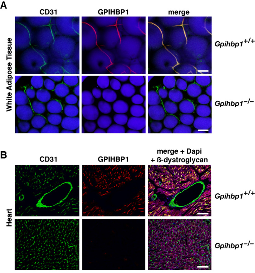
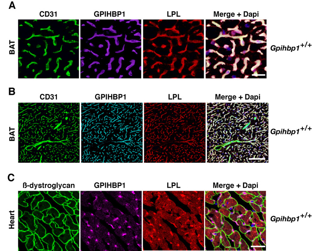
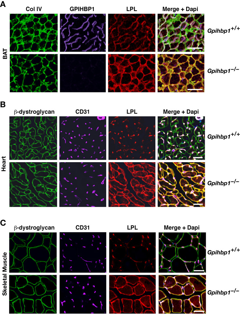
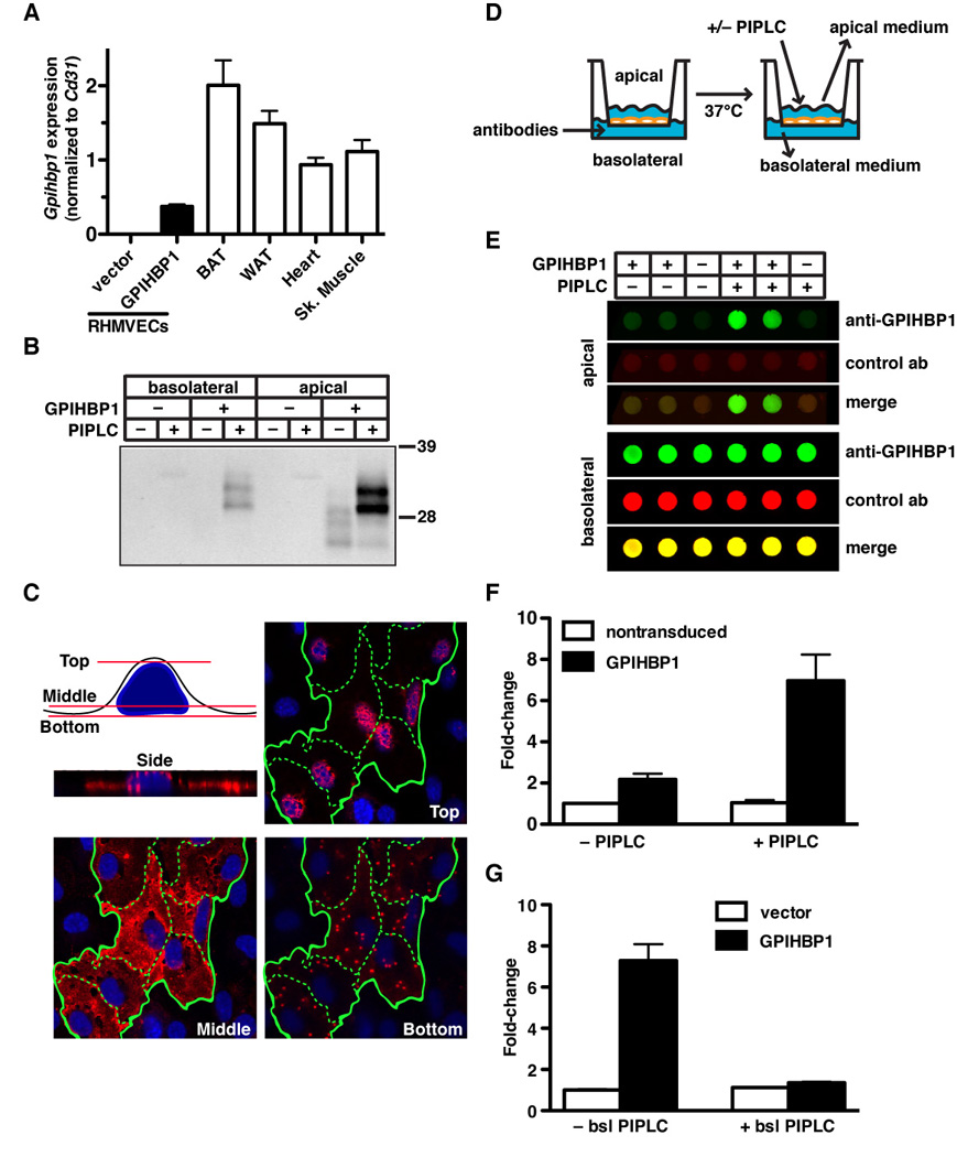
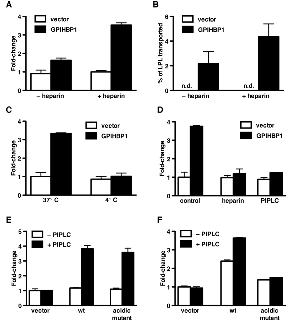
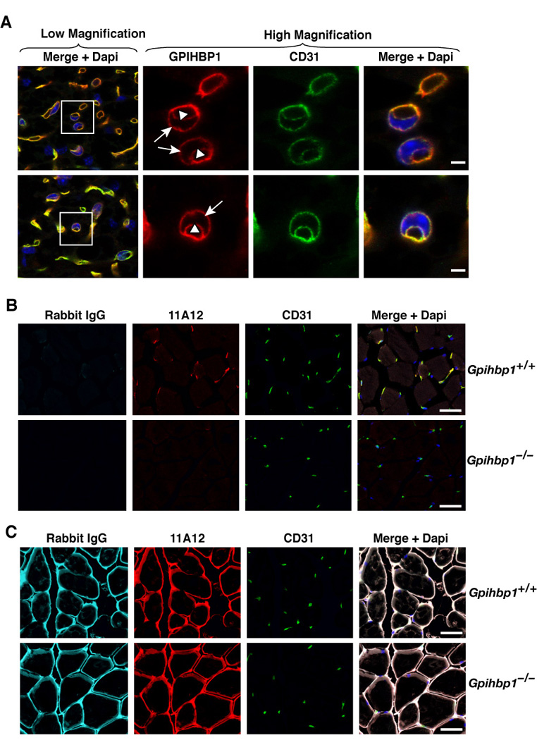
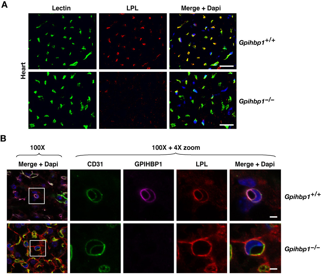
The lipolytic processing of triglyceride-rich lipoproteins by lipoprotein lipase (LPL) is the central event in plasma lipid metabolism, providing lipids for storage in adipose tissue and fuel for vital organs such as the heart. LPL is synthesized and secreted by myocytes and adipocytes, but then finds its way into the lumen of capillaries, where it hydrolyzes lipoprotein triglycerides. The mechanism by which LPL reaches the lumen of capillaries has remained an unresolved problem of plasma lipid metabolism. Here, we show that GPIHBP1 is responsible for the transport of LPL into capillaries. In Gpihbp1-deficient mice, LPL is mislocalized to the interstitial spaces surrounding myocytes and adipocytes. Also, we show that GPIHBP1 is located at the basolateral surface of capillary endothelial cells and actively transports LPL across endothelial cells. Our experiments define the function of GPIHBP1 in triglyceride metabolism and provide a mechanism for the transport of LPL into capillaries.


Conflict of interest statement

The authors have no conflicts of interest to declare.

Figures

Figure 1
Figure 1. GPIHBP1 is located within the lumen of capillaries
(A) Confocal images of white adipose tissue revealing GPIHBP1 in the capillaries of wild-type but not Gpihbp1−/− mice. Mice were injected intravenously with Alexa555-labeled anti-GPIHBP1 (red) and hamster anti-CD31 antibodies and then perfused extensively with PBS. Fixed adipose explants were stained with FITC-labeled anti-hamster antibodies (green) and BODIPY to label adipocytes (blue). The scale bar represents a distance of 100 µm. (B) Confocal images of heart tissue from the same antibody-injected wild-type and Gpihbp1−/− mice showing GPIHBP1 staining in the capillaries of wild-type but not Gpihbp1−/− mice. Tissues were stained with an antibody against β-dystroglycan (magenta), a plasma membrane protein of myocytes, to outline myocytes. The scale bar represents a distance of 100 µm. (See also Figure S1.)
Figure 2
Figure 2. GPIHBP1 and LPL colocalize within capillaries
(A and B) Confocal images showing the binding of GPIHBP1-, LPL-, and CD31-specific antibodies to brown adipose tissue (BAT) from a wild-type mouse. Images were taken with a 63× objective; (A) close-up (scale bar represents distance of 50 µm) and (B) wide-field (scale bar represents distance of 100 µm). (C) Confocal images showing the binding of GPIHBP1-, LPL-, and β-dystroglycan-specific antibodies to heart tissue from a wild-type mouse. The scale bar represents a distance of 25 µm. (See also Figure S2.)
Figure 3
Figure 3. Mislocalization of LPL in Gpihbp1−/− mice
(A) Confocal microscopy showing the binding of GPIHBP1-, LPL-, and collagen IV–specific antibodies to brown adipose tissue (BAT) from wild-type and Gpihbp1−/− mice. The scale bars represent a distance of 100 µm. (B and C) Confocal microscopy showing the binding of CD31-, LPL-, and β-dystroglycan-specific antibodies to heart (B) and skeletal muscle (C) of wild-type and Gpihbp1−/− mice. The scale bars represent a distance of 100 µm (skeletal muscle) or 50 µm (heart).
Figure 4
Figure 4. GPIHBP1 is present at the basolateral and apical surfaces of cultured endothelial cells and transports a GPIHBP1-specific monoclonal antibody across cells
(A) Gpihbp1 expression levels in lentivirus-transduced RHMVECs and mouse heart, skeletal muscle, brown adipose tissue (BAT), and white adipose tissue (WAT). Gpihbp1 expression levels were measured by quantitative RT-PCR and normalized to Cd31 (mean ± SEM). (B) Release of GPIHBP1 by PIPLC from apical and basolateral surfaces of GPIHBP1-transduced RHMVEC monolayers grown on filters. After incubating the cells with PIPLC (5 U/ml) for 1.5 h, the medium was harvested from the apical and basolateral chambers, precipitated with 10% TCA, and size-fractionated by SDS-PAGE. Western blots were performed with antibody 11A12. (C) Confocal microscopy showing the binding of antibody 11A12 to GPIHBP1-transduced RHMVECs. Alexa555-labeled antibody 11A12 was added to both the apical and basolateral chambers, and images of optical sections were obtained with a confocal microscope (63× objective) near the top, middle, and bottom of the cells. Green lines outline individual cells. (D) Schematic of the transport assay. RHMVECs were grown on filters (1-µm pore size) until they formed a tight monolayer. Antibodies were added to the basolateral chamber and cells were incubated at 37°C. After the incubation, PIPLC was added to the apical chamber to release GPIHBP1; medium from the apical and basolateral chambers was collected and analyzed. (E and F) Dot blots demonstrating the transport of antibody 11A12 from the basolateral to the apical surface of GPIHBP1-transduced RHMVEC monolayers. IRDye800-labeled antibody 11A12 (green) and an IRDye680-labeled anti-goat IgG control antibody (red) were added to the basolateral chamber and incubated for 3 h at 37°C. The apical surface was treated with PIPLC (1 U/ml; 30 min) or PBS, and both apical and basolateral media were dot blotted, scanned, and quantified with the Odyssey scanner. Bar graph in Panel F shows the fold change (mean ± SEM) compared to untreated vector-transduced cells from four independent experiments. (G) Analysis of the ability of basolateral PIPLC to block the transport of antibody 11A12 from the basolateral to the apical chamber. IRDye800-labeled antibody 11A12 and IRDye680-labeled anti-goat IgG antibody were added to the basolateral chamber in the presence or absence of PIPLC (1 U/ml) and incubated for 1.5 h at 37°C. The apical surface was treated with PIPLC (1 U/ml; 30 min) and the apical medium was dot blotted, scanned, and quantified with the Odyssey scanner. The bar graph shows the fold change (mean ± SEM) compared to untreated vector-transduced cells.
Figure 5
Figure 5. Transport of LPL across endothelial cell monolayers
(A) Dot blot analysis demonstrating the transport of V5-tagged human LPL (hLPL) from the basolateral to the apical chamber by GPIHBP1-transduced RHMVECs. After adding LPL to the basolateral chamber and incubating for 1 h at 37°C, either heparin (100 U/ml) or PBS was added to the apical chamber and the apical medium was dot blotted with an antibody against the V5 tag and quantified with the Odyssey scanner. Bar graph shows the fold change (mean ± SEM) compared to vector-transduced cells from four independent experiments. (B) Transport of bovine LPL from the basolateral to the apical chamber by GPIHBP1- or vector-transduced RHMVECs grown on filters. After adding bovine LPL to the basolateral chamber and incubating for 1 h at 37°C, the apical surface of cells was treated with heparin (100 U/ml) or PBS for 15 min at room temperature. The apical medium was collected, and the LPL concentration was determined with an ELISA. (C) GPIHBP1-mediated LPL transport across endothelial cells at 4° and 37°C. After adding V5-tagged hLPL to the basolateral chamber, GPIHBP1-transduced RHMVECs were incubated for 1 h at 4° or 37°C. Heparin was then added to the apical chamber (100 U/ml; 15 min at 4°C), and the amount of LPL in the apical medium was assessed by dot blot with an antibody against the V5 tag. The blot was scanned and quantified with the Odyssey scanner. Bar graph shows the fold change (mean ± SEM) compared to vector-transduced cells incubated at 37°C. (D) Ability of PIPLC or heparin in the basolateral medium to block GPIHBP1-mediated transport of the V5-tagged hLPL from the basolateral to the apical medium of RHMVECs. After adding LPL and either heparin (100 U/ml) or PIPLC (2.5 U/ml) to the basolateral chamber, the cells were incubated for 1 h at 37°C. Heparin was then added to the apical chamber (100 U/ml; 15 min at room temperature), and the amount of LPL in the apical medium was then assessed by dot blotting with an antibody against the V5 tag. The blot was scanned and quantified with the Odyssey scanner. Bar graph shows the fold change (mean ± SEM) compared to vector-transduced cells. (E) Dot blot analysis of antibody 11A12 transport in RHMVECs transduced with wild-type or mutant (D,E(24–48)A) GPIHBP1. IRDye800-labeled antibody 11A12 was added to the basolateral chamber and incubated for 1 h at 37°C. The apical surface was then treated with PIPLC (1 U/ml; 30 min) or PBS; the apical medium was dot blotted, scanned, and analyzed with the Odyssey scanner. Bar graph shows fold change (mean ± SEM) compared to vector-transduced control cells. (F) Dot blot analysis of LPL transport in RHMVECs transduced with either wild-type or mutant (D,E(24–48)A) GPIHBP1. After adding LPL to the basolateral chamber and incubating for 1 h at 37°C, the apical surface was treated with PIPLC (2.5 U/ml) or PBS (30 min at 37°C). The apical medium was then dot blotted with an antibody against the V5 tag, and the signal was quantified with the Odyssey scanner. Bar graph shows the fold change (mean ± SEM) compared to vector-transduced control cells. (See also Figure S4.)
Figure 6
Figure 6. GPIHBP1 is present at both the basolateral and apical surfaces of endothelial cells in vivo and transports monoclonal antibody 11A12 across capillary endothelial cells
(A) Confocal microscopy showing the binding of GPIHBP1- and CD31-specific antibodies to brown adipose tissue of a wild-type mouse. Images were taken with a 100× objective without optical zoom (low magnification) or with 4× optical zoom (high magnification). To visualize both the apical and basolateral surfaces of capillaries, cross sections of capillaries that cut through an endothelial cell nucleus (blue) were identified (boxed areas) and viewed at high magnification. Both GPIHBP1 (red) and CD31 (green) were expressed at the apical (arrowheads) and basolateral (arrows) surfaces of endothelial cells. The scale bar represents a distance of 2.5 µm. (B) Microscopy showing the transport of antibody 11A12 from the interstitial spaces to the lumens of capillaries of skeletal muscle. One quadriceps muscle of a wild-type and a Gpihbp1−/− mouse was injected with 10 µl of normal saline containing 15 µg each of the rat GPIHBP1-specific monoclonal antibody 11A12 and nonimmune rabbit IgG. After 30 min, the mice were injected intravenously with 200 µg each of fluorescent-labeled goat anti-rabbit IgG (pseudo-color cyan) and fluorescent-labeled goat anti-rat IgG (red). Three minutes later, the mice were extensively perfused with PBS and tissues fixed in situ with 3% PFA. Frozen sections were prepared and stained with antibodies against CD31 (green) to identify endothelial cells, and DAPI to identify nuclei. In wild-type mice, fluorescent-labeled anti-rat antibody was easily detectable within the capillaries of the injected muscle, whereas no staining was present in the skeletal muscle of Gpihbp1−/− mice. The scale bar represents a distance of 100 µm. (C) Microscopy showing the presence of injected antibodies in the interstitial spaces of skeletal muscle. Frozen sections adjacent to the sections shown in Panel B were stained with fluorescent-labeled anti-rat antibody (red) and anti-rabbit antibody (pseudo-color cyan) to document the presence of antibody 11A12 and rabbit IgG within the interstitial spaces of the injected muscles. As expected, antibody 11A12 and rabbit IgG were present in both wild-type and Gpihbp1−/− mice. The scale bar represents a distance of 100 µm.
Figure 7
Figure 7. LPL is absent from capillary lumens in Gpihbp1−/− mice
(A) Confocal images of heart showing the binding of FITC-labeled tomato lectin and a goat antibody against mouse LPL. Both the tomato lectin and the LPL-specific antibody were injected intravenously into a wild-type mouse and a Gpihbp1−/− mouse. Three min later, mice were perfused extensively with PBS and then perfusion-fixed with 3% PFA. The scale bars represent a distance of 50 µm. (B) Confocal microscopy showing the binding of CD31-, GPIHBP1- and LPL-specific antibodies to brown adipose tissue of a wild-type mouse and a Gpihbp1−/− mouse. Images were taken with a 100× objective without optical zoom or with 4× optical zoom. To visualize both the apical and basolateral surfaces of capillaries, cross sections of capillaries that cut through an endothelial cell nucleus (blue) were identified (boxed areas) and viewed at high magnification. GPIHBP1 (purple), LPL (red) and CD31 (green) were all expressed at the apical and basolateral surfaces of endothelial cells in wild-type mice, but no LPL was present on the apical (luminal) surface of the capillary endothelium in Gpihbp1−/− mice. The scale bar represents a distance of 2.5 µm. (See also Figure S5.)

Comment in

References

    1. Anderson RG. Plasmalemmal caveolae and GPI-anchored membrane proteins. Curr Opin Cell Biol. 1993;5:647–652. - PubMed
    1. Beigneux AP, Davies BS, Gin P, Weinstein MM, Farber E, Qiao X, Peale F, Bunting S, Walzem RL, Wong JS, Blaner WS, Ding ZM, Melford K, Wongsiriroj N, Shu X, de Sauvage F, Ryan RO, Fong LG, Bensadoun A, Young SG. Glycosylphosphatidylinositol-anchored high-density lipoprotein-binding protein 1 plays a critical role in the lipolytic processing of chylomicrons. Cell Metab. 2007;5:279–291. - PMC - PubMed
    1. Beigneux AP, Franssen R, Bensadoun A, Gin P, Melford K, Peter J, Walzem RL, Weinstein MM, Davies BS, Kuivenhoven JA, Kastelein JJ, Fong LG, Dallinga-Thie GM, Young SG. Chylomicronemia with a mutant GPIHBP1 (Q115P) that cannot bind lipoprotein lipase. Arterioscler Thromb Vasc Biol. 2009a;29:956–962. - PMC - PubMed
    1. Beigneux AP, Gin P, Davies BS, Weinstein MM, Bensadoun A, Fong LG, Young SG. Highly conserved cysteines within the Ly6 domain of GPIHBP1 are crucial for the binding of lipoprotein lipase. J Biol Chem. 2009b;284:30240–30247. - PMC - PubMed
    1. Brown DA, Rose JK. Sorting of GPI-anchored proteins to glycolipid-enriched membrane subdomains during transport to the apical cell surface. Cell. 1992;68:533–544. - PubMed

Publication types