. 2010 Sep;139(3):942-52.
doi: 10.1053/j.gastro.2010.05.083. Epub 2010 Jun 4.

Kitlow stem cells cause resistance to Kit/platelet-derived growth factor alpha inhibitors in murine gastrointestinal stromal tumors

Affiliations

Kitlow stem cells cause resistance to Kit/platelet-derived growth factor alpha inhibitors in murine gastrointestinal stromal tumors

Michael R Bardsley et al. Gastroenterology. 2010 Sep.

Abstract

Background & aims: Gastrointestinal stromal tumors (GIST) are related to interstitial cells of Cajal (ICC) and often contain activating stem cell factor receptor (Kit) or platelet-derived growth factor receptor alpha (Pdgfra) mutations. Kit/Pdgfra inhibitors such as imatinib mesylate have increased progression-free survival in metastatic GIST but are not curative. In mouse models we investigated whether Kit(low) ICC progenitors could represent an inherently Kit/Pdgfra inhibitor-resistant reservoir for GIST.

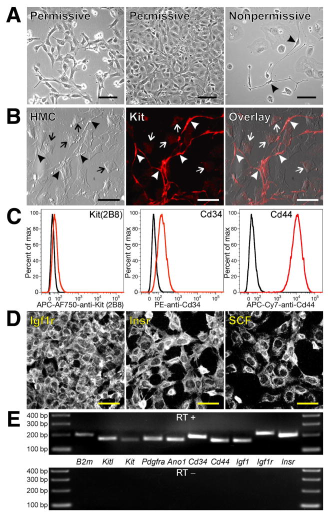
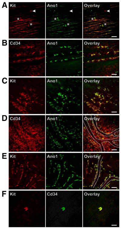
Methods: Isolated Kit(low)Cd44(+)Cd34(+) cells were characterized after serial cloning. The tumorigenic potential of spontaneously transformed cells was investigated in nude mice. The Kit(low)Cd44(+)Cd34(+) cells' responsiveness to Kit activation and blockade was studied by enumerating them in Kit(K641E) mice (a GIST model), in mice with defective Kit signaling, and pharmacologically.

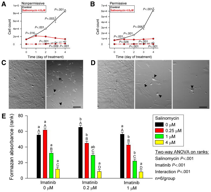
Results: Single isolated Kit(low)Cd44(+)Cd34(+) cells were clonogenic and capable of self-renewal and differentiation into ICC. In nude mice, spontaneously transformed cells formed malignant tumors expressing GIST markers. The Kit(low)Cd44(+)Cd34(+) cells were resistant to in vitro Kit blockade, including by imatinib, and occurred in normal numbers in mice with reduced Kit signaling. In Kit(K641E) mice, the mutant ICC stem cells were grossly hyperplastic but remained imatinib-resistant. In contrast, the cancer stem, cell-targeting drug salinomycin blocked the proliferation of Kit(low)Cd44(+)Cd34(+) cells and increased their sensitivity to imatinib.

Conclusions: Kit(low)Cd44(+)Cd34(+) progenitors are true stem cells for normal and hyperplastic ICC and give rise to GIST. Resistance to Kit/Pdgfra inhibitors is inherent in GIST and is caused by the native ICC stem cells' lack of dependence on Kit for survival, which is maintained after the acquisition of oncogenic Kit mutation. Cancer stem cell drugs may target these cells.


Figures

Figure 1
Figure 1. Clonally-derived, conditionally immortalized ICC progenitors maintain their undifferentiated phenotype in culture and give rise to ICC
(A) Morphology of D2211B cells grown under permissive or nonpermissive conditions. Arrowheads mark ICC-like cells. Scale bars, 100 μm. (B) Cell surface expression of Kit in the clonally-derived sub-line 2xSCS70; nonpermissive conditions; representative of 14 cultures from 4 passages). Scale bars, 50 μm. Arrowheads mark Kitbright ICC-like cells. Undifferentiated cells remained Kitlow (arrows). HMC, Hoffman modulation contrast. (C) Flow cytometry of proliferating D2211B cells. Black histograms show isotype controls. APC-AF750, allophycocyanin-Alexa Fluor 750 tandem conjugate; PE, phycoerythrin; APC-Cy7, allophycocyanin-cyanine 7 tandem conjugate. Isolated ICC progenitors maintained a KitlowCd34+Cd44+ phenotype. (D) Surface immunolabeling of D2211B cells under permissive conditions. Scale bars, 50 μm. (E) RT-PCR analysis of D2211B cells grown under permissive conditions. The reverse transcriptase was omitted from the control experiment (RT−). B2m, beta-2 microglobulin used as reference. Each reaction is a representative of at least two experiments performed in different passages.
Figure 2
Figure 2. Isolated, clonally-derived ICC stem cells transplanted into diabetic NOD/LtJ mice differentiate into ICC
(A) Expression of H-2Kb detected by flow cytometry in 2xSCS70 cells. PE-Cy5-SA, streptavidin coupled with PE-Cy5 tandem conjugate. Black histograms show controls stained with PE-Cy5-SA only. (B) Undifferentiated KitlowH-2Kb+ donor cells (arrowheads) on the serosal surface of the lesser curvature of the stomach of a transplanted mouse. All ICC (arrows) in this region were from the host. (C) An H-2Kb+Kit+ dividing cell with prominent ICC-like features in the same mouse. (D) Extensive incorporation of H-2Kb+ donor cells into Kit+ ICC networks in the distal corpus. Asterisks mark nonspecifically stained capillaries. (E) Quantitative estimate of colocalization of Kit and H-2Kb immunoreactivities in cell- and sham-transplanted mice. Overlap of red and green pixels was normalized to the number of green (Kit+) pixels in each confocal stack following equalization and binarization of the images (see Supplemental Methods). (F) Nonspecific staining of capillaries (asterisks) by mouse anti-H-2Kb monoclonal antibody and AF594-anti-mouse IgG in a sham-transplanted NOD/LtJ mouse. Background staining in the H-2Kb image was gamma-enhanced to demonstrate lack of nonspecific staining of Kit+ ICC. Scale bars are 50 μm.
Figure 3
Figure 3. Spontaneously transformed ICC stem cells transplanted into NCr-nu/nu mice give rise to malignant GIST-like tumors
(A) Tumor formed in a 2xSCS70-transplanted mouse 43 days after grafting. Scales are in centimeters. (B) Hematoxylin-eosin. Magnification is 400x. (C) Kit immunoreactivity (red; blue: DAPI) in tumors detected with a rabbit polyclonal antibody. The third panel from left is an enlargement of the area outlined in the preceding panel. Arrowhead indicates a bipolar, ICC-like cell. Arrows show dividing ICC-like cells. (D) Secondary-antibody-only control for C and F. (E) Kit immunoreactivity detected with rat monoclonal antibody 2B8. Right panel shows donor skin tissue lacking Kit immunostaining (asterisk) in the vicinity of the tumor as control. (F) Ano1 immunoreactivity. Middle panel is an enlargement of the area outlined in the left panel. Arrowheads indicate bipolar, ICC-like cells. Right panel shows Ano1+ tumor cells infiltrating host skeletal muscle (asterisks). White and yellow scale bars are 50 μm and 12.5 μm, respectively. Note moderate, generalized Kit and Ano1 immunostaining and high Kit and Ano1 expression in ICC-like cells and in some round cells.
Figure 4
Figure 4. ICC stem cells do not depend on SCF/Kit signaling for proliferation and maintenance
(A,B) D2211B cells were maintained with 5% fetal bovine serum (FBS) but without insulin for 6–10 days under the conditions indicated. Low FBS: 0.1%. End-point counts (mean+/− SEM) of PI-excluding (live) cells determined by flow cytometry are shown. Red horizontal lines indicate starting cell numbers. ACK2 (monoclonal anti-Kit antibody): 5 μg/mL; recombinant murine (rm)-SCF (Sigma): 20 ng/mL; bovine insulin (Invitrogen): 5 μg/mL, recombinant human IGF-I (Sigma): 100 ng/mL; polyclonal anti-SCF antibodies: 2 μg/mL. Note lack of effect of ACK2 and anti-SCF on basal proliferation and partial inhibition of IGF-I-induced growth. (C) Effects of imatinib mesylate. Solid lines, PI cells; dashed lines, PI+ (dead) cells. Note modest cytostatic effect under permissive conditions and lack of a cytocidal effect. Imatinib may have protected the cells from the effects of inactivating the immortalizing antigen. Groups not sharing the same label were different by post-hoc multiple comparisons. (D) Kit+ ICC in jejunal muscles from newborn BALB/c mice cultured for 3 days in the absence/presence of established inhibitors of SCF/Kit signaling. Representative confocal images collected from 2–3 tissues/group are shown. Scale bars, 50 μm. Inhibiton of SCF/Kit signaling resulted in the loss of differentiated ICC; order of efficacy: ACK2>imatinib 0.2 μM> imatinib 1.0 μM≫anti-SCF.
Figure 5
Figure 5. Gastric ICC stem cells are hyperplastic in mice with activating Kit mutation but unaffected by hypomorphic Kit
(W/Wv) and Kitl (Sl/Sld) mutations. Representative confocal images collected from whole-mounts of 3–6 stomachs/group are shown. Scale bars, 50 μm. (A) Solitary, Kit+Ano1+ ICC stem cells (asterisks) and a rare Kit+Ano1 mast cell (arrowhead) in the circular muscle layer of the antrum in a wild-type mouse. (B) ICC stem cells in the submucosa in the vicinity of the circular muscle layer of the antrum in a wild-type mouse. (C,D) Hyperplastic ICC stem cells in the submucosa close to the surface of the circular muscle in KitK641E mutant mice. These cells were particularly abundant in the Cd34+ perivascular connective tissue (D). Capillaries are marked by dotted lines. (E) ICC stem cells along capillary walls in the antrum of a W/Wv mouse and (F) in the circular muscle of the distal antrum of an adult Sl/Sld mouse lacking intramuscular ICC.
Figure 6
Figure 6. ICC stem cells are hyperplastic in KitK641E mice but unaffected by hypomorphic Kit (W/Wv) and Kitl (Sl/Sld) mutations or imatinib treatment
(A) 14–16-day-old KitK641E mice. (B) Adult W/Wv mice. (C) Adult Sl/Sld mice. +/+: age- and strain-matched, wild-type controls. Box plots show median and interquartile range; n=3–4/group. The frequency of ICC and ICC progenitors was higher in juvenile wild-type littermates of the KitK641E mice than in the adult wild-type controls for the hypomorphic mutants. (D) Effects of 28-day in vivo imatinib treatment on ICC and precursors in adult KitK641E/+ mice lacking the neomycin resistance cassette (n=3/group).
Figure 7
Figure 7. Salinomycin inhibits the proliferation of ICC stem cells and sensitizes them to imatinib
(A,B) Effects of salinomycin on the proliferation of D2211B cells. Solid lines, PI cells; dashed lines, PI+ (dead) cells. Note nearly complete inhibition. The number of PI+ dead cells was also reduced. (C) Morphology of D2211B cells cultured under nonpermissive conditions with vehicle (left panel) or salinomycin (right panel) for 4 days. Arrowhead, multipolar, ICC-like cells. Arrow, fibroblast-like cell filled with cytoplasmic vesicles. Scale bar, 25 μm. (D) Morphology of D2211B cells cultured under permissive conditions with vehicle (left panel) or salinomycin (middle and right panels) for 4 days. Note ICC-like phenotype of salinomycin-treated cells (arrowheads). Scale bar, 25 μm. (E) Dose-dependent effects of 5-day combined treatments with salinomycin and imatinib on the proliferation of D2211B cells under permissive conditions. The rank of 1 was assigned to the culture with the lowest cell number. Groups not sharing the same labels were different by post-hoc multiple comparisons. Lowercase and uppercase labels apply to groups receiving the same salinomycin and imatinib doses, respectively.

References

    1. Rubin BP, Heinrich MC, Corless CL. Gastrointestinal stromal tumour. Lancet. 2007;369:1731–1741. - PubMed
    1. Hirota S, Isozaki K, Moriyama Y, et al. Gain-of-function mutations of c-kit in human gastrointestinal stromal tumors. Science. 1998;279:577–580. - PubMed
    1. Huizinga JD, Zarate N, Farrugia G. Physiology, injury, and recovery of interstitial cells of Cajal: basic and clinical science. Gastroenterology. 2009;137:1548–1556. - PMC - PubMed
    1. Sanders KM, Ordog T, Koh SD, et al. Development and plasticity of interstitial cells of Cajal. Neurogastroenterol Motil. 1999;11:311–338. - PubMed
    1. Maeda H, Yamagata A, Nishikawa S, et al. Requirement of c-kit for development of intestinal pacemaker system. Development. 1992;116:369–375. - PubMed

Publication types

MeSH terms