. 2010 Sep;139(3):1019-29.
doi: 10.1053/j.gastro.2010.05.082. Epub 2010 Jun 2.

Therapeutic liver reconstitution with murine cells isolated long after death

Affiliations

Therapeutic liver reconstitution with murine cells isolated long after death

Laura Erker et al. Gastroenterology. 2010 Sep.

Abstract

Background & aims: Due to the shortage of donor organs, many patients needing liver transplantation cannot receive one. For some liver diseases, hepatocyte transplantation could be a viable alternative, but donor cells currently are procured from the same sources as whole organs, and thus the supply is severely limited.

Methods: Here, we investigated the possibility of isolating viable hepatocytes for liver cell therapy from the plentiful source of morgue cadavers. To determine the utility of this approach, cells were isolated from the livers of non-heart-beating cadaveric mice long after death and transplanted into fumarylacetoacetate hydrolase-deficient mice, a model for the human metabolic liver disease hereditary tyrosinemia type I and a stringent in vivo model for hepatic cell transplantation.

Results: Surprisingly, complete and therapeutic liver repopulation could be achieved with hepatocytes derived up to 27 hours post mortem.

Conclusions: Competitive repopulation experiments showed that cadaveric liver cells had a repopulation capacity similar to freshly isolated hepatocytes. Importantly, viable hepatocytes also could be isolated from cadaveric primate liver (monkey and human) efficiently. These data provide evidence that non-heart-beating donors could be a suitable source of hepatocytes for much longer time periods than previously thought possible.


Conflict of interest statement

There are no conflicts of interest to disclose for all authors.

Figures

Figure 1
Figure 1
Cadaveric mouse liver contains viable hepatocytes. (A) Cell yields from mice that were freshly killed (Fresh) or isolated 24, 27, 36, and 48 hours post-mortem. N=Number of mice measured. (B) Viability of cells isolated at different time points post-mortem; the number of mice tested is in parentheses on the x-axis. C-F; Comparison of hepatic marker expression in fresh and cadaveric cell preparations; immunocytochemistry for Fah in red (C), Albumin in green (D), G6PD in red (E) and HNF4a in red (F) and DAPI nuclear staining (blue). Controls for Fah, pre-immune sera (C), or a secondary antibody only for Alb (D), G6PD (E) and HNF4a (F) were negative.
Figure 2
Figure 2
Normal liver function in mice repopulated with cadaveric donor cells. (A-B) X-gal staining of tissue sections (A) and whole livers (B) of Fah-/- mice transplanted with fresh wild-type (Fresh WT) cells or cadaveric Rosa26. The presence of blue nodules demonstrates successful engraftment by cadaveric hepatocytes in Fah-/- mice. (C-E) Correction of hepatic functions after transplantation of cadaveric cells. Untransplanted (UT) controls are shown on the right. Blood levels of tyrosine (C), bilirubin (D), and aspartate aminotransferase (E) in Fah-/- mice transplanted with the following cell sources: freshly isolated donor (Fresh WT), 1:1 mix of Fresh:Cadaveric donor, and cadaveric cells alone (24 and 27 hours post-mortem).
Figure 3
Figure 3
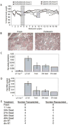
Cadaveric cells were capable of liver engraftment and repopulation. A; Representative weight curves after NTBC withdrawal of the recipients. Temporary reinstitution of NTBC treatment shown with gray over-lay; here two mice received fresh (solid lines, square and diamond) and two were transplanted with cadaveric cells (dashed lines, square and triangle). (B) Immuno-histochemistry for Fah on liver sections of transplanted Fah-/- mice, an Fah positive nodule is encircled in the left panel (Fresh), the cadaveric liver section is completely positive for Fah protein (right panel). (C) Fah enzyme activity; UT Fah-/- = untransplanted Fah mutants (n=2); UT WT= untransplanted wild-type controls (n=3); FRESH = Fah-/- mice transplanted with fresh donor cells; 24hr dead = Fah-/- mice transplanted with cadaveric cells isolated 24 hours post-mortem (n=3); 27hr dead = isolated 27 hours post-mortem (n=1). (D) Fah genomic qPCR to quantitate wild-type DNA in recipient livers. (E) Summary of cadaveric cell transplantation results for all time points.
Figure 4
Figure 4
Competitive repopulation between fresh and cadaveric donor cells. Cadaveric cells were from beta-galactosidase transgenic donors (Rosa26) and fresh cells were from non-transgenic wild-type donors (Fresh WT). (A) Fah immunohistochemistry (brown) demonstrates high levels of repopulation in all treatment groups. (B-C) The presence of cadaveric cells is confirmed by Xgal staining (blue) in tissue sections (B) and whole liver (C). (D) To quantify the relative repopulation levels from fresh and cadaveric cells qPCR for Rosa26 (black) and Fah (gray) was performed on genomic DNA from liver tissue of mice that received populations of fresh (Fah+/+) and cadaveric cells (Fah+/+ Rosa26+/-) in a 1:1 mix (n=5). Untransplanted controls are shown on the right.
Figure 5
Figure 5
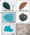
Mature hepatocytes survive death and are capable of liver engraftment. (A) Infection of R26R mice with the AAV-TTRcre virus results in widespread hepatic expression of beta-galactosidase (left panel). Negative controls are shown; the X-gal negative toe from the virus injected mouse (left panel-inset) and liver from a cadaveric R26R mouse that did not receive virus (right panel). (B) Beta-galactosidase positive binucleated hepatocytes were successfully isolated 24 hours post-mortem from R26R mice that received virus, white arrowheads mark the two nuclei of the binucleated hepatocyte (B-left panel). Successful engraftment by mature hepatocytes was confirmed by the presence of x-gal positive nodules in the recipient liver, visualized with X-gal stain in whole liver (Bright panel) and tissue sections (C-left panel), as well as by immunohistochemistry for Fah protein (C-right panel).
Figure 6
Figure 6
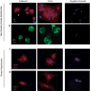
Viable hepatocytes are present in genomic DNA non-human primate and human liver. Non-human primate liver (A and B): Both cadaveric cells (left column) and fresh liver cells (right column) stain positively for hepatocyte markers Fah (A) and Albumin (B). The white arrow in the left panel (A, far left) denotes a Fah positive, binucleated, cell from cadaveric primate liver. Human liver (C and D): Both cadaveric and fresh hepatocytes from human liver were positive for G6PD (C) and HNF4a (D). Negative controls were run for each antibody, no primary antibody.

Comment in

References

    1. Fisher RA, Strom SC. Human hepatocyte transplantation: worldwide results. Transplantation. 2006;82:441–9. - PubMed
    1. Basma H, Soto-Gutierrez A, Yannam GR, Liu L, Ito R, Yamamoto T, Ellis E, Carson SD, Sato S, Chen Y, Muirhead D, Navarro-Alvarez N, Wong RJ, Roy-Chowdhury J, Platt JL, Mercer DF, Miller JD, Strom SC, Kobayashi N, Fox IJ. Differentiation and transplantation of human embryonic stem cell-derived hepatocytes. Gastroenterology. 2009;136:990–9. - PMC - PubMed
    1. Hay DC, Fletcher J, Payne C, Terrace JD, Gallagher RC, Snoeys J, Black JR, Wojtacha D, Samuel K, Hannoun Z, Pryde A, Filippi C, Currie IS, Forbes SJ, Ross JA, Newsome PN, Iredale JP. Highly efficient differentiation of hESCs to functional hepatic endoderm requires ActivinA and Wnt3a signaling. Proc Natl Acad Sci U S A. 2008;105:12301–6. - PMC - PubMed
    1. Cantz T, Zuckerman DM, Burda MR, Dandri M, Goricke B, Thalhammer S, Heckl WM, Manns MP, Petersen J, Ott M. Quantitative gene expression analysis reveals transition of fetal liver progenitor cells to mature hepatocytes after transplantation in uPA/RAG-2 mice. Am J Pathol. 2003;162:37–45. - PMC - PubMed
    1. Sharma AD, Cantz T, Vogel A, Schambach A, Haridass D, Iken M, Bleidissel M, Manns MP, Scholer HR, Ott M. Murine embryonic stem cell-derived hepatic progenitor cells engraft in recipient livers with limited capacity of liver tissue formation. Cell Transplant. 2008;17:313–23. - PubMed

Publication types

MeSH terms

LinkOut - more resources