Review
. 2010 Aug;47(13):2170-5.
doi: 10.1016/j.molimm.2010.05.009. Epub 2010 Jun 1.

Complement activation on platelets: implications for vascular inflammation and thrombosis

Affiliations
Review

Complement activation on platelets: implications for vascular inflammation and thrombosis

Ellinor I Peerschke et al. Mol Immunol. 2010 Aug.

Abstract

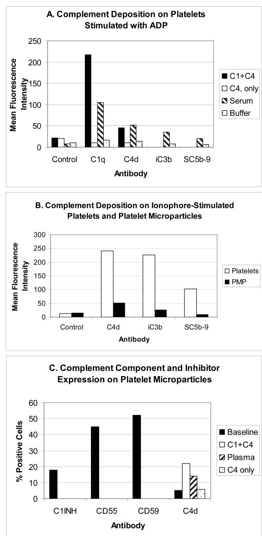
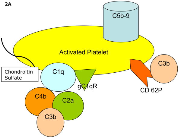
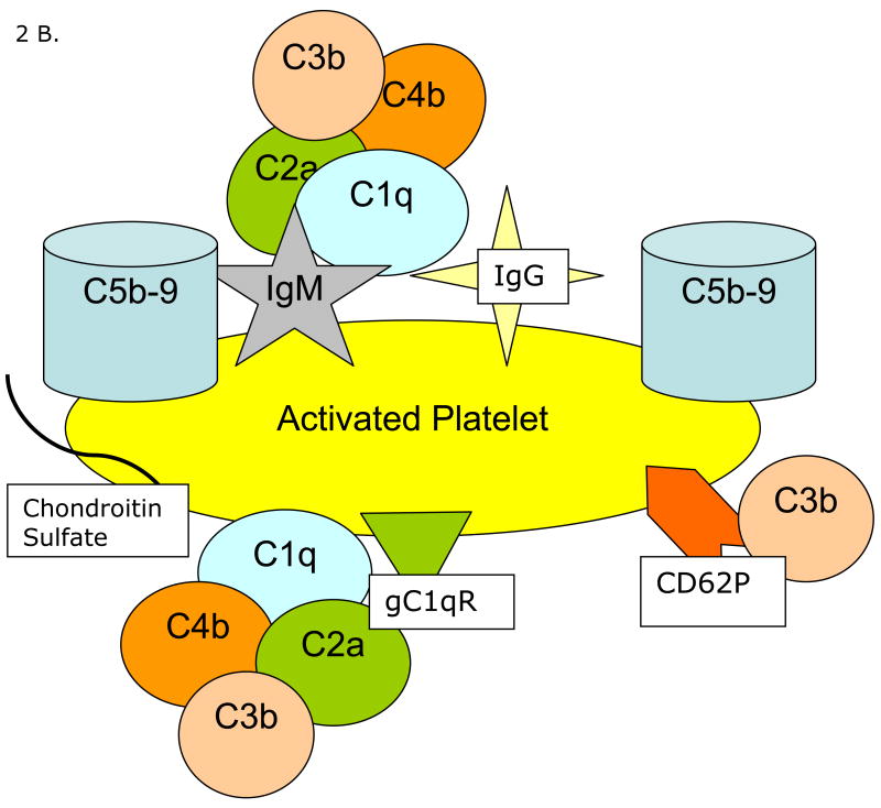
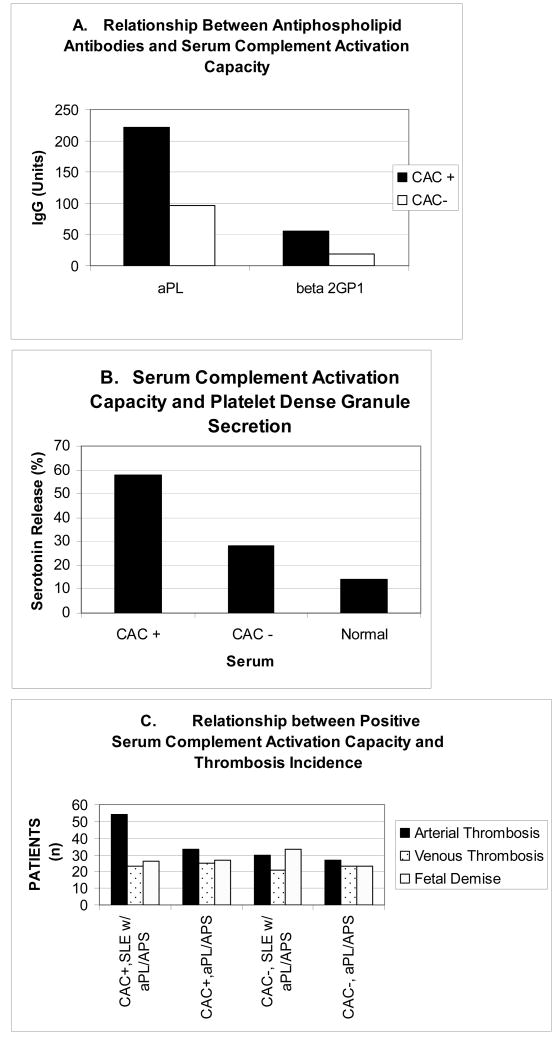
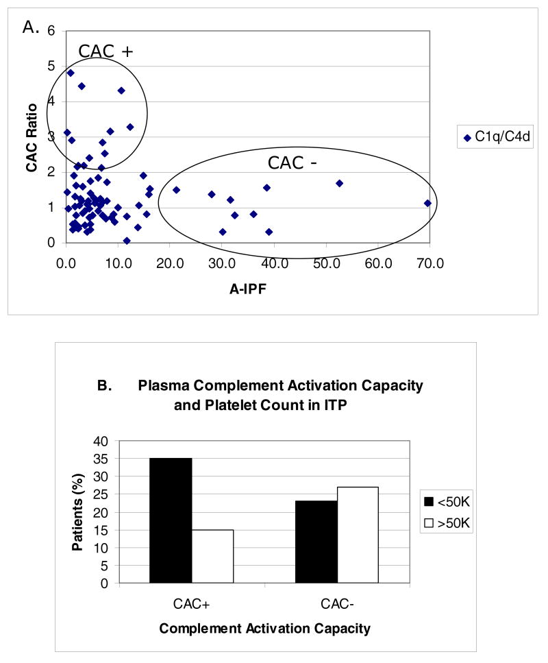
Platelets participate in a variety of responses of the blood to injury. An emerging body of evidence suggests that these cells express an intrinsic capacity to interact with and trigger both classical and alternative pathways of complement. This activity requires cell activation with biochemical agonists and/or shear stress, and is associated with the expression of P-selectin, gC1qR, and chondroitin sulfate. Platelet mediated complement activation measurably increases soluble inflammatory mediators (C3a and C5a). Platelets may also serve as targets of classical complement activation in autoimmune conditions such as antiphospholipid syndromes (APS) and immune thrombocytopenia purpura (ITP). Retrospective correlation with clinical data suggests that enhanced platelet associated complement activation correlates with increased arterial thrombotic events in patients with lupus erythematosus and APS, and evidence of enhanced platelet clearance from the circulation in patients with ITP. Taken together, these data support a role for platelet mediated complement activation in vascular inflammation and thrombosis.

Keywords: Platelets; antiphospholipid syndrome; complement; immune thrombocytopenia purpura; inflammation; systemic lupus erythematosus; thrombosis.


Figures

Figure 1
Figure 1
Complement activation on/by platelets and platelet microparticles. Results are of typical experiments. Deposition of activated complement components on platelets and microparticles was evaluated by flow cytometry using monoclonal antibodies to C1q, C4d, iC3b, and SC5b-9, and an Alexa 488-conjugated secondary goat anti rabbit antibody (F(ab)'2). Platelet and microparticle suspensions were exposed to purified complement components, normal serum (diluted 1/10 in buffer containing 1 mM CaCl2 and 1 mM MgCl2) or buffer, as a control. Following incubation (60 min, 37o C), platelets and microparticles were washed by centrifugation and resuspension, and probed with anti complement antibodies. Detailed methods are provided in Peerschke et al., 2005, and Yin et al., 2007. Panel A illustrates the deposition of complement components on platelets stimulated with 10 μM ADP. Note that detection of C4d on platelets is dependent on the presence of C1. Panel B compares complement component deposition on platelets stimulated with 5 μM ionophore (A23187) and resulting microparticles. Although complement deposition on microparticles is low compared to platelets, when results are interpreted in the context of microparticle size, complement component deposition on microparticles exceeds that on platelets. Panel C depicts complement inhibitor expression on platelet microparticles, as well as classical pathway C4d detection on microparticles following incubation with either purified complement components C1 and C4, or normal plasma (reconstituted with 1 mM CaCl2 and 1 mM MgCl2 and a selective thrombin inhibitor, 500 μM D-phenylalanyl-L-prolyl-L-arginine chloromethyl ketone, to prevent thrombin generation and fibrin formation).
Figure 2
Figure 2
Proposed mechanisms of complement activation on/by activated platelets. Panel A depicts the intrinsic capacity of platelets to activate the classical and alternative pathways of complement. Panel B demonstrates immune mediated enhanced complement activation requiring immune complex formation at the platelet surface (Panel B).
Figure 2
Figure 2
Proposed mechanisms of complement activation on/by activated platelets. Panel A depicts the intrinsic capacity of platelets to activate the classical and alternative pathways of complement. Panel B demonstrates immune mediated enhanced complement activation requiring immune complex formation at the platelet surface (Panel B).
Figure 3
Figure 3
Serum complement activating capacity (CAC) in patients with antiphospholipid antibodies (aPL), anti phospholipid syndrome (APS) and systemic lupus erythematosus (SLE). Panel A illustrates the correlation between antiphospholipid (aPL) and anti beta 2 glycoprotein 1 antibody titers expressed in GPL and SG units, respectively, and serum CAC. Sera were classified as CAC positive, if they produced a C1q and/or C4d deposition ratio on test platelets that was greater than 1.9 when compared to the assay normal serum standard. Panel B shows results of a typical experiment demonstrating the ability of CAC positive sera to activate platelets, as reflected by serotonin release from platelet dense bodies (Peerschke and Wainer, 1985). Panel C summarizes the relationship between serum complement CAC and incidence of arterial thrombosis in patients with SLE and aPL/APS, and patients with primary aPL or APS.
Figure 4
Figure 4
Plasma complement activating capacity (CAC) in patients with ITP. Panel A correlates CAC and the absolute immature platelet fraction (A-IPF) in peripheral blood. A-IPF values reflect number × 109/L. A low A-IPF suggests decreased or ineffective thrombopoiesis. Note that patients with the highest plasma CAC ratios had the lowest A-IPF, and that none of the patients with A-IPF >15 × 109/L had a positive plasma CAC. Panel B shows the relationship between CAC and thrombocytopenia, defined as circulating peripheral blood platelet counts less than 50K/μl. Plasma was classified as CAC positive, if it produced a C1q and/or C4d deposition ratio on test platelets that was greater than 1.9 when compared to the assay normal plasma standard.

References

    1. Arumugam TV, Shiels IA, Woodruff TM, Granger DN, Taylor SM. The role of the complement system in ischemia-reperfusion injury. Shock. 2004;21:401–409. - PubMed
    1. Choudhury A, Chung I, Blann AD, Lip GY. Elevated platelet microparticle levels in nonvalvular atrial fibrillation: relationship to p-selectin and antithrombotic therapy. Chest. 2007;131:809–815. - PubMed
    1. Cines DB, Bussel JB, Liebman HA, Luning Prak ET. The ITP syndrome: pathogenic and clinical diversity. Blood. 2009;113:6511. - PMC - PubMed
    1. Davis WD, Brey RL. Antiphospholipid antibodies and complement activation in patients with cerebral ischemia. Clin Exp Immunol. 1992;10:445–460. - PubMed
    1. DeJong A, Siboh V, Robbins D. Antiphospholipid antibodies and platelets. Curr Rheum Rep. 2000;2:238–245. - PubMed

Publication types

MeSH terms