

Curcumin decreases amyloid-beta peptide levels by attenuating the maturation of amyloid-beta precursor protein
- PMID: 20622013
- PMCID: PMC2937872
- DOI: 10.1074/jbc.M110.133520
Curcumin decreases amyloid-beta peptide levels by attenuating the maturation of amyloid-beta precursor protein
Abstract
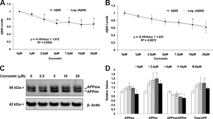
Alzheimer disease (AD) is a devastating neurodegenerative disease with no cure. The pathogenesis of AD is believed to be driven primarily by amyloid-beta (Abeta), the principal component of senile plaques. Abeta is an approximately 4-kDa peptide generated via cleavage of the amyloid-beta precursor protein (APP). Curcumin is a compound in the widely used culinary spice, turmeric, which possesses potent and broad biological activities, including anti-inflammatory and antioxidant activities, chemopreventative effects, and effects on protein trafficking. Recent in vivo studies indicate that curcumin is able to reduce Abeta-related pathology in transgenic AD mouse models via unknown molecular mechanisms. Here, we investigated the effects of curcumin on Abeta levels and APP processing in various cell lines and mouse primary cortical neurons. We show for the first time that curcumin potently lowers Abeta levels by attenuating the maturation of APP in the secretory pathway. These data provide a mechanism of action for the ability of curcumin to attenuate amyloid-beta pathology.
Figures


Comment in
-
Curcumin and Alzheimer disease: this marriage is not to be performed.J Biol Chem. 2011 Jan 21;286(3):le3; author reply le4. doi: 10.1074/jbc.L110.133520. J Biol Chem. 2011. PMID: 21239508 Free PMC article. No abstract available.
Similar articles
-
Curcumin and Alzheimer disease: this marriage is not to be performed.J Biol Chem. 2011 Jan 21;286(3):le3; author reply le4. doi: 10.1074/jbc.L110.133520. J Biol Chem. 2011. PMID: 21239508 Free PMC article. No abstract available.
-
A Curcumin Analog Reduces Levels of the Alzheimer's Disease-Associated Amyloid-β Protein by Modulating AβPP Processing and Autophagy.J Alzheimers Dis. 2019;72(3):761-771. doi: 10.3233/JAD-190562. J Alzheimers Dis. 2019. PMID: 31640096
-
Cerebrolysin decreases amyloid-beta production by regulating amyloid protein precursor maturation in a transgenic model of Alzheimer's disease.J Neurosci Res. 2006 May 15;83(7):1252-61. doi: 10.1002/jnr.20818. J Neurosci Res. 2006. PMID: 16511867
-
Alzheimer's disease.Subcell Biochem. 2012;65:329-52. doi: 10.1007/978-94-007-5416-4_14. Subcell Biochem. 2012. PMID: 23225010 Review.
-
Role of apoe/Abeta interactions in the pathogenesis of Alzheimer's disease and cerebral amyloid angiopathy.J Mol Neurosci. 2001 Oct;17(2):147-55. doi: 10.1385/JMN:17:2:147. J Mol Neurosci. 2001. PMID: 11816788 Review.
Cited by
-
Diverse effects of a low dose supplement of lipidated curcumin in healthy middle aged people.Nutr J. 2012 Sep 26;11:79. doi: 10.1186/1475-2891-11-79. Nutr J. 2012. PMID: 23013352 Free PMC article. Clinical Trial.
-
Upregulation of Alzheimer's Disease Amyloid-β Protein Precursor in Astrocytes Both in vitro and in vivo.J Alzheimers Dis. 2020;76(3):1071-1082. doi: 10.3233/JAD-200128. J Alzheimers Dis. 2020. PMID: 32597805 Free PMC article.
-
A Curcumin Analog Exhibits Multiple Biologic Effects on the Pathogenesis of Alzheimer's Disease and Improves Behavior, Inflammation, and β-Amyloid Accumulation in a Mouse Model.Int J Mol Sci. 2020 Jul 30;21(15):5459. doi: 10.3390/ijms21155459. Int J Mol Sci. 2020. PMID: 32751716 Free PMC article.
-
Lessons from two prevalent amyloidoses-what amylin and Aβ have in common.Front Aging Neurosci. 2013 Aug 8;5:38. doi: 10.3389/fnagi.2013.00038. eCollection 2013. Front Aging Neurosci. 2013. PMID: 23964237 Free PMC article.
-
Diets involved in PPAR and PI3K/AKT/PTEN pathway may contribute to neuroprotection in a traumatic brain injury.Alzheimers Res Ther. 2013 Sep 26;5(5):42. doi: 10.1186/alzrt208. eCollection 2013. Alzheimers Res Ther. 2013. PMID: 24074163 Free PMC article. Review.
References
-
- Cummings J. L. (2004) N. Engl. J. Med. 351, 56–67 - PubMed
-
- Bertram L., Tanzi R. E. (2008) Nat. Rev. Neurosci. 9, 768–778 - PubMed
-
- Tanzi R. E., Bertram L. (2005) Cell 120, 545–555 - PubMed
-
- Tanzi R. E., Gusella J. F., Watkins P. C., Bruns G. A., St George-Hyslop P., Van Keuren M. L., Patterson D., Pagan S., Kurnit D. M., Neve R. L. (1987) Science 235, 880–884 - PubMed
-
- Selkoe D. J. (2002) Science 298, 789–791 - PubMed
Publication types
MeSH terms
Substances
LinkOut - more resources
Full Text Sources
Molecular Biology Databases
