. 2010 Jul 27;107(30):13258-63.
doi: 10.1073/pnas.1006344107. Epub 2010 Jul 9.

High-throughput engineering and analysis of peptide binding to class II MHC

Affiliations

High-throughput engineering and analysis of peptide binding to class II MHC

Wei Jiang et al. Proc Natl Acad Sci U S A. .

Abstract

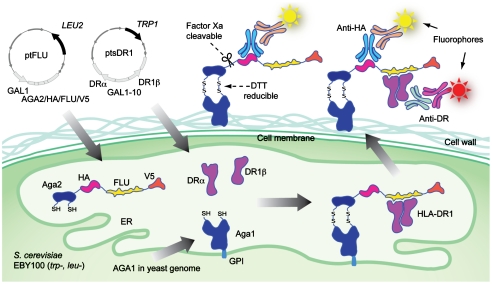
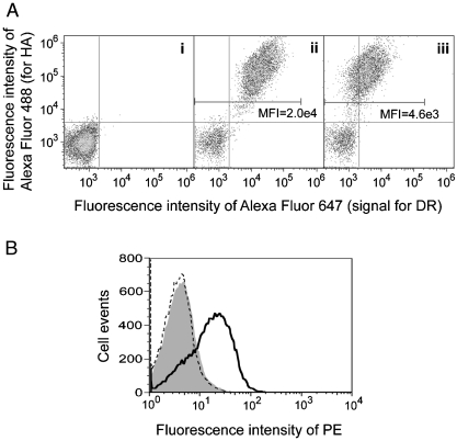
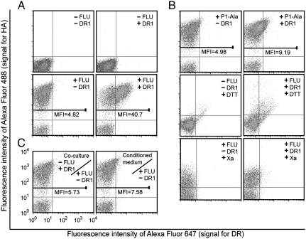
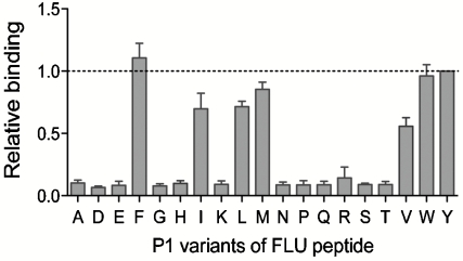
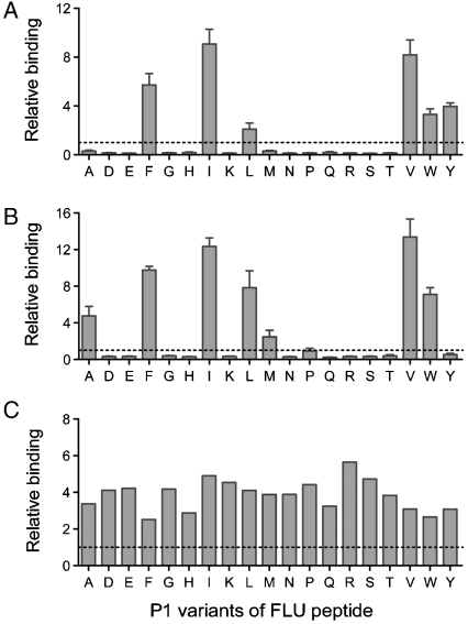
Class II major histocompatibility complex (MHC-II) proteins govern stimulation of adaptive immunity by presenting antigenic peptides to CD4+ T lymphocytes. Many allelic variants of MHC-II exist with implications in peptide presentation and immunity; thus, high-throughput experimental tools for rapid and quantitative analysis of peptide binding to MHC-II are needed. Here, we present an expression system wherein peptide and MHC-II are codisplayed on the surface of yeast in an intracellular association-dependent manner and assayed by flow cytometry. Accordingly, the relative binding of different peptides and/or MHC-II variants can be assayed by genetically manipulating either partner, enabling the application of directed evolution approaches for high-throughput characterization or engineering. We demonstrate the application of this tool to map the side-chain preference for peptides binding to HLA-DR1 and to evolve novel HLA-DR1 mutants with altered peptide-binding specificity.


Conflict of interest statement

The authors declare no conflict of interest.

Figures

Fig. 1.
Fig. 1.
Design of the yeast codisplay system for HLA-DR1. Aga2p-fused FLU peptide (PKYVKQNTLKLAT) is expressed flanked by the HA and V5 epitope tags, enabling detection of peptide levels by immunofluorescent staining with antibodies specific for either tag. The HLA-DRα and HLA-DR1β chain extracellular domains are expressed from separate cassettes. The FLU peptide is anchored to the cell surface via native processing and secretion of the assembled a-agglutinin protein (composed of the Aga1p and Aga2p subunits) as described (22), and the HLA-DR1 heterodimer is anchored by noncovalent binding to FLU. Relative fluorescence levels of different fluorophores coupled to antitag and anti-DR reagents indicate the level of saturation of available peptides by bound HLA-DR1. GPI: glycosylphosphatidylinositol.
Fig. 2.
Fig. 2.
Expression of functional HLA-DR1 heterodimer in yeast by surface display. (A). Aga2p and HLA-DR1 levels of yeast expressing (i) an irrelevant soluble protein, (ii) Aga2p-fused HLA-DR1 with FLU-covalently linked to the β chain N-terminus, or (iii) empty Aga2p-fused HLA-DR1. Cells were double-labeled with reagents specific for the HA epitope tag (Aga2p level) and HLA-DRα. Mean fluorescence intensity (MFI) of DR signal for HA positive cell population was indicated. (B) Peptide-binding capability of yeast-displayed HLA-DR1. Yeast displaying empty HLA-DR1 (solid curve) or an irrelevant protein (shaded) were incubated with 100 μM biotinylated FLU at pH 5.0; HLA-DR1-displaying yeast were also incubated with 100 μM biotinylated control peptide (dashed curve). All samples were stained with streptavidin-PE and analyzed by flow cytometry.
Fig. 3.
Fig. 3.
Yeast codisplay and surface detection of FLU peptide and FLU-bound soluble HLA-DR1. (A) The yeast parent strain or yeast expressing HLA-DR1, Aga2p-FLU, or both were double-labeled with anti-HA and anti-DR1 antibodies and analyzed by flow cytometry. (B) The yeast parent strain and yeast expressing Aga2p-P1-Ala alone or with HLA-DR1 or yeast expressing Aga2p-FLU with or without coexpressed HLA-DR1 treated with DTT or Factor Xa protease, as indicated, were double-labeled and analyzed by flow cytometry. (C) Yeast expressing Aga2p-FLU were cocultured at 1∶1 ratio with yeast expressing soluble HLA-DR1 or cultured in medium preconditioned by growing codisplaying yeast prior to labeling and analysis.
Fig. 4.
Fig. 4.
Peptide P1 anchor residue specificity of HLA-DR1 analyzed by yeast codisplay. Cells codisplaying HLA-DR1 and FLU peptides with the indicated P1 residue substitutions were analyzed by flow cytometry (Fig. S3) and normalized relative binding levels were calculated from fluorescence intensities (Materials and Methods). The dashed line represents binding to the wild-type FLU peptide with Tyr at P1. Error bars represent standard error of the mean determined from four independent experiments.
Fig. 5.
Fig. 5.
Peptide P1 anchor residue specificity of HLA-DR1 mutants generated by directed evolution. Relative binding to the P1 substituted FLU peptide panel were determined for (A) clone S4V1.5 selected against P1-Val, (B) clone S4A.19 selected against P1-Ala, and (C) S4E1.3 selected against P1-Glu. Dashed lines indicate the relative binding signal for wild-type HLA-DR1 codisplayed with wild-type FLU. Error bars represent standard error of the mean determined from four independent experiments.
Fig. 6.
Fig. 6.
Mutation sites conferring altered P1 anchor specificity on HLA-DR1. Positions mutated in S4V1.5 (cyan), S4A1.9 (orange), S4E1.3 (magenta), or all three clones (green) are highlighted on a top view of the α1 and β1 domains, with the peptide anchor residue positions P1, P4, P6, P7, and P9 also highlighted (blue). Images generated from coordinates in PDB file 1DHL.

References

    1. Brown JH, et al. Three-dimensional structure of the human class II histocompatibility antigen HLA-DR1. Nature. 1993;364:33–39. - PubMed
    1. Vyas JM, Van der Veen AG, Ploegh HL. The known unknowns of antigen processing and presentation. Nat Rev Immunol. 2008;8:607–618. - PMC - PubMed
    1. Watts C. The exogenous pathway for antigen presentation on major histocompatibility complex class II and CD1 molecules. Nat Immunol. 2004;5:685–692. - PubMed
    1. Hammer J, et al. Promiscuous and allele-specific anchors in HLA-DR-binding peptides. Cell. 1993;74:197–203. - PubMed
    1. Stern LJ, et al. Crystal structure of the human class II MHC protein HLA-DR1 complexed with an influenza virus peptide. Nature. 1994;368:215–221. - PubMed

Publication types

MeSH terms