. 2010 Sep 10;285(37):28540-52.
doi: 10.1074/jbc.M110.151779. Epub 2010 Jul 11.

Superinhibitory phospholamban mutants compete with Ca2+ for binding to SERCA2a by stabilizing a unique nucleotide-dependent conformational state

Affiliations

Superinhibitory phospholamban mutants compete with Ca2+ for binding to SERCA2a by stabilizing a unique nucleotide-dependent conformational state

Brandy L Akin et al. J Biol Chem. .

Abstract

Three cross-linkable phospholamban (PLB) mutants of increasing inhibitory strength (N30C-PLB < N27A,N30C,L37A-PLB (PLB3) < N27A,N30C,L37A,V49G-PLB (PLB4)) were used to determine whether PLB decreases the Ca(2+) affinity of SERCA2a by competing for Ca(2+) binding. The functional effects of N30C-PLB, PLB3, and PLB4 on Ca(2+)-ATPase activity and E1 approximately P formation were correlated with their binding interactions with SERCA2a measured by chemical cross-linking. Successively higher Ca(2+) concentrations were required to both activate the enzyme co-expressed with N30C-PLB, PLB3, and PLB4 and to dissociate N30C-PLB, PLB3, and PLB4 from SERCA2a, suggesting competition between PLB and Ca(2+) for binding to SERCA2a. This was confirmed with the Ca(2+) pump mutant, D351A, which is catalytically inactive but retains strong Ca(2+) binding. Increasingly higher Ca(2+) concentrations were also required to dissociate N30C-PLB, PLB3, and PLB4 from D351A, demonstrating directly that PLB antagonizes Ca(2+) binding. Finally, the specific conformation of E2 (Ca(2+)-free state of SERCA2a) that binds PLB was investigated using the Ca(2+)-pump inhibitors thapsigargin and vanadate. Cross-linking assays conducted in the absence of Ca(2+) showed that PLB bound preferentially to E2 with bound nucleotide, forming a remarkably stable complex that is highly resistant to both thapsigargin and vanadate. In the presence of ATP, N30C-PLB had an affinity for SERCA2a approaching that of vanadate (micromolar), whereas PLB3 and PLB4 had much higher affinities, severalfold greater than even thapsigargin (nanomolar or higher). We conclude that PLB decreases Ca(2+) binding to SERCA2a by stabilizing a unique E2.ATP state that is unable to bind thapsigargin or vanadate.


Figures
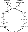
FIGURE 1.
FIGURE 1.
Reaction cycle of SERCA2a. E1 and E2 represent the high and low Ca2+ affinity conformations of SERCA2a, respectively. After sequential binding of two Ca2+ ions to E1, the enzyme is phosphorylated with the γ-phosphate of ATP at Asp351, forming the high energy intermediate, E1∼P. Ca2+ translocation across the SR membrane occurs during the E1 to E2 transition. TG inhibits Ca2+-ATPase activity by forming a dead-end complex with the enzyme in E2 (E2·TG) (18). E2·TG has a greatly reduced affinity for ATP relative to TG-free E2 (33, 34). PLB cross-linking studies indicate that PLB binds preferentially to E2 with bound ATP (E2·ATP·PLB). PLB does not bind to E2·TG or E2·cyclopiazonic acid (7, 10), E2-P (17), or to the Ca2+ pump with Ca2+ binding site 1 (12) or both sites (4, 17) occupied.
FIGURE 2.
FIGURE 2.
Complete amino acid sequences of the cross-linkable PLB mutants, N30C-PLB, PLB3, and PLB4. I and II designate cytoplasmic and transmembrane domains of PLB, respectively. Domain IA contains Ser16 and Thr17, the residues phosphorylated in response to β-adrenergic stimulation. The point mutations N27A in domain IB (15) and L37A (3, 4) and V49G (16, 17) in domain II are all supershifting mutations. The N30C mutation in domain IB allows PLB to be cross-linked to Lys328 of SERCA2a with KMUS (10). For consistency with previous publications, N30C-PLB was made on the Cys-less PLB background in which Cys residues 36, 41, and 46 were mutated to Ala (7, 10–12, 17).
FIGURE 3.
FIGURE 3.
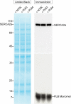
Amido Black staining and immunoblot of SERCA2a co-expressed with N30C-PLB, PLB3, and PLB4. SERCA2a and N30C-PLB, PLB3, or PLB4 were co-expressed in Sf21 insect cells. Membrane samples (11 μg) were then subjected to SDS-PAGE, transferred to nitrocellulose, and the nitrocellulose sheet stained with Amido Black (left panel). The nitrocellulose sheet was then cut in half, and the upper portion was probed with the anti-SERCA2a antibody, 2A7-A1, and the lower half was probed with the anti-PLB antibody, 2D12, followed by 125I-protein A (right panel). Control experiments showed that the 2D12 antibody bound with equal strength to all three PLB mutants (data not shown).
FIGURE 4.
FIGURE 4.
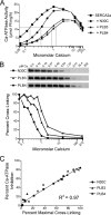
Ca2+ activation of Ca2+-ATPase activity and Ca2+ inhibition of cross-linking. SERCA2a was expressed alone or co-expressed with N30C-PLB, PLB3, or PLB4 in Sf21 cells and SERCA2a and PLB expression levels were determined by Western blotting. Panel A depicts Ca2+-ATPase activities of membrane fractions measured as described under “Experimental Procedures.” Enzyme activities were normalized to expression levels of SERCA2a expressed alone. The gray line intersecting the ordinate indicates the 50% Vmax value determined for SERCA2a expressed alone. Panel B shows cross-linking of the PLB mutants to SERCA2 determined under identical conditions as the Ca2+-ATPase assay. Aliquots were taken from the Ca2+-ATPase assay and cross-linked for 15 s with 1 mm KMUS at 37 °C. Samples were then subjected to SDS-PAGE and immunoblotting with the anti-PLB antibody, 2D12. Protein bands in the upper panel show SERCA2a cross-linked with the PLB monomer. PLB cross-linking is quantified in the graph below. The graph in panel C was derived from the data in panels A and B. The percent of maximal PLB cross-linking to SERCA2a (determined in the absence of Ca2+) was calculated at each Ca2+ concentration for each PLB mutant, and then plotted against the percent inhibition of Ca2+-ATPase activity by PLB obtained at the same Ca2+ concentration. The percent inhibition of Ca2+-ATPase activity by PLB at each Ca2+ concentration was calculated by dividing the Ca2+-ATPase activity of membranes expressing SERCA2a plus PLB by the Ca2+-ATPase activity of membranes expressing SERCA2a alone, and multiplying by 100.
FIGURE 5.
FIGURE 5.
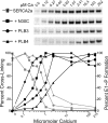
PLB effect on formation of the phosphorylated enzyme intermediate. Ca2+ stimulation of phosphoenzyme formation by [γ-32P]ATP was determined for SERCA2a expressed alone, and co-expressed with each PLB mutant. The upper panel is the autoradiograph depicting the radioactive phosphoenzyme, and results are plotted below (dark lines). For comparison, Ca2+ effects on N30C-PLB (squares), PLB3 (circles), and PLB4 (triangles) cross-linking to SERC2a are also displayed (gray lines), taken from Fig. 4B.
FIGURE 6.
FIGURE 6.
Effect of 2D12 on Ca2+-ATPase activity (A–C) and PLB cross-linking to SERCA2a (D–F). Ca2+-ATPase activities were determined in the presence (2D12) and absence (Con) of 2D12 as described under “Experimental Procedures” for membranes co-expressing SERCA2a and each PLB mutant. Gray lines show results obtained for membranes expressing SERCA2a alone. Cross-linking of PLB to SERCA2a was determined under identical conditions as the Ca2+-ATPase assay.
FIGURE 7.
FIGURE 7.
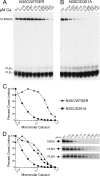
Ca2+ effect on PLB cross-linking to D351A. Ca2+ inhibition of N30C-PLB cross-linking to wild-type SERCA2a (A) and D351A (B) was determined under Ca2+-ATPase assay conditions, as described under “Experimental Procedures.” PLB/SER designates the PLB monomer cross-linked to the Ca2+ pump at 110 kDa, and free PLB monomers (PLB1) and dimers (PLB2) are visible below at 6 and 12 kDa, respectively. The full autoradiographs are shown, demonstrating the highly specific cross-linking reaction; PLB cross-linked exclusively to expressed wild-type SERCA2a or D351A in Sf21 membranes. C, graph of Ca2+ inhibition of N30C-PLB cross-linking to wild-type SERCA2a and D351A. D, Ca2+ inhibition of N30C-PLB, PLB3, and PLB4 cross-linking to D351A. Ki values for Ca2+ inhibition of cross-linking to D351A are listed under “Results.”
FIGURE 8.
FIGURE 8.
TG effect on PLB cross-linking. A, autoradiographs showing concentration dependence of TG inhibition N30C-PLB, PLB3, and PLB4 cross-linking to SERCA2a, measured in the absence (−ATP) and presence (+ATP) of 3 mm ATP. B and C, graphs of TG inhibition of cross-linking, determined in the absence and presence of 3 mm ATP, respectively.
FIGURE 9.
FIGURE 9.
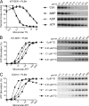
Nucleotide effect on PLB4 cross-linking to wild-type SERCA2a and D351A. A, effect of 3 mm AMP, ADP, ATP, and no added nucleotide (Con) on PLB4 cross-linking to wild-type SERCA2a. TG concentrations were varied as indicated. B, ATP stimulation of PLB4 cross-linking to wild-type SERCA2a, determined at different TG concentrations. ATP concentrations were varied as indicated. C, ATP stimulation of PLB4 cross-linking to D351A, determined at different TG concentrations.
FIGURE 10.
FIGURE 10.
Vanadate effect on PLB cross-linking to SERCA2a. A, autoradiographs showing concentration dependence of vanadate inhibition N30C-PLB, PLB3, and PLB4 cross-linking to SERCA2a, measured in the absence (−ATP) and presence (+ATP) of 36 μm ATP. B and C, graphs of vanadate inhibition of cross-linking, determined in the absence and presence of 36 μm ATP, respectively.

References

    1. Simmerman H. K., Jones L. R. (1998) Physiol. Rev. 78, 921–947 - PubMed
    1. MacLennan D. H., Kranias E. G. (2003) Nat. Rev. Mol. Cell Biol. 4, 566–577 - PubMed
    1. Kimura Y., Kurzydlowski K., Tada M., MacLennan D. H. (1997) J. Biol. Chem. 272, 15061–16064 - PubMed
    1. Autry J. M., Jones L. R. (1997) J. Biol. Chem. 272, 15872–15880 - PubMed
    1. Cornea R. L., Jones L. R., Autry J. M., Thomas D. D. (1997) Biochemistry 36, 2960–2967 - PubMed

Publication types

MeSH terms

LinkOut - more resources