Review
. 2010 Sep;32(3):275-87.
doi: 10.1007/s00281-010-0216-x. Epub 2010 Jul 11.

Knocking at the brain's door: intravital two-photon imaging of autoreactive T cell interactions with CNS structures

Affiliations
Review

Knocking at the brain's door: intravital two-photon imaging of autoreactive T cell interactions with CNS structures

Naoto Kawakami et al. Semin Immunopathol. 2010 Sep.

Abstract

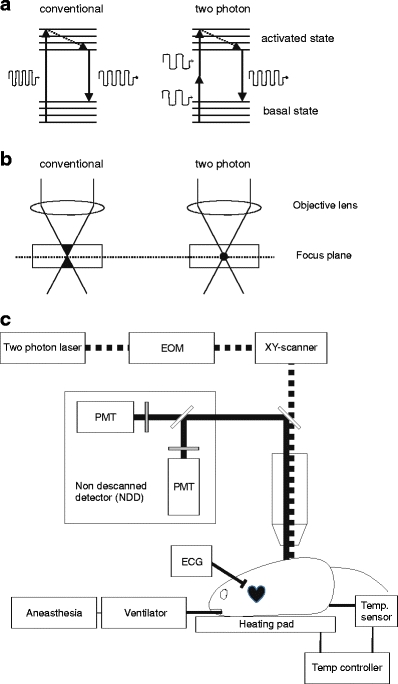
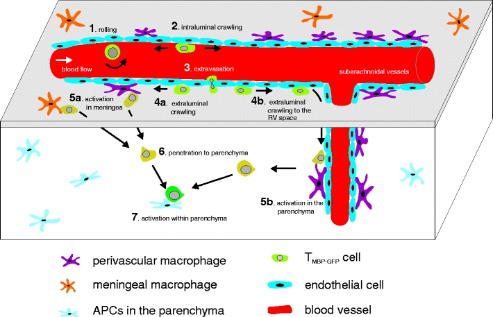
Since the first applications of two-photon microscopy in immunology 10 years ago, the number of studies using this advanced technology has increased dramatically. The two-photon microscope allows long-term visualization of cell motility in the living tissue with minimal phototoxicity. Using this technique, we examined brain autoantigen-specific T cell behavior in experimental autoimmune encephalitomyelitis, the animal model of human multiple sclerosis. Even before disease symptoms appear, the autoreactive T cells arrive at their target organ. There they crawl along the intraluminal surface of central nervous system (CNS) blood vessels before they extravasate. In the perivascular environment, the T cells meet phagocytes that present autoantigens. This contact activates the T cells to penetrate deep into the CNS parenchyma, where the infiltrated T cells again can find antigen, be further activated, and produce cytokines, resulting in massive immune cell recruitment and clinical disease.


Figures

Fig. 1
Fig. 1
a, b Comparison between conventional one-photon excitation versus two-photon excitation. a During conventional excitation, a photon activates a fluorescence molecule (left), whereas during two-photon excitation, two photons can sum up their energy to activate a fluorescence molecule (right). After nonradiative relaxation (dotted line), a fluorescence molecule emits fluorescence, which is common in both conventional and two-photon excitation. b Conventional laser can excite a fluorescence molecule on its way (indicated as black area in left). However, fluorescent molecules can be activated only at the focus point since two-photon effect needs energy accumulation (indicated as black circle in right). c CNS intravital imaging setup. The animal is anesthetized and respiration is controlled during the entire imaging session. Additionally, body temperature is kept at 37°C and ECG is monitored. Two-photon laser is routed to xy-scanner via EOM to control laser power. Thereafter, laser is focused by objective lens and reaches the sample. Emission is collected by the objective lens and detected by NDDs which are located immediately behind it
Fig. 2
Fig. 2
Intravital recording of TMBP-GFP cells (green) in vessels of the CNS (a) and the ear (b). Vessels were marked by i.v. injection of Texas Red dextran conjugates (red). Thirty-minute movies, consisting of 60 time points, were time-projected. Crawling T cells appear as continuous lines, whereas rolling cells appear as round-shaped dots. Scale bar 30 μm
Fig. 3
Fig. 3
Schema of the invasion steps of TMBP-GFP cells into the CNS parenchyma. TMBP-GFP cells arrive at CNS leptomeningeal vessels and roll on endothelial cells (1). This is followed by intraluminal crawling directed both upstream and downstream of blood flow (2). After the extravasation (3), TMBP-GFP cells continue to crawl to the abluminal surface of vessels (4a). A part of T cells dive to the Robin Virchow space (RV; 4b). T cells meet local antigen-presenting cells at the perivascular space of leptomeningeal vessels (5a) or parenchyma (5b) and are activated. Activated T cells are allowed to penetrate within the CNS parenchyma (6). Finally, T cells can be further activated by antigen-presenting cells in the CNS parenchyma (7)

References

    1. Hickey WF, Osborn JP, Kirby WM. Expression of Ia molecules by astrocytes during acute experimental allergic encephalomyelitis in the Lewis rat. Cell Immunol. 1985;91:528–535. doi: 10.1016/0008-8749(85)90251-5. - DOI - PubMed
    1. Traugott U, Scheinberg LC, Raine CS. On the presence of Ia-positive endothelial cells and astrocytes in multiple sclerosis lesions and its relevance to antigen presentation. J Neuroimmunol. 1985;8:1–14. doi: 10.1016/S0165-5728(85)80043-6. - DOI - PubMed
    1. Gold R, Linington C, Lassmann H. Understanding pathogenesis and therapy of multiple sclerosis via animal models: 70 years of merits and culprits in experimental autoimmune encephalomyelitis research. Brain. 2006;129:1953–1971. doi: 10.1093/brain/awl075. - DOI - PubMed
    1. Freund J, Stern ER, Pisani TM. Isoallergic encephalomyelitis and radiculitis in Guinea pigs after one injection of brain and mycobacteria in water-in-oil emulsion. J Immunol. 1947;57:179–195. - PubMed
    1. Ben-Nun A, Wekerle H, Cohen IR. The rapid isolation of clonable antigen-specific T lymphocyte lines capable of mediating autoimmune encephalomyelitis. Eur J Immunol. 1981;11:195–199. doi: 10.1002/eji.1830110307. - DOI - PubMed

Publication types

MeSH terms

Substances

LinkOut - more resources