Review
. 2011 Jan 1;14(1):137-67.
doi: 10.1089/ars.2010.3153. Epub 2010 Oct 26.

Heme oxygenase in the regulation of vascular biology: from molecular mechanisms to therapeutic opportunities

Affiliations
Review

Heme oxygenase in the regulation of vascular biology: from molecular mechanisms to therapeutic opportunities

Young-Myeong Kim et al. Antioxid Redox Signal. .

Abstract

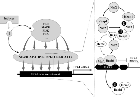
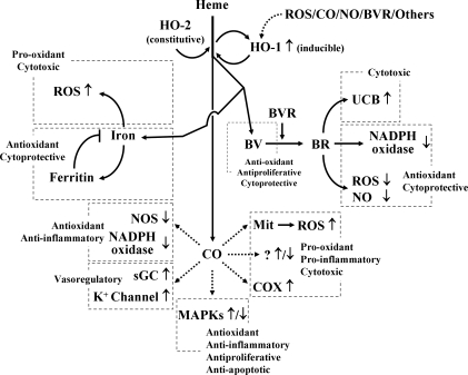
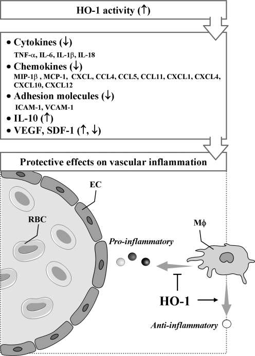
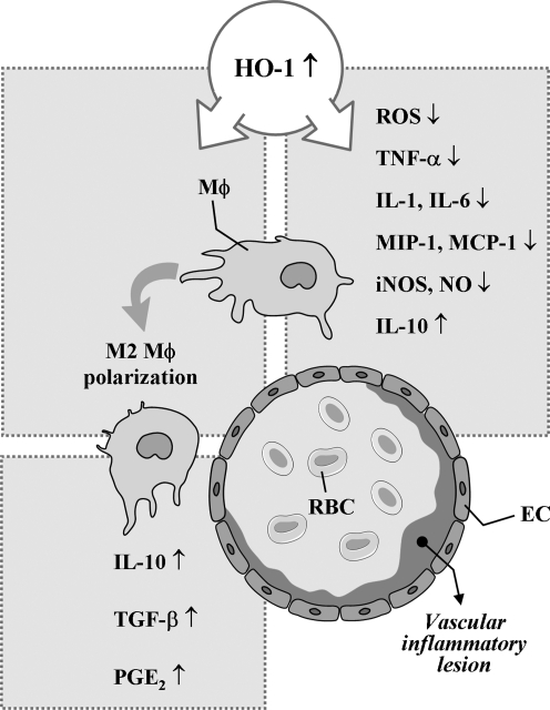
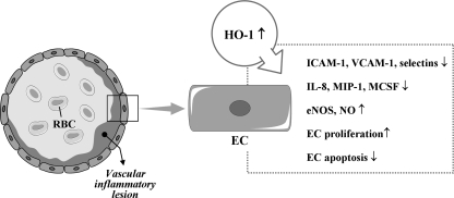
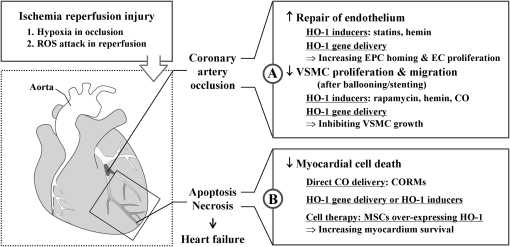
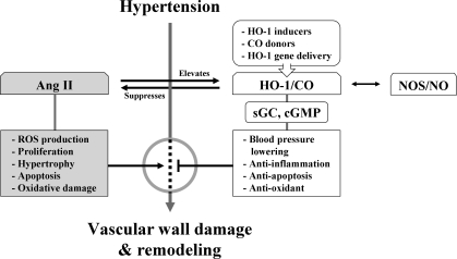
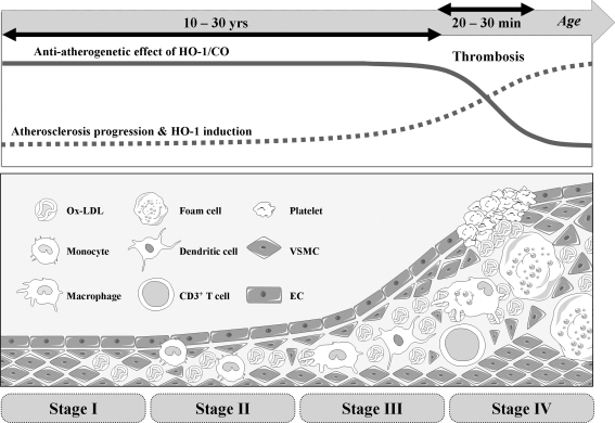
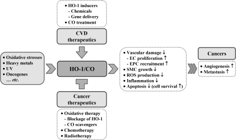
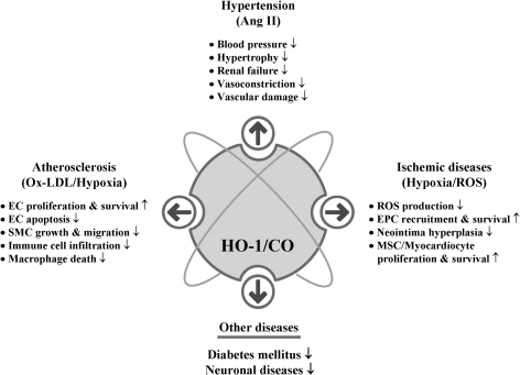
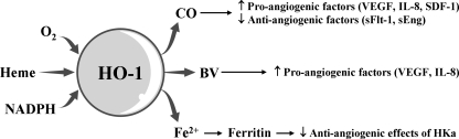
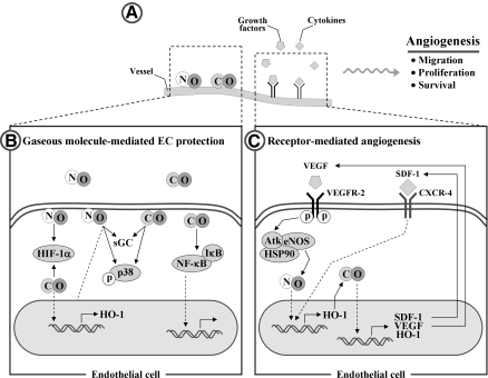
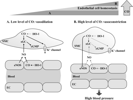
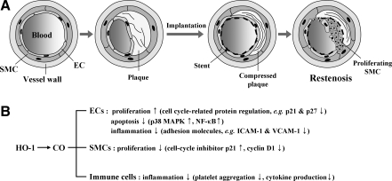
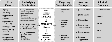
Heme oxygenases (HOs) are the rate-limiting enzymes in the catabolism of heme into biliverdin, free iron, and carbon monoxide. Two genetically distinct isoforms of HO have been characterized: an inducible form, HO-1, and a constitutively expressed form, HO-2. HO-1 is a kind of stress protein, and thus regarded as a sensitive and reliable indicator of cellular oxidative stress. The HO system acts as potent antioxidants, protects endothelial cells from apoptosis, is involved in regulating vascular tone, attenuates inflammatory response in the vessel wall, and participates in angiogenesis and vasculogenesis. Endothelial integrity and activity are thought to occupy the central position in the pathogenesis of cardiovascular diseases. Cardiovascular disease risk conditions converge in the contribution to oxidative stress. The oxidative stress leads to endothelial and vascular smooth muscle cell dysfunction with increases in vessel tone, cell growth, and gene expression that create a pro-thrombotic/pro-inflammatory environment. Subsequent formation, progression, and obstruction of atherosclerotic plaque may result in myocardial infarction, stroke, and cardiovascular death. This background provides the rationale for exploring the potential therapeutic role for HO system in the amelioration of vascular inflammation and prevention of adverse cardiovascular outcomes.


Figures

FIG. 1.
FIG. 1.
Regulation of HO-1 induction by transcription factors and kinases. A HO-1 inducer may activate at least one or more of kinases (e.g., MAPKs, PKC, PKA, and PI3K) and/or one of transcription factors (e.g., NF-κB, activator protein-1, Nrf2, CREB, BVR, and activating transcription factor 2). Under normal conditions, the transcription factors are located in cytosol or nucleus. Upon activation by external stimuli, the active forms of the transcription factors may translocate to the nucleus where they bind to the specific DNA sequence leading to the transcription of ho-1 gene. The dotted box shows a typical example in which Nrf2, Keap1, and Bach1 may interact with each other in response to free heme or ROS. Under normal conditions, Bach1/small Maf complexes bind constitutively MARE in the ho-1 gene promoter and inhibit ho-1 gene transcription. Nrf2 is ubiquitinated by forming a complex with the Keap1 (see text). In response to heme and/or ROS, Bach1 is exported from the nucleus, ubiquitinated (circled U), and degraded, releasing transcriptional repression. ROS caused by free heme may also induce Keap1 ubiquitination and degradation, allowing Nrf2 accumulation in the nucleus. Nrf2/small Maf complexes bind to MARE and promote ho-1 gene transcription. Bach1, bric-à-brac, tramtrack and broad complex and cap ’n’ collar homology 1; BVR, biliverdin reductase; CREB, cyclic adenosine monophosphate-responsive element-binding protein; HO, heme oxygenase; Keap1, Kelch-like ECH-associated protein 1; Maf, Musculo-aponeurotic fibrosarcoma; MAPK, mitogen-activated protein kinase; MARE, Maf protein recognition elements; NF-κB, nuclear factor-κB; Nrf2, nuclear factor E2-related factor 2; PI3K, phosphatidyl inositol 3-kinase; PKA, protein kinase A; PKC, protein kinase C; ROS, reactive oxygen species.
FIG. 2.
FIG. 2.
Heme degradation products and their effects. HO-1 and HO-2 catalyze the stereospecific degradation of heme to BV, with the concurrent release of Fe2+ and CO. BV is converted to BR by BVR. Iron is sequestered by ferritin, reducing its toxic effects, but it may cause ROS. CO confers cytoprotection by inhibiting apoptosis, proliferation, and inflammation and also by increasing cGMP levels. However, CO may increase the formation of ROS and pro-inflammatory molecules. BV and BR are known to have potent antioxidant effects, but an increase in unconjugated BR may cause BR-induced neurologic damage by deposition of UCB in the central nervous system. HO-1 is induced in response to free heme, ROS, BVR, CO, and NO. BR, bilirubin; BV, biliverdin; cGMP, cyclic guanine monophosphate; CO, carbon monoxide; NO, nitric oxide.
FIG. 3.
FIG. 3.
Protective effects of HO-1 on vascular inflammation through modulation of cytokines, chemokines, and mediators. EC, endothelial cell; RBC, red blood cell; MΦ, macrophage;↑, increased;↓, decreased; ⊣, attenuated.
FIG. 4.
FIG. 4.
HO-1 in macrophage at vascular inflammation. ↑, increased; ↓, decreased.
FIG. 5.
FIG. 5.
HO-1 and ECs in vascular inflammation. ↑, increased; ↓, decreased.
FIG. 6.
FIG. 6.
Roles of HO-1/CO in ischemia-reperfusion injury. Ischemia is caused by blockage of blood supply to tissues. Heart, brain, and kidneys are most vulnerable to hypoxia, but secondary damage by ROS after reperfusion leads to more severe damage. (A) Occlusion of coronary artery is usually caused by thrombosis from atherosclerotic plaque rupture. Ballooning and/or stenting can open the blood vessel at the occlusion site, but this treatment can also damage endothelial layer by physical stress. HO-1/CO can stimulate ECs to proliferate in the lesions and enhance EPCs recruitment at the injury site. In contrast, HO-1/CO inhibits SMC proliferation and migration, which is important for the prevention of restenosis. Recovery of damaged myocardium after ischemia/reperfusion injury can be enhanced by treatment with HO-1 inducers, vector containing HO-1 gene, and CORMs. (B) Hypoxia-induced HO-1 expression helps the tissue to resist oxidative stress. Direct delivery of CO into the heart or cardiac injection of vector containing HO-1 induces proliferation and survival of cardiomyocytes and mesenchymal stem cells. The cells overexpressing HO-1 decreases tissue necrosis and repairs injured tissues. ↑, increased; ↓, decreased; CORM, CO-releasing molecule; EPC, endothelial precursor cell; SMC, smooth muscle cell.
FIG. 7.
FIG. 7.
Role of HO-1/CO in hypertension. As Ang II in blood stream causes hypertension, chronic exposure of Ang II in spontaneously hypertensive rats induces hypertension. CO, like NO, activates sGC and cGMP pathway and can open potassium channels in VSMCs. This reduces vascular contractility and BP. Hypertension increases soluble VEGFs, soluble endoglins, and tumor necrosis factor-α, accompanied by low level of HO-1 activity. Upregulating HO-1 expression in myofibroblasts and infiltrated inflammatory cells reduces BP. Upregulation of aortic HO-1 protects tissue damage from high BP by reducing inflammatory response. HO-1 inducers or HO-1 gene/CO delivery may efficiently protect tissue damages by vascular inflammation and vascular remodeling. Ang II, angiotensin II; BP, blood pressure; sGC, soluble guanylyl cyclase; VEGF, vascular endothelial growth factor; VSMC, vascular SMC.
FIG. 8.
FIG. 8.
Role of HO-1/CO in atherosclerosis. Atherosclerosis initiates from the oxidation of accumulated lipoprotein in subendothelial layer of blood vessel. Oxidized LDL induces production of inflammatory cytokines and metalloproteinases, which can breakdown extracellular matrix network. As disease progresses, inflammatory cells are infiltrated and especially monocytes are differentiated to foam cells. SMCs in media are proliferated and migrated to make intima. As the atherosclerotic lesion progress, ECs and SMCs gradually express HO-1, which is mainly induced by oxidized LDL and hypoxia, whereas cells at early stage barely express HO-1. Delivering HO-1/CO to the atherosclerotic lesion helps stabilizing the lesion by enhancing EPC recruitment, EC proliferation/survival, and inhibition of VSMC growth/migration. However, delivering HO-1/CO in advanced lesions does not show much protective effects. LDL, low-density lipoprotein.
FIG. 9.
FIG. 9.
Role of HO-1 in cardiovascular disease and cancer. HO-1 is induced by many different stimuli and has protective effect on vascular cell damage. Most of tumor cells express high levels of HO-1. Increase of angiogenesis, inflammation, and decrease of apoptosis in cancer cells facilitate metastasis. High level of HO-1 expression hampers chemotherapy or radiotherapy in patients with cancer, whereas HO-1 deficient cancer cells can be killed efficiently by the same therapies. This figure summarizes different treatment effects of HO-1 between cardiovascular disease and cancer. ↑, increased; ↓, decreased.
FIG. 10.
FIG. 10.
HO-1/CO roles in vascular diseases. HO-1/CO has been shown to have antiinflammatory, antiapoptotic, and antioxidant effects on various cell types and its functions are further proven in a number of animal disease models. HO-1/CO is intimately involved in the vascular disease. HO-1/CO increases EPC recruitment and proliferation as well as EC proliferation and survival. HO-1/CO reduces BP by inhibiting vasoconstriction. HO-1/CO enhances proliferation and survival of myocardiocytes and mesenchymal stem cells. ↑, increased; ↓, decreased; MSC, mesenchymal stem cell.
FIG. 11.
FIG. 11.
Potential pro-angiogenic effects of HO-1-mediated reaction products. HO-1 catalyzes the oxidative degradation of heme to CO, BV, and Fe2+. These three products possess the potential pro-angiogenic effects by inducing the pro-angiogenic mediators or by antagonizing the antiangiogenic factor. ↑, increased; ↓, decreased.
FIG. 12.
FIG. 12.
HO-1-induced angiogenesis. (A) HO-1 is induced by NO and CO, hypoxia-induced growth factors (e.g., VEGF), and cytokines (e.g., SDF-1) in ECs. (B) Signaling pathways affected by gaseous molecules such as NO and CO. NO and CO can activate sGC, modulate p38 MAPK activities, stabilize HIF-1α protein, and increase expression of HO-1. CO is also involved in the activation of NF-κB transcription factor. (C) VEGF-induced angiogenesis is initiated through binding of VEGF to its cognate receptor VEGFR-2, which leads to the activation of eNOS and production of NO in ECs. NO upregulates HO-1 with production of CO, resulting in the increase in levels of SDF-1, VEGF, and HO-1 in ECs. SDF-1-induced angiogenesis is initiated through binding of SDF-1 to its receptor CXCR-4, which increases the HO-1 level, consequently inducing SDF-1, VEGF, and HO-1. This positive regulation of VEGF and SDF-1 promotes angiogenesis via HO-1. eNOS, endothelial NO synthase; HIF, hypoxia inducible factor; SDF-1, stromal cell–derived factor 1; VEGFR, VEGF receptor.
FIG. 13.
FIG. 13.
Crosstalk between CO and NO. Low level of CO stimulates NO release, whereas higher level of CO inhibits NO synthase. Coordinated crosstalk between CO and NO contributes to the EC homeostasis. (A) Low level of CO activates sGC to increase intracellular cGMP levels, and also activates calcium-activated K+ channels. The activation of K+ channels leads to membrane hyperpolarization, causing SMC relaxation. EC-derived NO may facilitate the vasodilatory action of CO by stimulating sGC in SMCs. (B) In cases in which CO level is significantly high in SMCs and ECs, CO can act as a negative regulator of eNOS in ECs, reducing NO production and inducing vasoconstriction.
FIG. 14.
FIG. 14.
Pathological sequences of restenosis. (A) In some cases, surgery to widen or unblock a blood vessel can cause the induction of VSMC proliferation, consequently resulting in the even more narrowed vessels than before the surgery, which is called restenosis. (B) Protective effects of HO-1 and its product CO. CO protects ECs by stimulating proliferation, inhibiting apoptosis, and reducing inflammatory responses. CO inhibits SMC proliferation, and decreases the production of inflammatory cytokines from immune cells. ↑, increased; ↓, decreased.
FIG. 15.
FIG. 15.
Critical roles of oxidative stress and inflammation in the progression of vascular diseases, and their attenuation by the HO system. CHF, congestive heart failure; CVD, cardiovascular disease; DM, diabetes mellitus; MI, myocardial infarcton; ↑, increased; ↓, decreased; ⊣, attenuated.

References

    1. Abraham NG. Kappas A. Pharmacological and clinical aspects of heme oxygenase. Pharmacol Rev. 2008;60:79–127. - PubMed
    1. Abraham NG. Kushida T. McClung J. Weiss M. Quan S. Lafaro R. Darzynkiewicz Z. Wolin M. Heme oxygenase-1 attenuates glucose-mediated cell growth arrest and apoptosis in human microvessel endothelial cells. Circ Res. 2003;93:507–514. - PubMed
    1. Abraham NG. Rezzani R. Rodella L. Kruger A. Taller D. Li Volti G. Goodman AI. Kappas A. Overexpression of human heme oxygenase-1 attenuates endothelial cell sloughing in experimental diabetes. Am J Physiol Heart Circ Physiol. 2004;287:H2468–H2477. - PubMed
    1. Abraham NG. Tsenovoy PL. McClung J. Drummond GS. Heme oxygenase: a target gene for anti-diabetic and obesity. Curr Pharm Des. 2008;14:412–421. - PubMed
    1. Abuarqoub H. Foresti R. Green CJ. Motterlini R. Heme oxygenase-1 mediates the anti-inflammatory actions of 2'-hydroxychalcone in RAW 264.7 murine macrophages. Am J Physiol Cell Physiol. 2006;290:C1092–C1099. - PubMed

Publication types

Substances