. 2010;11(7):R72.
doi: 10.1186/gb-2010-11-7-r72. Epub 2010 Jul 12.

Long noncoding RNA genes: conservation of sequence and brain expression among diverse amniotes

Affiliations

Long noncoding RNA genes: conservation of sequence and brain expression among diverse amniotes

Rebecca A Chodroff et al. Genome Biol. 2010.

Abstract

Background: Long considered to be the building block of life, it is now apparent that protein is only one of many functional products generated by the eukaryotic genome. Indeed, more of the human genome is transcribed into noncoding sequence than into protein-coding sequence. Nevertheless, whilst we have developed a deep understanding of the relationships between evolutionary constraint and function for protein-coding sequence, little is known about these relationships for non-coding transcribed sequence. This dearth of information is partially attributable to a lack of established non-protein-coding RNA (ncRNA) orthologs among birds and mammals within sequence and expression databases.

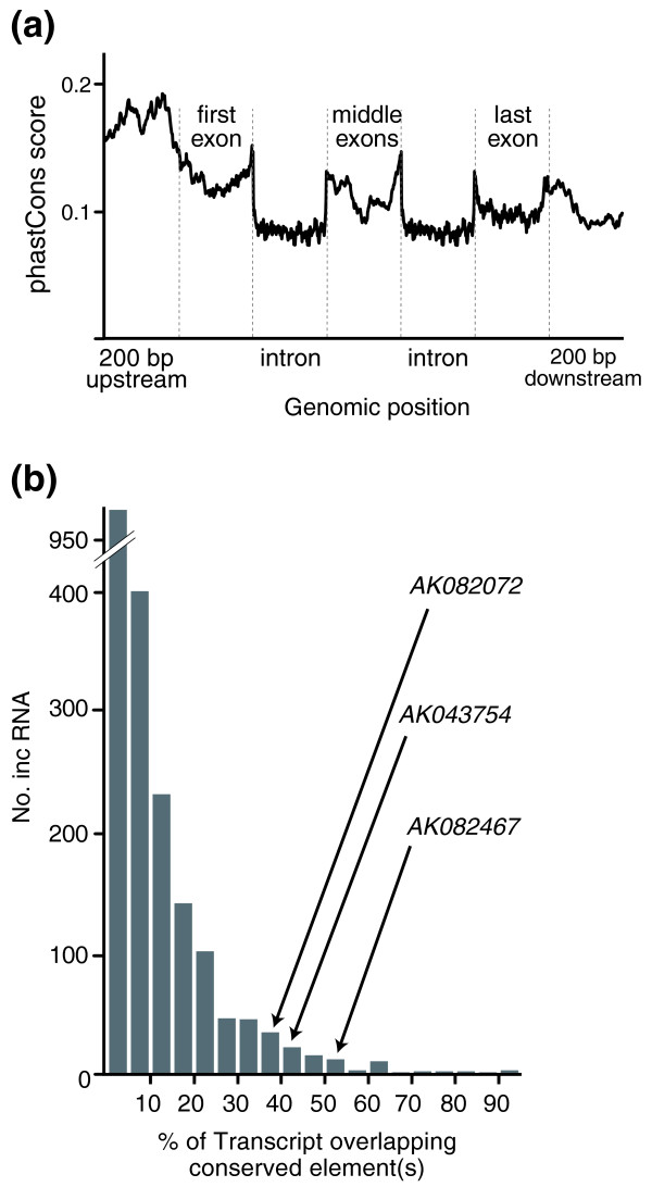
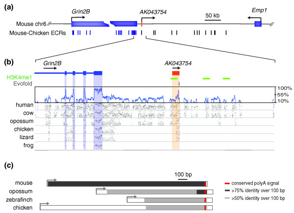
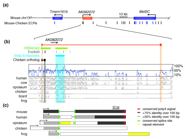
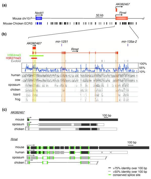
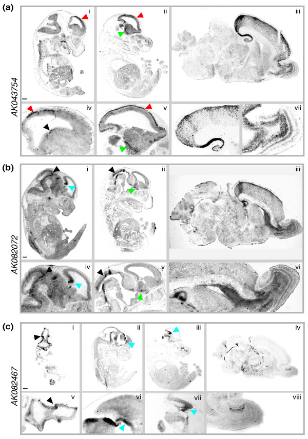
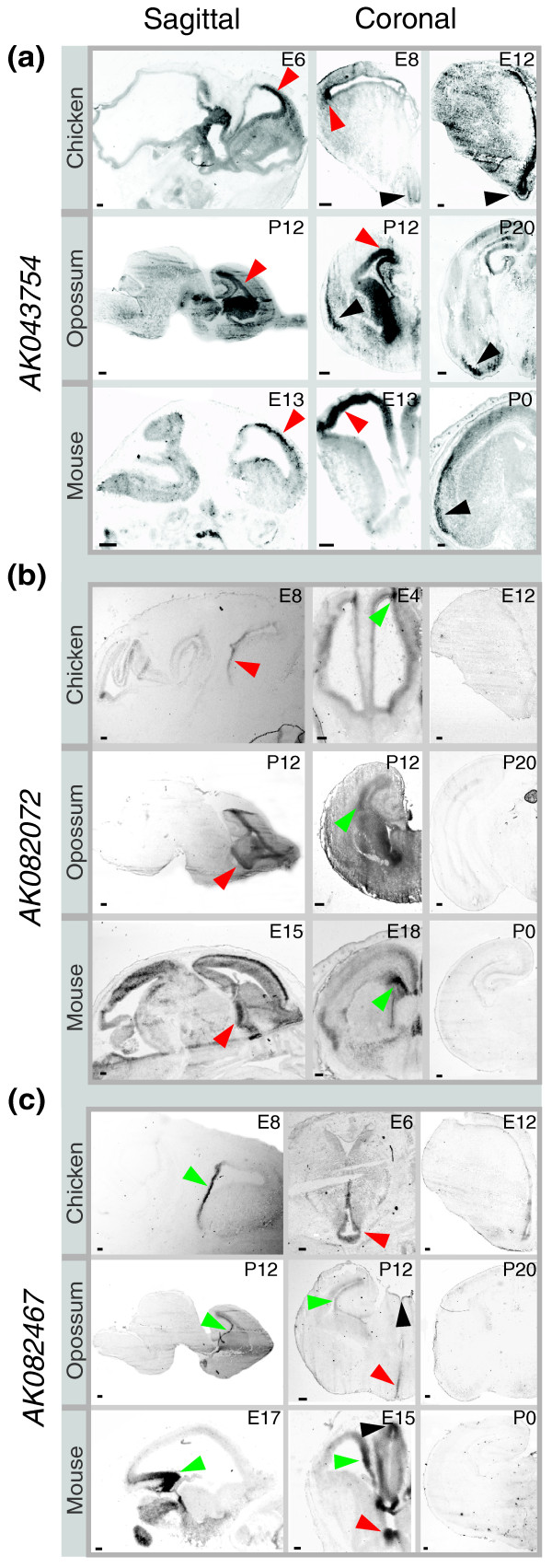
Results: Here, we performed a multi-disciplinary study of four highly conserved and brain-expressed transcripts selected from a list of mouse long intergenic noncoding RNA (lncRNA) loci that generally show pronounced evolutionary constraint within their putative promoter regions and across exon-intron boundaries. We identify some of the first lncRNA orthologs present in birds (chicken), marsupial (opossum), and eutherian mammals (mouse), and investigate whether they exhibit conservation of brain expression. In contrast to conventional protein-coding genes, the sequences, transcriptional start sites, exon structures, and lengths for these non-coding genes are all highly variable.

Conclusions: The biological relevance of lncRNAs would be highly questionable if they were limited to closely related phyla. Instead, their preservation across diverse amniotes, their apparent conservation in exon structure, and similarities in their pattern of brain expression during embryonic and early postnatal stages together indicate that these are functional RNA molecules, of which some have roles in vertebrate brain development.


Figures

Figure 1
Figure 1
Sequence conservation among lncRNAs. (a) Conservation across a generic lncRNA locus, based on 877 mouse multi-exon lncRNAs. We sampled 200 evenly spaced bases across each region listed, with regions containing fewer than 200 bases sampled entirely. The graph shows the average vertebrate phastCons score at each genomic position across all multi-exon lncRNA loci. Note phastCons score peaks within the putative promoter region (200 bp upstream) and near donor and acceptor splice sites (analysis inspired by Figure 25a in [31]). (b) Overlap between vertebrate phastCons-predicted conserved elements and mouse lncRNA exons. Of 2,055 lncRNAs with signatures of purifying selection initially identified in mouse [18], 1,095 contain exons that overlap phastCons-predicted vertebrate conserved elements (log-odds score range 1 to 1,000) [30]. Depicted is a histogram showing the percentage of each lncRNA transcript that overlaps a phastCons-predicted vertebrate conserved element. The relative positions of three selected lncRNAs (AK082072, AK043754, and AK082467 with overlaps of 36.7, 44.8, and 51.7%, respectively) are shown.
Figure 2
Figure 2
Evolutionary constraint of AK043754. (a) The genomic region of mouse chromosome 6 (chr6) encompassing the lncRNA locus AK043754 (1.7 kb) is depicted. Note the locations of flanking protein-coding genes: Grin2B (glutamate receptor, ionotropic, NMDA2B (N-methyl-D-aspartic acid)) and Emp1 (epithelial membrane protein 1). Also shown are the positions of mouse-chicken ECRs (evolutionarily conserved regions at least 100 bp in size with 70% sequence identity between the mouse and chicken genomes); ECRs within protein-coding regions are shown in blue. (b) A more detailed representation of AK043754 (single exon highlighted in orange) and its immediate flanking regions, including the 3' end of Grin2B. Below the gene structures are the positions of H3K4me1 chromatin marks (green) detected in mouse embryonic stem cells (obtained from UCSC Genome Browser), EvoFold predictions of RNA secondary structures (grey), a SinicView conservation plot [68] based on a 21-vertebrate multispecies sequence alignment (using Threaded Blockset Aligner) generated with mouse as the reference sequence, and Gmaj [66] views of alignments between mouse and the indicated species' sequences (note the detected homology with the orthologous lizard and chicken, but not frog, sequences). (c) Conservation and relative sizes of AK043754 orthologs in various species. The TSSs (arrows) and transcript lengths are depicted in each case. Note the conserved position of a polyA signal (red) and increased sequence conservation (relative to the mouse sequence) towards the 3' end. ECR, evolutionarily conserved region.
Figure 3
Figure 3
Evolutionary constraint of AK082072. (a) The genomic region of mouse chromosome 13 (chr13) encompassing lncRNA AK082072 (523 bp) is depicted. Note the locations of the flanking protein-coding genes: Tmem161b (transmembrane protein 161b) and Mef2C (myocyte enhancer factor 2C). (b) A more detailed representation of AK082072 (exons highlighted in orange) and its immediate flanking regions. Below the gene structures are the positions of H3K4me3 chromatin marks (green) detected in mouse brain, VISTA conserved non-coding midbrain enhancer element 268 (obtained from the UCSC Genome Browser), and a BLAT alignment of the chicken AK082072 ortholog, as well as similar tracks as those in Figure 2b. Note the detected homology with orthologous frog sequence in exon 1. (c) Conservation and relative sizes of AK082072 orthologs in various species. Note the sequence conservation (relative to the mouse sequence) at both the 5' and 3' ends and the conserved position of splice sites (green). Unlike the other vertebrate genomes considered, the zebra finch genome did not align to the proximal promoter or first exon of mouse AK082072. This apparent lack of sequence identity might reflect either an unannotated gap in its genome assembly or rapidly evolving sequence within its orthologous genomic region. Other details are provided in the legend to Figure 2. ECR, evolutionarily conserved region.
Figure 4
Figure 4
Evolutionary constraint of AK082467 and Rmst. (a) The genomic region of mouse chromosome 10 (chr 10) encompassing lncRNAs AK082467 (2.7 kb) and Rmst (2.7 kb) is depicted. Note the presence of the protein-coding gene Nedd1 (neural precursor expressed developmentally down-regulated protein 1) upstream of AK082467 and Rmst. (b) A more detailed representation of AK082467 and Rmst (exons highlighted in yellow and orange, respectively), microRNAs mir-1251 and mir-135a-2, and their immediate flanking regions. Below the gene structures are the positions of H3K4me3 (green) and H3K27me3 (red) chromatin marks detected in mouse brain (obtained from the UCSC Genome Browser) as well as similar tracks as those in Figure 2b. Note the detected homology with orthologous frog sequence in Rmst exons 1, 2, 4, and 11. (c) Conservation and relative sizes of AK082467 and Rmst orthologs in various species. Note the conserved splice sites (green bars) in mouse Rmst exons 1, 4, and 11 as well as the sequence conservation (relative to mouse sequence) in exons 1 and 11, but differences in total exon number among species. The 3' ends of opossum and chicken orthologs have not been experimentally verified. Other details are provided in the legend to Figure 2. ECR, evolutionarily conserved region.
Figure 5
Figure 5
lncRNAs are specifically expressed and developmentally regulated in the mouse brain. (a-c) Digoxigenin-labeled riboprobes complementary to AK043754 (a), AK082072 (b), and AK082467 (c) were hybridized to sagittal sections of C57BL/6J mouse brains at different development stages (E9, E13, E17, and P3). (a) The AK043754 probe hybridized to the first generated cell layer of the preplate or primordial plexiform zone (red arrowheads) at E13 (i, iv) and E17 (ii, v), the ventricular zone of the medial and lateral ganglionic eminences (black arrowhead) at E13, the latero-caudal migratory path from the basal telencephalon to the striatum (green arrowhead) at E17 (ii, v), and the hippocampus (iii, vi) and the olfactory bulb (iii, vii) at P3. Scale bar (shown in (i)) is 500 μm in (i), 543 μm in (ii), 322 μm in (iii), 292 μm in (iv), 300 μm in (v), 167 μm in (vi), and 214 μm in (vii). (b) The AK082072 probe hybridized to the hem of the embryonic cerebral cortex (blue arrowheads) and the roof of the midbrain (black arrowheads) at E13 (i, iv) and E17 (ii, v), and to the hippocampus (iii, vi), rostral migratory stream (iii, vi), and internal plexiform and granule cell layer of the olfactory bulb (iii, vi) at P3. Scale bar (shown in (i)) is 500 μm in (i), 595 μm in (ii), 422 μm in (iii), 357 μm in (iv), 386 μm in (v), and 311 μm in (vi). (c) The AK082467 probe hybridized to the optic stalk (black arrowheads) at E9 (i, v), the cortical hem (blue arrowheads) at E13 (ii, vi) and E17 (ii, vii), and the accessory olfactory bulb (iii, viii) at P3. Scale bar (shown in i)) is 500 μm in (i), 637 μm in (ii), 684 μm in (iii), 522 μm in (iv), 182 μm in (v), 177 μm in (vi), 176 μm in (vii), and 110 μm in (viii).
Figure 6
Figure 6
Conservation of lncRNA expression in developing avian and mammalian brains. (a-c) Digoxigenin-labeled riboprobes complementary to lncRNAs AK043754 (a), AK082072 (b), and AK082467 (c) were hybridized to chicken (E4, 6, 8,12), opossum (P12, 20), and mouse (E13, 15, 17, 18 and P0) brain sections. (a) AK043754: strong hybridization seen in the germinal zone of the telencephalic cortex at early developmental time points (red arrowheads) and then concentrated within the piriform (olfactory) cortex at later stages (black arrowheads). (b) AK082072: hybridization signals seen in the stria terminalis (red arrowheads) and the telencephalic ventricular zone (green arrowheads). Signal was undetectable at later developmental stages. (c) AK082467: hybridization signals seen in the ventricular zone of the hippocampal formation (green arrowheads), the preoptic area of the hypothalamus (red arrowheads), and the epithalamus (black arrowheads). Signal was undetectable at later developmental stages. Scale bars = 200 μm.

References

    1. Birney E, Stamatoyannopoulos JA, Dutta A, Guigo R, Gingeras TR, Margulies EH, Weng Z, Snyder M, Dermitzakis ET, Thurman RE, Kuehn MS, Taylor CM, Neph S, Koch CM, Asthana S, Malhotra A, Adzhubei I, Greenbaum JA, Andrews RM, Flicek P, Boyle PJ, Cao H, Carter NP, Clelland GK, Davis S, Day N, Dhami P, Dillon SC, Dorschner MO, Fiegler H. Identification and analysis of functional elements in 1% of the human genome by the ENCODE pilot project. Nature. 2007;447:799–816. doi: 10.1038/nature05874. - DOI - PMC - PubMed
    1. Church DM, Goodstadt L, Hillier LW, Zody MC, Goldstein S, She X, Bult CJ, Agarwala R, Cherry JL, DiCuccio M, Hlavina W, Kapustin Y, Meric P, Maglott D, Birtle Z, Marques AC, Graves T, Zhou S, Teague B, Potamousis K, Churas C, Place M, Herschleb J, Runnheim R, Forrest D, Amos-Landgraf J, Schwartz DC, Cheng Z, Lindblad-Toh K, Eichler EE. Lineage-specific biology revealed by a finished genome assembly of the mouse. PLoS Biol. 2009;7:e1000112. doi: 10.1371/journal.pbio.1000112. - DOI - PMC - PubMed
    1. Bertone P, Gerstein M, Snyder M. Applications of DNA tiling arrays to experimental genome annotation and regulatory pathway discovery. Chromosome Res. 2005;13:259–274. doi: 10.1007/s10577-005-2165-0. - DOI - PubMed
    1. Cheng J, Kapranov P, Drenkow J, Dike S, Brubaker S, Patel S, Long J, Stern D, Tammana H, Helt G, Sementchenko V, Piccolboni A, Bekiranov S, Baily DK, Ganesh M, Ghosh S, Bell I, Gerhard DS, Gingeras TR. Transcriptional maps of 10 human chromosomes at 5-nucleotide resolution. Science. 2005;308:1149–1154. doi: 10.1126/science.1108625. - DOI - PubMed
    1. Kapranov P, Willingham AT, Gingeras TR. Genome-wide transcription and the implications for genomic organization. Nat Rev Genet. 2007;8:413–423. doi: 10.1038/nrg2083. - DOI - PubMed

Publication types

Substances

LinkOut - more resources