. 2011 Jan 1;54(1):439-46.
doi: 10.1016/j.neuroimage.2010.07.004. Epub 2010 Jul 17.

Flow of affective information between communicating brains

Affiliations

Flow of affective information between communicating brains

Silke Anders et al. Neuroimage. .

Abstract

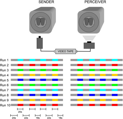
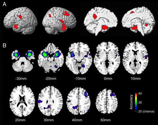
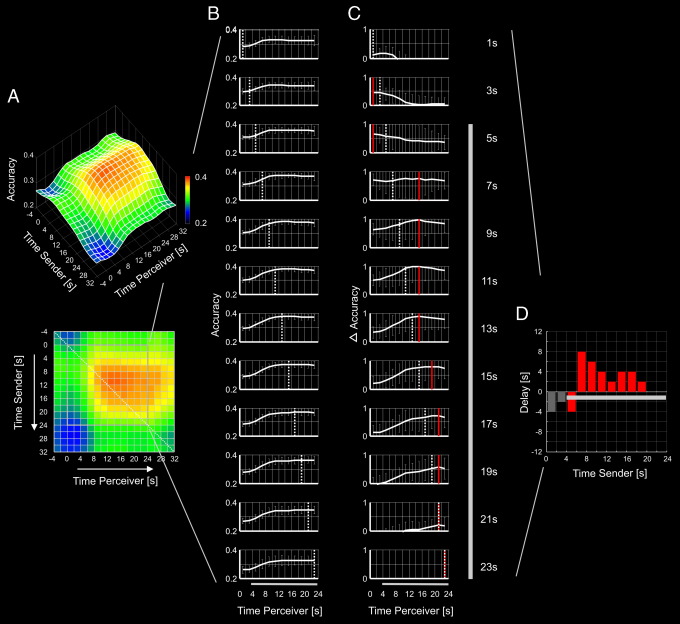
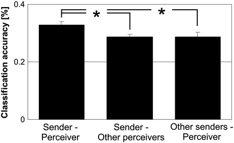
When people interact, affective information is transmitted between their brains. Modern imaging techniques permit to investigate the dynamics of this brain-to-brain transfer of information. Here, we used information-based functional magnetic resonance imaging (fMRI) to investigate the flow of affective information between the brains of senders and perceivers engaged in ongoing facial communication of affect. We found that the level of neural activity within a distributed network of the perceiver's brain can be successfully predicted from the neural activity in the same network in the sender's brain, depending on the affect that is currently being communicated. Furthermore, there was a temporal succession in the flow of affective information from the sender's brain to the perceiver's brain, with information in the perceiver's brain being significantly delayed relative to information in the sender's brain. This delay decreased over time, possibly reflecting some 'tuning in' of the perceiver with the sender. Our data support current theories of intersubjectivity by providing direct evidence that during ongoing facial communication a 'shared space' of affect is successively built up between senders and perceivers of affective facial signals.


Figures

Fig. 1
Fig. 1
Experimental design. Senders and perceivers participated in 10 runs of fMRI. Colours indicate emotion periods; each colour indicates a different emotion (joy, anger, disgust, fear, or sadness), grey indicates resting periods. The order of runs was chosen by the sender with the restriction that each emotion had to be chosen once before an emotion could occur a second time. The sender's facial expression was video-taped throughout scanning and shown to the perceiver while he was scanned immediately after scanning of the sender had been completed.
Fig. 2
Fig. 2
The ‘shared network of affect’. A. Clusters in which the perceiver’s brain activity could successfully be predicted from the level of the sender’s brain activity, depending on the communicated affect (p = .01, corrected for multiple comparisons at cluster level). Significant clusters are projected onto the surface of a standard brain (MNI). B. Average voxel-wise decoding accuracies within each cluster, projected onto axial slices of the same brain as in A. Slices are shown in neurological convention (left is left). Numbers below slices indicate z coordinates.
Fig. 3
Fig. 3
Dynamics of information flow. Each row of the time-resolved matrix of decoding accuracies (A) represents the time course (B) with which information from the sender's brain in a specific time window was encoded in the perceiver's brain. Subtracting the average from these time courses reveals the temporal dynamics of information flow (C). Time courses of delta accuracy in C are scaled to the overall maximum of delta accuracy. Red lines indicate the peak of each individual time course; dashed lines and numbers on the right indicate the time window the sender's brain activity was taken from. The bar chart (D) shows the delay with which information from the sender's brain was reflected in the perceiver's brain. Dark grey bars in C and D represent an approximation of the interval covered by the predicted time course of the hemodynamic response during affective communication.
Fig. 4
Fig. 4
Specificity of information within the ‘shared network of affect’. Bar charts represent average classification accuracies when information was combined across all voxels within the ‘shared network of affect’. Classification accuracy was significantly lower within sender – other-perceiver dyads and other-sender – perceiver dyads than within true sender–perceiver pairs. Error bars represent standard errors of the mean.

References

    1. Adolphs R., Damasio H., Tranel D., Cooper G., Damasio A.R. A role for somatosensory cortices in the visual recognition of emotion as revealed by three-dimensional lesion mapping. J. Neurosci. 2000;20(7):2683–2690. - PMC - PubMed
    1. Anders S., Lotze M., Erb M., Grodd W., Birbaumer N. Brain activity underlying emotional valence and arousal: a response-related fMRI study. Hum. Brain Mapp. 2004;23(4):200–209. - PMC - PubMed
    1. Anders S., Eippert F., Wiens S., Birbaumer N., Lotze M., Wildgruber D. When seeing outweighs feeling: a role for prefrontal cortex in passive control of negative affect in blindsight. Brain. 2009;132(Pt 11):3021–3031. - PMC - PubMed
    1. Andersson J.L., Hutton C., Ashburner J., Turner R., Friston K. Modeling geometric deformations in EPI time series. Neuroimage. 2001;13(5):903–919. - PubMed
    1. Bastiaansen J.A., Thioux M., Keysers C. Evidence for mirror systems in emotions. Philos. Trans. R. Soc. Lond. B Biol. Sci. 2009;364(1528):2391–2404. - PMC - PubMed

Publication types