Comparative Study
. 2010 Sep 1;588(Pt 17):3277-93.
doi: 10.1113/jphysiol.2010.192823. Epub 2010 Jul 12.

Participation of KCNQ (Kv7) potassium channels in myogenic control of cerebral arterial diameter

Affiliations
Comparative Study

Participation of KCNQ (Kv7) potassium channels in myogenic control of cerebral arterial diameter

Xi Zoë Zhong et al. J Physiol. .

Abstract

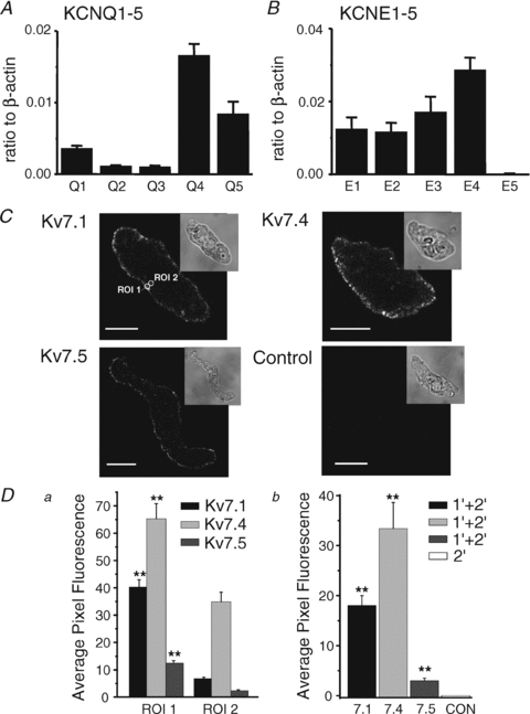
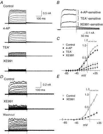
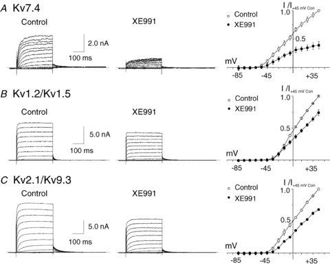
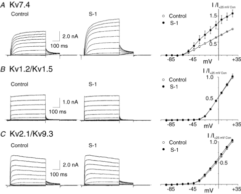
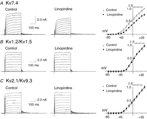
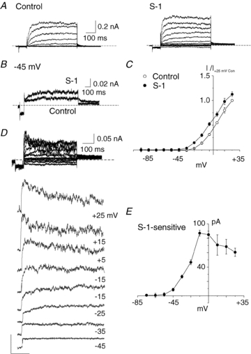
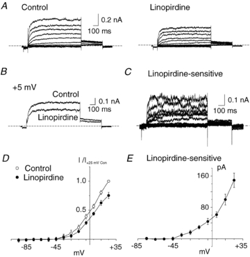
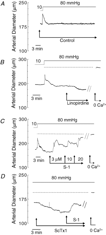
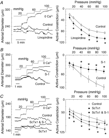
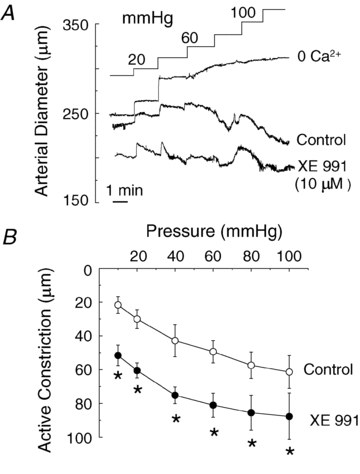
KCNQ gene expression was previously shown in various rodent blood vessels, where the products of KCNQ4 and KCNQ5, Kv7.4 and Kv7.5 potassium channel subunits, respectively, have an influence on vascular reactivity. The aim of this study was to determine if small cerebral resistance arteries of the rat express KCNQ genes and whether Kv7 channels participate in the regulation of myogenic control of diameter. Quantitative reverse transcription polymerase chain reaction (QPCR) was undertaken using RNA isolated from rat middle cerebral arteries (RMCAs) and immunocytochemistry was performed using Kv7 subunit-specific antibodies and freshly isolated RMCA myocytes. KCNQ4 message was more abundant than KCNQ5 = KCNQ1, but KCNQ2 and KCNQ3 message levels were negligible. Kv7.1, Kv7.4 and Kv7.5 immunoreactivity was present at the sarcolemma of freshly isolated RMCA myocytes. Linopirdine (1 microm) partially depressed, whereas the Kv7 activator S-1 (3 and/or 20 microm) enhanced whole-cell Kv7.4 (in HEK 293 cells), as well as native RMCA myocyte Kv current amplitude. The effects of S-1 were voltage-dependent, with progressive loss of stimulation at potentials of >15 mV. At the concentrations employed linopirdine and S-1 did not alter currents due to recombinant Kv1.2/Kv1.5 or Kv2.1/Kv9.3 channels (in HEK 293 cells) that are also expressed by RMCA myocytes. In contrast, another widely used Kv7 blocker, XE991 (10 microm), significantly attenuated native Kv current and also reduced Kv1.2/Kv1.5 and Kv2.1/Kv9.3 currents. Pressurized arterial myography was performed using RMCAs exposed to intravascular pressures of 10-100 mmHg. Linopirdine (1 microm) enhanced the myogenic response at 20 mmHg, whereas the activation of Kv7 channels with S-1 (20 microm) inhibited myogenic constriction at >20 mmHg and reversed the increased myogenic response produced by suppression of Kv2-containing channels with 30 nm stromatoxin (ScTx1). These data reveal a novel contribution of KCNQ gene products to the regulation of myogenic control of cerebral arterial diameter and suggest that Kv7 channel activating drugs may be appropriate candidates for the development of an effective therapy to ameliorate cerebral vasospasm.


Figures

Figure 1
Figure 1. Expression of KCNQ genes in rat cerebral arteries
A and B, quantitative, real-time PCR detection of KCNQ (KCNQ15) and KCNE (KCNE1–5) subunit transcript expression relative to β-actin in mRNA extracted from RMCAs. All expression data are expressed as means ±s.e.m. (n= 4 for each). C, immunostaining of freshly isolated myocytes using anti-Kv7.1, -Kv7.4, or -Kv7.5, and a control experiment in which primary antibody was omitted. The transmitted light image for each cell is shown in upper right inset images. Circles indicate regions of interest: ROI 1 and ROI 2, which were used to analyse the localization of fluorescence (see Methods for details). The calibration bars in panel C are 10 μm. D, summarized data for localization of Kv7.1, Kv7.4 and Kv7.5 immunofluorescence in RMCA myocytes. There was significantly more fluorescence in the region of plasma membrane (ROI 1) than in deep cytoplasm (ROI 2) (Da, n= 10 myocytes for each antibody). Db shows summarized data for average pixel fluorescence in a whole-cell confocal plane compared to control myocyte that demonstrated a lack of immunofluorescence in the absence of primary antibody (n= 10 myocytes; 1′, primary antibody; 2′, secondary antibody). **Statistical significance at P < 0.01.
Figure 2
Figure 2. Suppression of RMCA myocyte Kv current by XE991
A, representative families of Kv currents of an RMCA myocyte evoked by 300 ms steps to voltages between −95 and +45 mV prior to repolarization to −55 mV (a similar protocol was used for all recordings unless indicated otherwise) in the absence (Control) and presence of 100 μm 4-AP, 4-AP plus 4 mM TEA+, and 4-AP, TEA+ and 10 μm XE991. B, representative traces from panel A for voltage steps to +25 mV showing the 4-AP-, TEA+- and XE991-sensitive components of net Kv current. C, mean ±s.e.m. normalized (to peak current at +45 mV in control condition) I–V relations for Kv current of RMCA myocytes (n= 3) sequentially treated with 4-AP, TEA+ and XE991 as in panel A. D, representative families of Kv currents from an RMCA myocyte in the absence and presence of 10 μm XE991 followed by drug washout. Voltage protocol as in panel A but from −95 to +25 mV only. E, mean ±s.e.m. normalized (to peak current at +25 mV in control condition) I–V relations for Kv current of RMCA myocytes (n= 5) in the absence (Control) and presence of 10 μm XE991. Note that XE991 caused a significant suppression of net Kv current in panels D and E that was larger than that predicted by the results of panels A–C.
Figure 3
Figure 3. Suppression of recombinant homomeric Kv7.4 and heteromeric Kv1.2/Kv1.5 and Kv2.1/Kv9.3 currents by XE991
A, representative families and mean ±s.e.m. (n= 3) I–V relations for whole-cell Kv7.4 currents (HEK 293 cells) in the absence (Control) and presence of 10 μm XE991. B, representative families and mean ±s.e.m. (n= 3) normalized (to peak current at +45 mV in control condition) I–V relations for whole-cell Kv1.2/Kv1.5 currents (HEK 293 cells) in the absence (Control) and presence of 10 μm XE991 (note voltage steps were to between −95 and +25 mV only). C, representative families and mean ±s.e.m. (n= 3) normalized (to peak current at +45 mV in control condition) I–V relations for whole-cell Kv2.1/Kv9.3 currents (HEK 293 cells) in the absence (Control) and presence of 10 μm XE991. Note the suppression of Kv1.2/Kv1.5 and Kv2.1/Kv9.3 currents by XE991 at 10 μm.
Figure 5
Figure 5. Stimulation of recombinant homomeric Kv7.4, but not heteromeric Kv1.2/Kv1.5 and Kv2.1/Kv9.3 currents by S-1
A, representative families and mean ±s.e.m. (n= 4) normalized (to peak current at +25 mV in control condition) I–V relations for whole-cell Kv7.4 currents (HEK 293 cells) in the absence (Control) and presence of 3 μm S-1. B, representative families and mean ±s.e.m. (n= 3) normalized (to peak current at +25 mV in control condition) I–V relations for whole-cell Kv1.2/Kv1.5 currents (HEK 293 cells) in the absence (Control) and presence of 3 μm S-1. C, representative families and mean ±s.e.m. (n= 3) normalized (to peak current at +25 mV in control condition) I–V relations for whole-cell Kv2.1/Kv9.3 currents (HEK 293 cells) in the absence (Control) and presence of 3 μm S-1. Note the lack of stimulation of Kv1.2/Kv1.5 and Kv2.1/Kv9.3 currents by S-1 at 3 μm.
Figure 4
Figure 4. Suppression of recombinant homomeric Kv7.4, but not heteromeric Kv1.2/Kv1.5 and Kv2.1/Kv9.3 currents by linopirdine
A, representative families and mean ±s.e.m. (n= 4) normalized (to peak current at +45 mV in control condition) I–V relations for whole-cell Kv7.4 currents (HEK 293 cells) in the absence (Control) and presence of 1 μm linopirdine. B, representative families and mean ±s.e.m. (n= 3) normalized (to peak current at +45 mV in control condition) I–V relations for whole-cell Kv1.2/Kv1.5 currents (HEK 293 cells) in the absence (Control) and presence of 1 μm linopirdine. C, representative families and mean ±s.e.m. (n= 3) normalized (to peak current at +45 mV in control condition) I–V relations for whole-cell Kv2.1/Kv9.3 currents (HEK 293 cells) in the absence (Control) and presence of 1 μm linopirdine. Note the lack of suppression of Kv1.2/Kv1.5 and Kv2.1/Kv9.3 currents by linopirdine at 1 μm.
Figure 7
Figure 7. Stimulation of native RMCA myocyte Kv current by S-1
A, representative families of whole-cell RMCA myocyte Kv current in the absence (Control) and presence of 3 μm S-1 (note voltage clamp protocol included a 80 ms step to −95 mV prior to test steps). B, representative expanded traces for currents ± S-1 at −45 mV from cell in panel A. C, mean ±s.e.m. (n= 3) normalized (to peak current at +25 mV in control condition) I–V relations for Kv current in the absence (Control) and presence of S-1. D, representative S-1-sensitive Kv current obtained by digital subtraction of control from S-1 current in panel A. Expanded versions of each current recording for voltage steps to between −45 and +25 are shown below for clarity. Note the increased apparent inactivation of the S-1-sensitive current at potential positive to −5 mV. E, mean ±s.e.m. (n= 3) I–V relation of S-1-sensitive current determined from end pulse difference current amplitude. Note the decline in S-1-sensitive current positive to −15 mV, and y-axis is at −5 mV..
Figure 6
Figure 6. Suppression of native RMCA myocyte Kv current by linopirdine
A, representative families of whole-cell RMCA myocyte Kv currents in the absence (Control) and presence of 1 μm linopirdine (note voltage clamp protocol included a 80 ms step to −95 mV prior to test steps to between −80 and +25 mV in 10 mV increments followed by repolarization to −45 mV). B, representative recordings of Kv current at +5 mV in the absence and presence of linopirdine (1 μm). C, representative family of linopirdine-sensitive Kv current obtained by digital subtraction of currents in linopirdine from those in control condition in panel A. D, mean ±s.e.m. (n= 3) normalized (to peak current at +25 mV in control condition) I–V relations for Kv current in the absence (Control) and presence linopirdine. E, mean ±s.e.m. (n= 3) I–V relation for linopirdine-sensitive Kv current determined from end-pulse difference current amplitude.
Figure 8
Figure 8. Vasoconstriction and dilatation of RMCAs by linopirdine and S-1 at a constant pressure of 80 mmHg
A, representative recording of RMCA diameter during a pressure step from 10 to 80 mmHg in control conditions. B, representative recording of RMCA diameter during a pressure step from 10 to 80 mmHg followed by subsequent sequential exposure to 1 μm linopirdine and zero Ca2+ solution (i.e. no added Ca2+ and 2 mm EGTA). Dashed line represents the passive diameter of the vessel in 0 Ca2+ solution at 80 mmHg. C, representative recording of RMCA diameter during a pressure step from 10 to 80 mmHg followed by subsequent sequential exposure to 3, 10 and 20 μm S-1 and zero Ca2+ solution. Dashed line represents the passive diameter of the vessel in 0 Ca2+ solution at 80 mmHg. Note concentration-dependent vasodilatation by S-1. D, representative recording of RMCA diameter at 80 mmHg during sequential exposure to 30 nm ScTx1 followed by S-1 (20 μm) and zero Ca2+ solution. Dashed line represents the passive diameter of the vessel in 0 Ca2+ solution at 80 mmHg. Note the reversal of ScTx1-induced vasoconstriction by S-1.
Figure 9
Figure 9. Stimulation and suppression of RMCA myogenic response by linopirdine and S-1
A, representative recordings of RMCA diameter during a series of pressure steps from 10 to between 20 and 100 mmHg in 20 mmHg increments in control conditions (Control), linopirdine (1 μm) and in zero Ca2+ solution, and mean ±s.e.m. (n= 5) active constriction in control and linopirdine (i.e. difference between diameter in zero Ca2+ and control or linopirdine). Note increase in active constriction in linopirdine at ≥20 mmHg. B, representative recordings of RMCA diameter during a series of pressure steps from 10 to between 20 and 100 mmHg in 20 mmHg increments in control conditions (Control), S-1 (20 μm) and in zero Ca2+ solution, and mean ±s.e.m. (n= 5) active constriction in control and S-1 (i.e. difference between diameter in zero Ca2+ and control or S-1). Note suppression of active constriction in S-1 at >20 mmHg. C, representative recordings of RMCA diameter during a series of pressure steps from 10 to between 20 and 100 mmHg in 20 mmHg increments in control conditions (Control), ScTx1 (30 nm), ScTx1 and S-1 (20 μm) and in zero Ca2+ solution, and mean ±s.e.m. (n= 3) active constriction in control, ScTx1, and ScTx1 and S-1. Note decrease in the ScTx1-enhanced active constriction in the presence of S-1 at 80 and 100 mmHg. *Significant difference at P < 0.05.
Figure 10
Figure 10. Stimulation of RMCA myogenic response by XE991
A, representative recordings of RMCA diameter during a series of pressure steps from 10 to between 20 and 100 mmHg in 20 mmHg increments in control conditions (Control), XE991 (10 μm) and in zero Ca2+ solution, and mean ±s.e.m. (n= 5) active constriction in control and XE991. Note increase in active constriction over the entire pressure range from 10 to 100 mmHg in XE991. *Significant difference at P < 0.05.

References

    1. Albarwani S, Nemetz LT, Madden JA, Tobin AA, England SK, Pratt PF, Rusch NJ. Voltage-gated K+ channels in rat small cerebral arteries: molecular identity of the functional channels. J Physiol. 2003;551:751–763. - PMC - PubMed
    1. Amberg GC, Santana LF. Kv2 channels oppose myogenic constriction of rat cerebral arteries. Am J Physiol Cell Physiol. 2006;291:C348–C356. - PubMed
    1. Bal M, Zhang J, Zaika O, Hernandez CC, Shapiro MS. Homomeric and heteromeric assembly of KCNQ (Kv7) K+ channels assayed by total internal reflection fluorescence/fluorescence resonance energy transfer and patch clamp analysis. J Biol Chem. 2008;283:30668–30676. - PMC - PubMed
    1. Bentzen BH, Schmitt N, Calloe K, Dalby Brown W, Grunnet M, Olesen SP. The acrylamide (S)-1 differentially affects Kv7 (KCNQ) potassium channels. Neuropharmacology. 2006;51:1068–1077. - PubMed
    1. Brueggemann LI, Moran CJ, Barakat JA, Yeh JZ, Cribbs LL, Byron KL. Vasopressin stimulates action potential firing by protein kinase C-dependent inhibition of KCNQ5 in A7r5 rat aortic smooth muscle cells. Am J Physiol Heart Circ Physiol. 2007;292:H1352–H1363. - PMC - PubMed

Publication types

LinkOut - more resources