. 2010 Jul 12;190(1):73-87.
doi: 10.1083/jcb.201001036.

Dissecting the role of MPS1 in chromosome biorientation and the spindle checkpoint through the small molecule inhibitor reversine

Affiliations

Dissecting the role of MPS1 in chromosome biorientation and the spindle checkpoint through the small molecule inhibitor reversine

Stefano Santaguida et al. J Cell Biol. .

Abstract

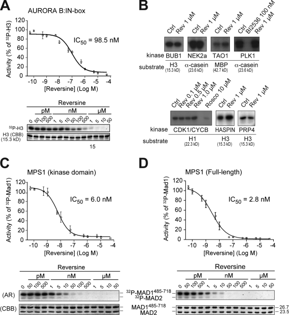
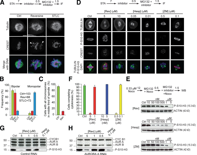
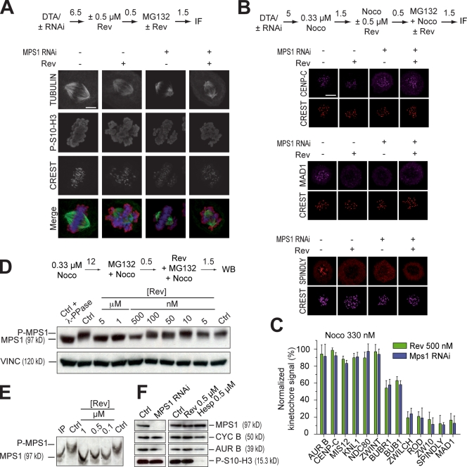
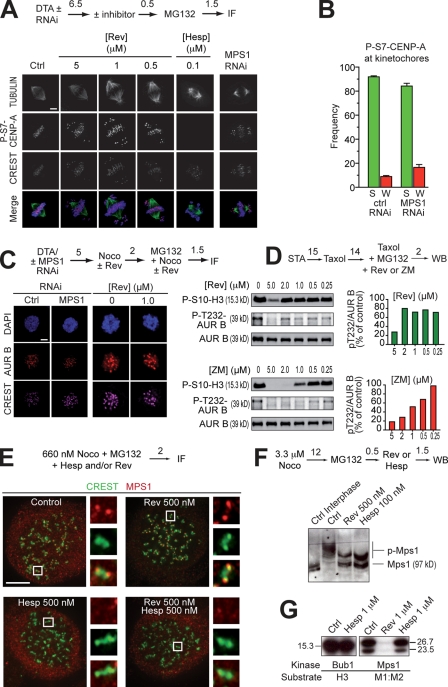
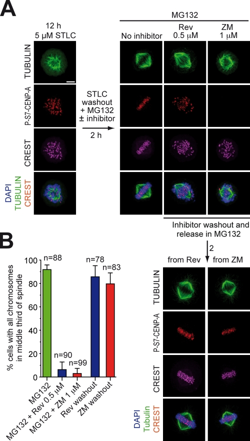
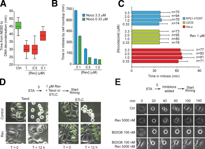
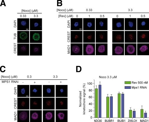
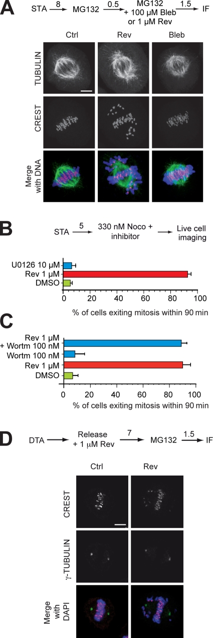
The catalytic activity of the MPS1 kinase is crucial for the spindle assembly checkpoint and for chromosome biorientation on the mitotic spindle. We report that the small molecule reversine is a potent mitotic inhibitor of MPS1. Reversine inhibits the spindle assembly checkpoint in a dose-dependent manner. Its addition to mitotic HeLa cells causes the ejection of Mad1 and the ROD-ZWILCH-ZW10 complex, both of which are important for the spindle checkpoint, from unattached kinetochores. By using reversine, we also demonstrate that MPS1 is required for the correction of improper chromosome-microtubule attachments. We provide evidence that MPS1 acts downstream from the AURORA B kinase, another crucial component of the error correction pathway. Our experiments describe a very useful tool to interfere with MPS1 activity in human cells. They also shed light on the relationship between the error correction pathway and the spindle checkpoint and suggest that these processes are coregulated and are likely to share at least a subset of their catalytic machinery.


Figures

Figure 1.
Figure 1.
Reversine inhibits MPS1 in vitro. (A) A kinase assay on the human AURORA B1–344–INCENP835–903 complex with the indicated concentrations of reversine. The substrate is histone H3. (B) The indicated recombinant, purified mitotic kinases were tested with the indicated substrates for their sensitivity to 1 µM reversine (Rev). None of the kinases were significantly inhibited. Specific inhibitors against PLK1 (BI2536) and CDK1 (roscovitine) were used as positive controls. (C and D) A reversine titration experiment on the kinase domain of MPS1 (C) or full-length MPS1 (D). The substrate is the MAD1–MAD2 complex (Sironi et al., 2001). Molecular mass is indicated in kilodaltons. (A, C, and D) Error bars are mean ± SEM. AR, autoradiography; CBB, Coomassie brilliant blue staining; Ctrl, control; CYCB, CYCLIN B.
Figure 2.
Figure 2.
Submicromolar reversine does not inhibit Aurora B in living cells. (A) Reversine does not prevent spindle bipolarization, but several chromosomes fail to congress (arrowheads). 7 h (numbers above arrows indicate time in hours) after a single thymidine arrest (STA), STLC (an Eg5 inhibitor causing spindle monopolarization) or reversine was added, and after an additional 0.5 h, MG132 was added to prevent mitotic exit. After 1.5 h, cells were then processed for immunofluorescence (IF). (B) Quantification of the experiment in A on the indicated number of cells. (C) Quantification of alignment defects in the same experiment. (D) After release from a single thymidine arrest, cells entered mitosis in the presence of reversine (Rev), hesperadin (Hesp), and ZM447439 (ZM) and were fixed and subjected to immunofluorescence to monitor the levels of P-S10-H3. (E) A comparison of the effects of the inhibitors tested in D on the levels of P-S10-H3 in total cell lysates of mitotic HeLa cells. (F) The effects of the same three inhibitors on cytokinesis were evaluated in a time-lapse experiment. (B, C, and F) Error bars are mean ± SEM. (G and H) AURORA A (AUR A) does not contribute to the generation of P-S10-H3. Under conditions of RNAi of AURORA A, the disappearance of P-S10-H3 in the presence of reversine (H) followed the same pattern as in the control experiments (G). A similar experiment testing the effects on P-S7–CENP-A is reported in Fig. S4 B. Dashed lines highlight the disappearance of AURORA A in H as the result of RNAi. Ctrl, control; CYC B1, CYCLIN B1; Noco, nocodazole; WB, Western blot. Bars, 5 µm.
Figure 3.
Figure 3.
Reversine inhibits MPS1 in living cells. (A) Chromosome alignment phenotypes of mitotic HeLa cells that were depleted of MPS1 by RNAi or treated with 0.5 µM reversine (Rev), or both. The levels of P-S10-H3 appeared unaltered in all three experiments. A representative RNAi-based depletion of MPS1 is shown in F. (B) Representative localization experiments on different kinetochore proteins including CREST, CENP-C, MAD1, and SPINDLY (Griffis et al., 2007; Chan et al., 2009). Results from the complete analysis are summarized in C and in Fig. S3. (A and B) Numbers above arrows indicate time in hours. (C) The RZZ subunits ROD, ZWILCH, and ZW10, as well as the RZZ-associated protein SPINDLY and MAD1 are all largely evicted from kinetochores when the spindle checkpoint is triggered with 330 nM nocodazole (Noco), with no significant difference between MPS1 RNAi or reversine treatment. The effects on localization are expressed as ratios of the fluorescence value of the indicated protein to the value of CREST (both background subtracted) normalized to the equivalent ratio in control cells. Error bars are mean ± SEM. (D) Dose-dependent inhibition of MPS1 phosphorylation in the presence of reversine. Vinculin (VINC) was used as a loading control. (E) Dose-dependent inhibition of MPS1. Samples were separated on an 8% gel with the PHOS tag method (Kinoshita et al., 2006). (F) Western blotting demonstrates that P-S10-H3 levels are untouched upon MPS1 RNAi or inhibition with reversine. The hesperadin control illustrates the effects from inhibiting AURORA B (AUR B). Ctrl, control; CYC B, CYCLIN B; DTA, double thymidine arrest; IF, immunofluorescence; WB, Western blot. Bars, 5 µm.
Figure 4.
Figure 4.
MPS1 acts downstream from Aurora B. (A) P-S7–CENP-A in mitotic HeLa cells is unaltered even at 5 µM reversine (Rev). The antigen is present on centromeres/kinetochores of chromosomes near the poles, as well as of chromosomes at the equator. The antigen is invisible in the presence of 100 nM hesperadin (Hesp). No compensation from Aurora A was observed (Fig. S4 B). (B) A quantification of the results in A. “S” and “W” indicate strong and weak binding, respectively. These criteria are indicated in Fig. S4 A. Error bars are mean ± SEM. (C) Kinetochore localization of AURORA B (AUR B) in HeLa cells is unaffected after MPS1 RNAi or the addition of reversine. (D, top) Phosphorylation of the activation loop of AURORA B (P-T232) is not affected by reversine until above 2 µM. The pattern of loss of activation loop phosphorylation follows the pattern of loss of P-S10-H3 phosphorylation. (bottom) The same experiment with ZM447439 (ZM) as a positive control. (E) Kinetochore localization of MPS1 in 660 nM nocodazole (Noco) is enhanced by 0.5 µM reversine. If AURORA B is inhibited with 0.5 µM hesperadin, reversine-induced localization of MPS1 is abrogated. Images were taken on a Delta Vision microscope. The insets represent 10× zooms of the boxed areas interpolated using SoftWoRx. (F) Both MPS1 and AURORA B inhibitors reduce the phosphorylation of mitotic MPS1, as visualized through the PHOS tag method (Kinoshita et al., 2006). (A and C–F) Numbers above arrows indicate time in hours. (G) Hesperadin does not inhibit BUB1 or MPS1 in an in vitro kinase assay (see also Table S1). Molecular mass is indicated in kilodaltons. Ctrl, control; DTA, double thymidine arrest; IF, immunofluorescence; WB, Western blot. Bars, 5 µm.
Figure 5.
Figure 5.
MPS1 is involved in error correction. (A) Cycling HeLa cells were treated with STLC for 12 h. Most cells arrest in mitosis with a monopolar spindle. After STLC washout in MG132, control cells bipolarize and form a normal metaphase. If STLC washout is performed in the presence of reversine (Rev) or ZM447439 (ZM), the spindle bipolarizes normally, but a large fraction of improper attachments are visible. P-S7–CENP-A, a bona fide AURORA B substrate, appears unaltered in reversine-treated cells but disappears in ZM447439. After removal of the inhibitors, a metaphase plate forms. P-S7–CENP-A reappears after washout of ZM447439. In vitro, 2 µM ZM447439 does not inhibit MPS1 (Table S1 and not depicted). (B) Quantification of results with number of cells monitored in the experiment. Error bars are mean ± SEM. Bar, 5 µm.
Figure 6.
Figure 6.
Reversine is a spindle checkpoint inhibitor. (A) Reversine (Rev) causes normally cycling HeLa cells to exit mitosis prematurely, which is a consequence of spindle checkpoint inactivation. The plot is a quantification of a time-lapse video microscopy experiment. (B) As in A, the experiment quantifies the behavior of cells in time-lapse video microscopy experiments in which HeLa cells were treated with two concentrations of nocodazole (Noco). Additional values (including controls) are collected in Table S2. (C) The ability of reversine to drive HeLa cells out of mitosis extends to at least two additional cell types. (A–C) Error bars are mean ± SEM. (D) The ability of HeLa cells to arrest in mitosis in the presence of 500 nM Taxol or 10 µM STLC was tested in a time-lapse experiment in the presence of reversine. 12 h after the beginning of the video, control cells treated with Taxol or STLC were still in mitosis, whereas the presence of reversine caused a spindle assembly checkpoint override and mitotic exit. (E) Similar experiments were performed in the presence of the Polo kinase inhibitor BI2536, which causes a mitotic arrest (Lénárt et al., 2007). In this case, time 0 refers to the last video frame before mitotic rounding up. (D and E) Numbers above arrows indicate time in hours. Ctrl, control; NEBD, nuclear envelope breakdown. Bars, 10 µm.
Figure 7.
Figure 7.
MPS1 is required for kinetochore recruitment of the RZZ and MAD1 even in high nocodazole. (A) Immunofluorescence analysis of the distribution of TUBULIN (TUB) in the presence of 0.33 and 3.3 µM nocodazole (Noco). Residual foci of polymerized TUBULIN are visible in 0.33 µM nocodazole but not in 3.3 µM nocodazole. HeLa cells were incubated in the presence of nocodazole for 15 min before fixation for immunofluorescence. (B) 5 h after release from a double thymidine arrest, reversine (Rev; at the indicated concentrations) and nocodazole (0.33 or 3.3 µM) were added. MAD1 failed to localize to kinetochores at either nocodazole concentration. (C) The same experimental scheme as in B was used under conditions of RNAi-based depletion of MPS1. (D) The histogram summarizes results on localization experiments equivalent to those in B and C on MAD1 and the additional indicated kinetochore proteins. Localization data were quantified as in Fig. 3 C. Error bars are mean ± SEM. Bars, 5 µm.
Figure 8.
Figure 8.
Reversine does not inhibit NMMII, MEK1, PI3K, or centrosome duplication. (A) At 100 µM, blebbistatin (Bleb) does not cause evident perturbations of chromosome alignment, contrarily to 1 µM reversine (Rev). (B) At 10 µM, the MEK1 inhibitor U0126 does not affect the duration of the spindle checkpoint. (C) At 100 nM, the PI3K inhibitor wortmannin (Wortm) does not affect the duration of the spindle checkpoint. (B and C) Error bars are mean ± SEM. (D) Cells arrested with a double thymidine arrest (DTA) procedure were released in the cell cycle, and the number of centrosomes was measured in the subsequent mitosis. In all cases, two centrosomes were measured, indicating that centrosome duplication takes place normally in the presence of reversine. (A, B, and D) Numbers above arrows indicate time in hours. Ctrl, control; IF, immunofluorescence; STA, single thymidine arrest. Bars, 5 µm.

References

    1. Amabile G., D’Alise A.M., Iovino M., Jones P., Santaguida S., Musacchio A., Taylor S., Cortese R. 2009. The Aurora B kinase activity is required for the maintenance of the differentiated state of murine myoblasts. Cell Death Differ. 16:321–330 10.1038/cdd.2008.156 - DOI - PubMed
    1. Andrews P.D., Ovechkina Y., Morrice N., Wagenbach M., Duncan K., Wordeman L., Swedlow J.R. 2004. Aurora B regulates MCAK at the mitotic centromere. Dev. Cell. 6:253–268 10.1016/S1534-5807(04)00025-5 - DOI - PubMed
    1. Bayliss R., Sardon T., Vernos I., Conti E. 2003. Structural basis of Aurora-A activation by TPX2 at the mitotic spindle. Mol. Cell. 12:851–862 10.1016/S1097-2765(03)00392-7 - DOI - PubMed
    1. Biggins S., Murray A.W. 2001. The budding yeast protein kinase Ipl1/Aurora allows the absence of tension to activate the spindle checkpoint. Genes Dev. 15:3118–3129 10.1101/gad.934801 - DOI - PMC - PubMed
    1. Brito D.A., Yang Z., Rieder C.L. 2008. Microtubules do not promote mitotic slippage when the spindle assembly checkpoint cannot be satisfied. J. Cell Biol. 182:623–629 10.1083/jcb.200805072 - DOI - PMC - PubMed

Publication types

MeSH terms