

Review
doi: 10.1074/jbc.R110.155341.
Epub 2010 Jul 12.
Role of epithelial sodium channels and their regulators in hypertension
Affiliations
- PMID: 20624922
- PMCID: PMC2945528
- DOI: 10.1074/jbc.R110.155341
Item in Clipboard
Review
Role of epithelial sodium channels and their regulators in hypertension
J Biol Chem.
.
Abstract
The kidney has a central role in the regulation of blood pressure, in large part through its role in the regulated reabsorption of filtered Na(+). Epithelial Na(+) channels (ENaCs) are expressed in the most distal segments of the nephron and are a target of volume regulatory hormones. A variety of factors regulate ENaC activity, including several aldosterone-induced proteins that are present within an ENaC regulatory complex. Proteases also regulate ENaC by cleaving the channel and releasing intrinsic inhibitory tracts. Polymorphisms or mutations within channel subunits or regulatory pathways that enhance channel activity may contribute to an increase in blood pressure in individuals with essential hypertension.
Figures
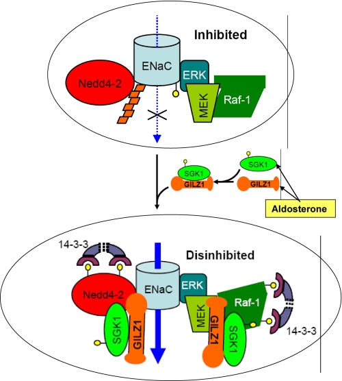
Aldosterone regulation of ENaC activity: role of the ERC. In the “basal/no-hormone” state, ENaC is associated with and inhibited by an ERC, which contains Nedd4-2, as well as Raf-1, MEK, and ERK. ERK phosphorylates the channel (small yellow circles), which in turn stimulates recruitment of Nedd4-2 and hence channel ubiquitination (orange trapezoids). ENaC internalization, endocytic trafficking, and lysosome-mediated degradation are thus augmented. Aldosterone coordinately induces the expression of SGK1 and GILZ1. In turn, GILZ1 (a) recruits SGK1 to the ERC; (b) increases interaction of SGK1 with its substrates Nedd4-2 and Raf-1; (c) augments SGK1 inhibition of Nedd4-2 and Raf-1 within the ERC, which results in the recruitment of inhibitory 14-3-3 proteins; and (d) synergizes with SGK1 to selectively stimulate its ENaC-specific functions (90). The net effect is “dual disinhibition” of ENaC (by SGK1 and GILZ1), resulting in increased channel surface expression and activity. Blue arrows represent Na+ movement through the channel, whereas black arrows represent proteins recruited to the complex. The top ellipse shows ENaC in the “inhibited” state (endocytosis and degradation favored), whereas the bottom ellipse shows the activated or “disinhibited” state (favoring accumulation at the plasma membrane).
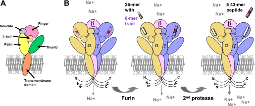
Activation of ENaC by proteases: the second-hit hypothesis. A, the five domains within the extracellular region of an ENaC subunit are illustrated. B, ENaC α-, β-, and γ-subunits are shown within a trimeric complex. Both the N and C termini are in the cytoplasm. Inhibitory tracts are present within the α- and γ-subunits and are released by proteases. The α-subunit is cleaved twice in its finger domain by the protease furin in the biosynthetic pathway, releasing a 26-residue fragment containing a key 8-mer inhibitory tract (shown in purple) (99, 100) and partially activating ENaC. Furin cleaves the γ-subunit at one site in the finger domain, leaving the inhibitory tract in place. A second cleavage of the γ-subunit in a post-biosynthetic compartment releases a 43-residue or larger peptide containing a minimal inhibitory tract (shown in red) and fully activates ENaC.
References
-
- Weinberger M. H. (1996) Hypertension 27, 481–490 - PubMed
-
- Meneton P., Jeunemaitre X., de Wardener H. E., MacGregor G. A. (2005) Physiol. Rev. 85, 679–715 - PubMed
-
- Stokes J. B. (1999) Kidney Int. 56, 2318–2333 - PubMed
-
- Bhalla V., Soundararajan R., Pao A. C., Li H., Pearce D. (2006) Am. J. Physiol. Renal Physiol. 291, F714–F721 - PubMed
-
- Lifton R. P., Gharavi A. G., Geller D. S. (2001) Cell 104, 545–556 - PubMed
Publication types
MeSH terms
Substances
Grants and funding
LinkOut - more resources
Full Text Sources
Medical
Molecular Biology Databases
