. 2010 Aug 15;185(4):2089-98.
doi: 10.4049/jimmunol.1000818. Epub 2010 Jul 12.

Tc17 cells are capable of mediating immunity to vaccinia virus by acquisition of a cytotoxic phenotype

Affiliations

Tc17 cells are capable of mediating immunity to vaccinia virus by acquisition of a cytotoxic phenotype

Norman Yeh et al. J Immunol. .

Abstract

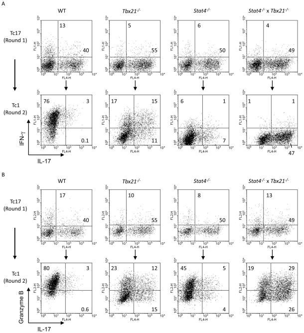
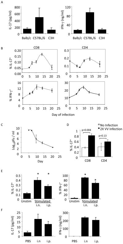
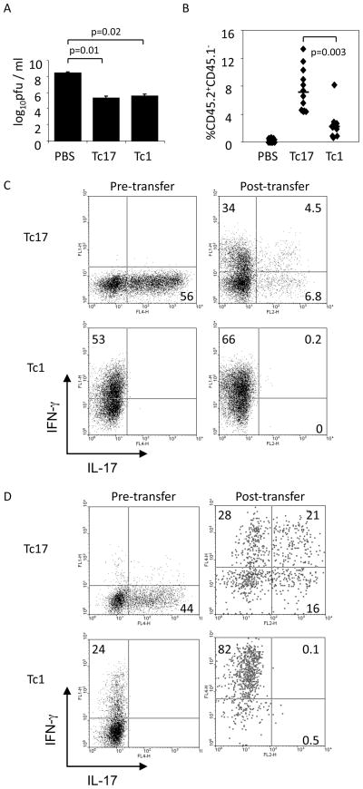
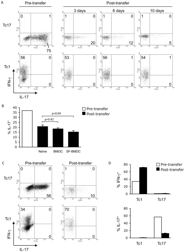
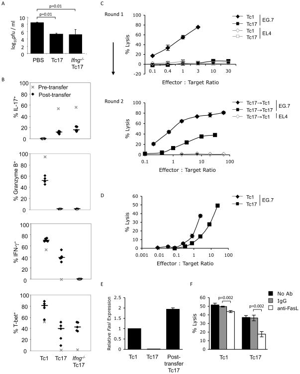
CD8 T cells can acquire cytokine-secreting phenotypes paralleling cytokine production from Th cells. IL-17-secreting CD8 T cells, termed Tc17 cells, were shown to promote inflammation and mediate immunity to influenza. However, most reports observed a lack of cytotoxic activity by Tc17 cells. In this study, we explored the anti-viral activity of Tc17 cells using a vaccinia virus (VV) infection model. Tc17 cells expanded during VV infection, and TCR transgenic Tc17 cells were capable of clearing recombinant VV infection. In vivo, adoptively transferred Tc17 cells lost the IL-17-secreting phenotype, even in the absence of stimulation, but they did not acquire IFN-gamma-secreting potential unless stimulated with a virus-encoded Ag. However, examination of cells following infection demonstrated that these cells acquired cytotoxic potential in vivo, even in the absence of IFN-gamma. Cytotoxic potential correlated with Fasl expression, and the cytotoxic activity of postinfection Tc17 cells was partially blocked by the addition of anti-FasL. Thus, Tc17 cells mediate VV clearance through expression of specific molecules associated with cytotoxicity but independent of an acquired Tc1 phenotype.


Figures

Figure 1
Figure 1
Tc17 cells are unstable and have increased Granzyme B production in a second round of culture. WT (A) or Ifng−/− (B) CD8+ T cells were primed under Tc17 conditions. After five days, Tc17 cells were restimulated in the presence of α-IFNγ alone, or with the addition of the indicated cytokines, or IL-12 alone for an additional five days. IL-17-, IFN-γ-, and Granzyme B-positive cells were detected using intracellular cytokine staining after the first round or second round of stimulation as indicated.
Figure 2
Figure 2
Tc17 instability is dependent on both Stat4 and T-bet. WT, Tbx21−/−, Stat4−/−, or Tbx21−/− x Stat4−/− CD8+ T cells were cultured under Tc17 conditions. After five days, Tc17 cells were restimulated in the presence of IL-12 (Tc1 conditions) for an additional five days. IL-17, IFN-γ, and Granzyme B were detected using intracellular cytokine staining after the first round of stimulation (A, B-top panel) and also at the end of the second round of stimulation (A, B-bottom panel).
Figure 3
Figure 3
Expression of cytotoxicity-associated genes in Tc17 cultures. OT-l CD8+ T cells were cultured under Tc1 or Tc17 conditions. After five days, Tc17 cells were re-stimulated under Tc1 or Tc17 conditions for an additional five days. RNA was isolated from differentiated cells after 4 h of re-stimulation with SIINFEKL peptide. Expression for the indicated genes was measured using quantitative PCR with samples being normalized to the expression of β2-microglobulin mRNA and are relative to levels in Tc1 cells, with the exception of Il17a. A logarithmic scale was used to display the relative expression of all genes with the exception of Il17a and Tnf.
Figure 4
Figure 4
Tc17 cells are induced in response to infection with vaccinia virus. (A) CD8+ T cells were isolated from the spleens of Balb/c, C57BL/6, or C3H/HeJ mice and stimulated for 48 h before cell free supernatants were used to measure IL-17 and IFN-γ protein levels using ELISA. Results are presented as the mean ± SEM of CD8+ T cells from 3 mice. (B, C) C57BL/6 mice were infected i.p. with 5x10⁶ pfu VV per mouse. Spleens and ovaries were respectively isolated for cytokine analysis following re-stimulation (B) or viral titers (C) at 5, 7, 10, 14, and 21 days postinfection. The data are presented as an average of values from 4 mice per time point ± SEM. (D) C57BL/6 mice were infected on days 0 and 21 with 1x10⁶ pfu VV, i.p., per mouse at each time point. Five days after the second infection, CD8+ and CD4+ cells were isolated from the spleens and re-stimulated before analyzing IL-17 and IFN-γ production using ICS. The data shown are an average of values from 2–4 mice ± SEM. (E–F) BoyJ homozygous (CD45.1+) or heterozygous (CD45.1+CD45.2+) mice were injected i.v. with 1x10⁶ OT-I / Rag1−/− CD8+ (CD45.2+) T cells. One day later, mice were infected with either 2x10⁶ pfu VV-SIINFEKL i.n., or 2x10⁷ pfu VV-SIINFEKL, i.p. (E) Five days after infection, splenocytes were stimulated with SIINFEKL peptide or left unstimulated. CD45.1-CD45.2+ IL-17+ and IFN-γ+ cells were detected using ICS. The data are the average ± SEM of 5 mice. (F) Splenocytes were stimulated with SIINFEKL peptide for 48 h and IL-17 and IFN-γ protein levels in cell free supernatants were measured using ELISA. Statistics in B and D were performed using a Student’s T test. *, p<0.05 compared to unstimulated condition.
Figure 5
Figure 5
Adoptively transferred Tc17 cells promote VV-SIINFEKL clearance and convert to an IFN-γ-secreting phenotype. (A–C) BoyJ (CD45.1+) mice were infected with 2x10⁷ pfu VV-SIINFEKL, i.p., and injected i.v. with differentiated OT-l Tc17 or Tc1 cells (1x10⁶, CD45.2+) or PBS after 24 h. Six days later ovaries were harvested for viral titer determination (A), and splenocytes were surface stained with antibodies to CD45.1 and CD45.2 to identify transferred cells (B). (C) IL-17+ and IFN-γ+ cells were identified in differentiated OT-l Tc17 or Tc1 cells immediately before adoptive transfer (left panels) and in splenocytes six days after adoptive transfer (right panels). Transferred cells in the right panels are gated on CD45.1CD45.2+ cells. Data are the average of 4–5 mice ± SEM (A) or are representative experiments (B, C). (D) Differentiated OT-I Tc17 or Tc1 cells (1x10⁶) or PBS were injected i.v. into BoyJ mice. One day later, mice were infected with 2x10⁶ pfu VV-SIINFEKL intranasally. IL-17+ and IFN-γ+ cells were determined in Tc17 or Tc1 cells immediately before adoptive transfer (left panels) and five days after adoptive transfer in isolated splenocytes (right panels). Transferred cells in the right panels are gated on the CD45.1CD45.2+ cells. The data are representative of 6–7 mice. Statistics in A and B were performed using a Student’s T test.
Figure 6
Figure 6
VV-encoded antigen is required for Tc17 cells to convert to an IFN-γsecreting phenotype in vivo. (A) Naïve BoyJ (CD45.1+) mice were injected i.v. with five day differentiated OT-l Tc17 or Tc1 cells (1x10⁶, CD45.2+) or PBS. After an additional 3, 6, or 10 days, spleens were isolated and total splenocytes were stimulated with SIINFEKL (SF) peptide. IL-17+ and IFN-γ+ cells were identified in differentiated Tc17 or Tc1 cells immediately before adoptive transfer (left panels) and 3, 6, and 10 days after adoptive transfer from splenocytes (right panels). Transferred cells in the right panels are gated on the CD45.1CD45.2+ cells. The data are representative of two experiments. (B) BoyJ mice were injected i.v. with BMDCs untreated or pulsed with SIINFEKL peptide (1x10⁶, CD45.1+) or PBS-treated and injected i.v. with differentiated OT-l Tc17 cells (1x10⁶, CD45.2+) 48 h later. After an additional 5 days, spleens were isolated and total splenocytes were stimulated with SIINFEKL peptide. IL-17+ cells were identified in differentiated Tc17 cells immediately before adoptive transfer and 5 days after adoptive transfer from total splenocytes. Transferred cells are gated on CD45.1CD45.2+ cells. The data are the average of 3–4 mice ± SEM and are representative of 2 or more experiments. (C, D) BoyJ mice were infected i.p. with 2x10⁷ pfu VV-WR and injected i.v. with differentiated OT-l Tc17 or Tc1 cells (1x10⁶) or PBS one day later. After an additional six days, spleens were isolated and total splenocytes were stimulated with SIINFEKL peptide. (C) IL-17+ and IFN-γ+ cells were analyzed in differentiated OT-l Tc17 or Tc1 cells immediately before adoptive transfer (left panels) and six days after adoptive transfer in spleen (right panels). The transferred cells were gated on CD45.1CD45.2+ cells. Data are representative of 5 mice (C), or are the average ± SEM of 5 mice (D) and are representative of 2 or more experiments. Statistics in B were performed using a Student’s T test.
Figure 7
Figure 7
Adoptively transferred Tc17 cells acquire cytotoxic potential in vivo. (A, B) BoyJ (CD45.1+) mice were infected with 2x10⁷ pfu VV-SIINFEKL i.p. and injected i.v. with differentiated OT-l Tc17, Ifng−/− Tc17 and Tc1 cells (1x10⁶, CD45.2+) or PBS 24 h later. After six days, ovaries were harvested for viral titer determination (A), and IL-17+, IFN-γ+, Granzyme B+, and T-bet+ CD45.1CD45.2+ splenic cells were analyzed using ICS (B). Median values are displayed as horizontal lines and symbols represent individual mice. (C) After culture for one or two rounds (as indicated) under Tc17, Tc1 or Tc17 switched to Tc1 conditions, OT-I CD8+ T cells were tested for cytotoxic potential using a 51Cr-release assay. Effector cells were co-cultured with 51Cr-labeled, Ova-expressing EG.7 target cells for 6 h at the indicated effector to target ratios. (D–F) Mice were infected with VV-SIINFEKL and injected with OT-I Tc17 or Tc1 cells as described in (A). Six days later, injected cells were isolated using anti-CD45.2 antibodies and magnetic selection. (D) A 51Cr release assay was performed as in (C). E:T ratios were normalized to the percentages of CD45.1CD45.2+ cells in the purified populations. Cytotoxicity against EL4 targets was less than 1.5%. (E) Expression of Fasl mRNA was determined in post-transfer Tc17 cells and compared to in vitro derived Tc1 and Tc17 cells using quantitative PCR. (F) A 51Cr release assay was performed as in (D) with the addition of control antibodies or anti-FasL. E:T ratios for post-transfer Tc1 were 2.5:1 and for post-transfer Tc17 were 20:1. Statistics in A and F were performed using a Student’s T test.

References

    1. Intlekofer AM, Takemoto N, Wherry EJ, Longworth SA, Northrup JT, Palanivel VR, Mullen AC, Gasink CR, Kaech SM, Miller JD, Gapin L, Ryan K, Russ AP, Lindsten T, Orange JS, Goldrath AW, Ahmed R, Reiner SL. Effector and memory CD8+ T cell fate coupled by T-bet and eomesodermin. Nat Immunol. 2005;6:1236–1244. - PubMed
    1. Li Q, Eppolito C, Odunsi K, Shrikant PA. IL-12-programmed long-term CD8+ T cell responses require STAT4. J Immunol. 2006;177:7618–7625. - PubMed
    1. Pearce EL, Mullen AC, Martins GA, Krawczyk CM, Hutchins AS, Zediak VP, Banica M, DiCioccio CB, Gross DA, Mao CA, Shen H, Cereb N, Yang SY, Lindsten T, Rossant J, Hunter CA, Reiner SL. Control of effector CD8+ T cell function by the transcription factor Eomesodermin. Science. 2003;302:1041–1043. - PubMed
    1. Takemoto N, Intlekofer AM, Northrup JT, Wherry EJ, Reiner SL. Cutting Edge: IL-12 inversely regulates T-bet and eomesodermin expression during pathogen-induced CD8+ T cell differentiation. J Immunol. 2006;177:7515–7519. - PubMed
    1. Huber M, Heink S, Grothe H, Guralnik A, Reinhard K, Elflein K, Hunig T, Mittrucker HW, Brustle A, Kamradt T, Lohoff M. A Th17-like developmental process leads to CD8(+) Tc17 cells with reduced cytotoxic activity. Eur J Immunol. 2009;39:1716–1725. - PubMed

Publication types

MeSH terms