. 2010 Aug 10;107(32):14286-91.
doi: 10.1073/pnas.1008300107. Epub 2010 Jul 12.

Kinetic phases of distribution and tumor targeting by T cell receptor engineered lymphocytes inducing robust antitumor responses

Affiliations

Kinetic phases of distribution and tumor targeting by T cell receptor engineered lymphocytes inducing robust antitumor responses

Richard C Koya et al. Proc Natl Acad Sci U S A. .

Abstract

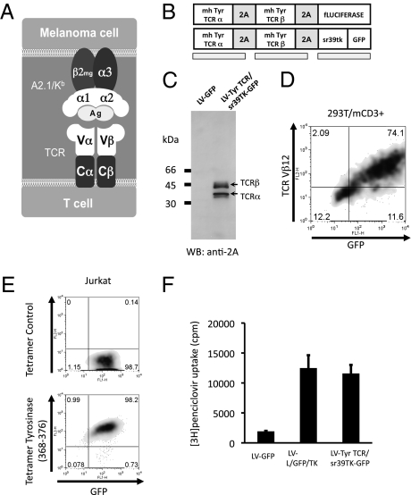
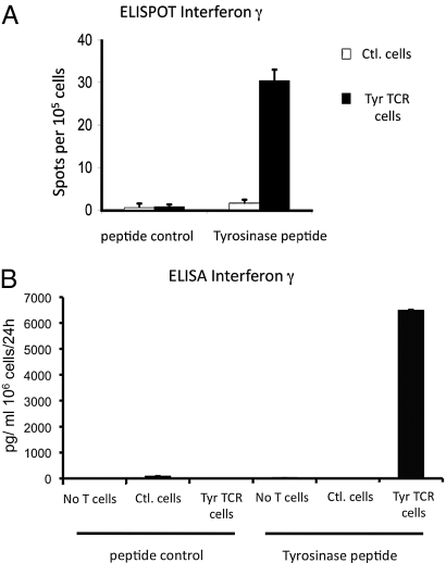
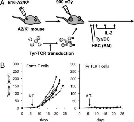
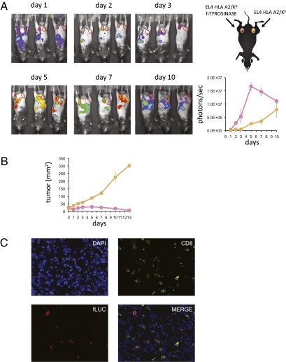
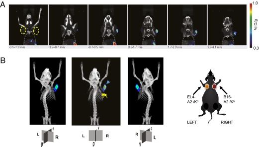
A key issue in advancing the use of adoptive cell transfer (ACT) of T cell receptor (TCR) engineered lymphocytes for cancer therapy is demonstrating how TCR transgenic cells repopulate lymphopenic hosts and target tumors in an antigen-specific fashion. ACT of splenocytes from fully immunocompetent HLA-A2.1/K(b) mice transduced with a chimeric murine/human TCR specific for tyrosinase, together with lymphodepletion conditioning, dendritic cell (DC)-based vaccination, and high-dose interleukin-2 (IL-2), had profound antitumor activity against large established MHC- and antigen-matched tumors. Genetic labeling with bioluminescence imaging (BLI) and positron emitting tomography (PET) reporter genes allowed visualization of the distribution and antigen-specific tumor homing of TCR transgenic T cells, with trafficking correlated with antitumor efficacy. After an initial brief stage of systemic distribution, TCR-redirected and genetically labeled T cells demonstrated an early pattern of specific distribution to antigen-matched tumors and locoregional lymph nodes, followed by a more promiscuous distribution 1 wk later with additional accumulation in antigen-mismatched tumors. This approach of TCR engineering and molecular imaging reporter gene labeling is directly translatable to humans and provides useful information on how to clinically develop this mode of therapy.


Conflict of interest statement

The authors declare no conflict of interest.

Figures

Fig. 1.
Fig. 1.
Model system, vector schematic, and transgene expression. (A) Schematic of the chimeric murine/human interaction between the transgenic TCR and MHC molecules in the A2.1/Kb mouse model. In gray are the proximal murine sequences, and in white the distal human sequences allowing the presentation and recognition of peptide antigen with human restriction. (B) Tyrosinase (Tyr)-TCR/sr39TK-GFP and firefly luciferase vector schematic representation. (C) Immunoblotting of 293T cells transduced with control lentiviral vector expressing GFP (LV-GFP) or the tyrosinase TCR and sr39tk (LV-TCR/sr39TK-GFP) vectors, incubation with rabbit anti-2A primary antibody. Arrows indicate cleaved products with sizes corresponding to the TCR α and β chains. (D) Flow-cytometric analysis of 293T cells expressing CD3 transduced with LV-TCR/sr39TK-GFP vector, stained with specific clonotypic anti-Vβ12 antibody. (E) Tyrosinase368–376 specific and negative control peptide HLA-A2.1 tetramer assay of Jurkat cells transduced with LV-TCR/sr39TK-GFP vector. (F) Penciclovir uptake assay of Jurkat cells transduced with negative control LV-GFP, positive control LV-L/GFP/TK, or LV-TCR/sr39TK-GFP vectors.
Fig. 2.
Fig. 2.
In vitro functional analysis of murine primary T cells transduced with tyrosinase TCR retroviral supernatants. (A) ELISPOT assay for cellular IFN-γ secretion of control T cells and tyrosinase TCR transduced T cells incubated with control scrambled or tyrosinase368–376 peptides. (B) ELISA for total IFN-γ secretion of 24 h-collection supernatants of EL4-A2/Kb pulsed with control or tyrosinase peptides coincubated with control T cells or tyrosinase TCR transduced T cells.
Fig. 3.
Fig. 3.
In vivo function of murine primary T cells transduced with tyrosinase TCR retrovirus. (A) HLA-A2/Kb transgenic mice with s.c. B16-A2/Kb tumors received the full protocol of myelodepletion with hematopoietic stem cells (HSC)/ bone marrow (BM) transplantation, adoptive cell transfer (ACT) of control T cells or tyrosinase TCR-transduced T cells followed by tyrosinase368–376 peptide-pulsed dendritic cell (DC) vaccination and high-dose IL-2. (B) Mice were followed for tumor growth measurements (product of two diameters as mm2), P < 0.01.
Fig. 4.
Fig. 4.
Bioluminescence imaging (BLI) of T cell trafficking in vivo. (A) HLA-A2/Kb transgenic mice with inguinal s.c. EL4-A2/Kb-expressing tyrosinase (Left) and control EL4-A2/Kb (Right) tumors received the full protocol of adoptive cell transfer (ACT) with tyrosinase TCR/fLuciferase-transduced T cells. Mice were followed from day 1 to 10 post-ACT and bioluminescence signal of ventral views were recorded and quantified on region of interest (ROI) drawn on tumor sites. Representative animals are shown. Pink, EL4-A2/Kb-tyrosinase+; yellow, control EL4-A2/Kb. (B) These mice were followed for tumor growth measurements (product of two diameters as mm2). (C) EL4-A2/Kb-tyrosinase+ tumors were costained with DAPI (nucleus localization), anti-CD8, and anti-fLuciferase (40×).
Fig. 5.
Fig. 5.
PET CT imaging of T cell trafficking in vivo in mice with control EL4-A2/Kb tumors or with contralateral tyrosinase-positive B16-A2/Kb tumors at day 5 post-ACT. (A) HLA-A2/Kb transgenic mice with thoracic dorsal s.c. B16-A2/Kb (Right) and control EL4-A2/Kb (Left) tumors were adoptively transferred with tyrosinase TCR/sr39TK/GFP transduced T cells. Representative animals are shown. Specific signal quantification ratio above background: Left ROI = 1.05 ± 0.59%ID/g; Right ROI = 3.00 ± 0.61%ID/g. (B) Schematic representation of tumor location and reconstructed tridimensional PET CT scan image with the nonspecific signal from abdominal excretion of [18F]FHBG subtracted from the final image. Yellow arrows, signals on axillary lymph nodes (specific signal quantification ratio above background: Right LND = 2.21 ± 0.52%ID/g; Left LND = 2.05 ± 0.63%ID/g). Blue arrow, signal in B16-A2/Kb tumor. R, right; L, left; D, dorsal; V, ventral sides.

Comment in

References

    1. Yee C, et al. Adoptive T cell therapy using antigen-specific CD8+ T cell clones for the treatment of patients with metastatic melanoma: In vivo persistence, migration, and antitumor effect of transferred T cells. Proc Natl Acad Sci USA. 2002;99:16168–16173. - PMC - PubMed
    1. Rosenberg SA, Restifo NP, Yang JC, Morgan RA, Dudley ME. Adoptive cell transfer: A clinical path to effective cancer immunotherapy. Nat Rev Cancer. 2008;8:299–308. - PMC - PubMed
    1. Dembić Z, et al. Transfection of the CD8 gene enhances T-cell recognition. Nature. 1987;326:510–511. - PubMed
    1. Schumacher TN. T-cell-receptor gene therapy. Nat Rev Immunol. 2002;2:512–519. - PubMed
    1. Morgan RA, et al. Cancer regression in patients after transfer of genetically engineered lymphocytes. Science. 2006;314:126–129. - PMC - PubMed

Publication types

Substances