. 2010 Jul 27;107(30):13438-43.
doi: 10.1073/pnas.1002423107. Epub 2010 Jul 12.

Notch2 is required for progression of pancreatic intraepithelial neoplasia and development of pancreatic ductal adenocarcinoma

Affiliations

Notch2 is required for progression of pancreatic intraepithelial neoplasia and development of pancreatic ductal adenocarcinoma

Pawel K Mazur et al. Proc Natl Acad Sci U S A. .

Abstract

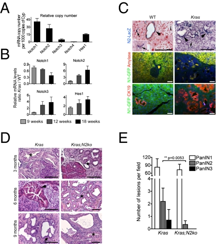
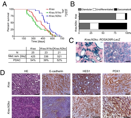
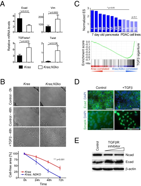
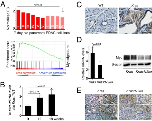
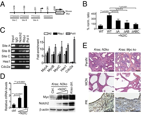
Pancreatic cancer is one of the most fatal malignancies lacking effective therapies. Notch signaling is a key regulator of cell fate specification and pancreatic cancer development; however, the role of individual Notch receptors and downstream signaling is largely unknown. Here, we show that Notch2 is predominantly expressed in ductal cells and pancreatic intraepithelial neoplasia (PanIN) lesions. Using genetically engineered mice, we demonstrate the effect of conditional Notch receptor ablation in KrasG12D-driven pancreatic carcinogenesis. Deficiency of Notch2 but not Notch1 stops PanIN progression, prolongs survival, and leads to a phenotypical switch toward anaplastic pancreatic cancer with epithelial-mesenchymal transition. By expression profiling, we identified increased Myc signaling regulated by Notch2 during tumor development, placing Notch2 as a central regulator of PanIN progression and malignant transformation. Our study supports the concept of distinctive roles of individual Notch receptors in cancer development.


Conflict of interest statement

The authors declare no conflict of interest.

Figures

Fig. 1.
Fig. 1.
Expression analysis of Notch receptors in WT and KrasG12D-induced pancreata. (A) Transcript levels of Notch receptors and Hes1 in relation to cyclophilin gene expression in WT pancreata (n = 3). (B) Quantification of Notch receptor and Hes1 gene expression at indicated time points in Kras pancreatic tissue. Values represent WT-to-Kras tissue ratios of relative expression levels (n = 4). (C) Expression of Notch1 and Notch2 in distinct compartments of 18-wk-old WT and Kras pancreas using Notch1 and Notch2 reporter mice. Arrows indicate centroacinar cells, and arrowheads point to X-Gal+ ducts and PanINs. i, islets. (D) H&E staining of 3-, 6-, and 9-mo-old Kras and Kras;N2ko pancreata. Asterisks indicate PanIN1, arrowhead points to PanIN2, and arrow indicates PanIN3 lesions. Note the absence of PanIN2/3 in Kras;N2ko mice. (Scale bars: 50 μm.) (E) Quantification of PanINs in 9-mo-old Kras (n = 4) and Kras;N2ko (n = 5) mice shows a significant reduction in PanIN2 and absence of PanIN3 lesions in Kras;N2ko mice.
Fig. 2.
Fig. 2.
Deficiency of Notch2 prolongs survival and delays development of anaplastic PDAC. (A) Kaplan–Meier survival data and PDAC development of Kras, Kras;N1ko, and Kras;N2ko mice. Kras;N2ko mice have significantly prolonged survival compared with Kras and Kras;N1ko mice (P < 0.02). n.s., not significant. (B) Tumor differentiation analysis reveals more anaplastic PDAC in Kras;N2ko mice compared with Kras mice. (C) Positive X-Gal staining shows Cre-induced recombination in cells of MCN-like cysts and anaplastic PDAC in Kras;N2ko;Rosa26R+/LSL-lacZ mice. (D) Histological and immunohistochemical analysis of Kras and Kras;N2ko tumors. Expression of E-cadherin in Kras PDAC and low to absent expression in Kras;N2ko tumors. The Notch targets HES1 and PDX1 are expressed in tumors derived from both genotypes. (Scale bars: 50 μm.)
Fig. 3.
Fig. 3.
EMT is a prominent feature in Kras;N2ko PDAC. (A) Quantitative RT-PCR analysis of EMT-associated genes expressed by cancer cells from Kras and Kras;N2ko PDAC (n = 4 for each genotype). (B) Assessment of cell migration in wound closure assays performed in Kras and Kras;N2ko cells treated with TGF-β. Wound closure is delayed in Kras cells compared with Kras;N2ko cells. Quantification of wound closure is plotted as the percentage of the cell-free area over time. (C) Comparison of TGF-β gene sets by GSEA reveals significantly up-regulated TGF-β signatures in Kras;N2ko pancreata isolated from 7-d-old mice (dark blue, n = 2 and 4) and cancer cells (light blue, n = 6 each). A positive normalized enrichment score indicates elevated TFG-β–associated gene expression. Roman numbers refer to the detailed analysis in Tables S10 and S11. (D) Kras;N2ko cells reveal morphological and molecular responses characteristic of EMT in response to TGF-β, including loss of E-cadherin expression and nuclear translocation of SMAD4. (Scale bars: 50 μm.) (E) Treatment with the TGF-β receptor inhibitor SB431542 is sufficient to reverse the EMT-associated cadherin switch, suggesting that EMT in Kras;N2ko cells is dependent on a TGF-β autocrine loop.
Fig. 4.
Fig. 4.
Myc is up-regulated during pancreatic carcinogenesis and down-regulated in Kras;N2ko mice. (A) GSEA shows significantly enriched Myc signatures in Kras vs. Kras;N2ko pancreata isolated from 7-d-old mice (dark red, n = 2 and 4) and primary cancer cells (light red, n = 6 each). Roman numbers refer to detailed analysis in Tables S12 and S13. (B) Myc transcript levels increase during carcinogenesis in Kras pancreata at indicated time points. Values represent WT-to-Kras ratio of relative expression levels (n = 3 for each time point). (C) Expression of Myc is low in the normal pancreas and increases in PanIN lesions of Kras mice. (D) Kras;N2ko cancer cells (n = 4) show decreased Myc mRNA and protein expression compared with Kras cells (n = 5). (E) Immunohistochemical staining in Kras;N2ko-derived anaplastic PDAC shows lower expression of Myc compared with Kras PDAC. (Scale bar: 50 μm.)
Fig. 5.
Fig. 5.
Myc is a downstream target of Notch, and its ablation resembles features of the Notch2-deficient phenotype. (A) Analysis of Notch/Rbpj binding sites in the mouse Myc promoter using the consensus RTGGGAA motif reveals three sites: A, B, and C. (B) Activity of a Myc promoter fragment containing binding regions A, B, and C was analyzed using luciferase reporter assays. Kras;N2ko cells were cotransfected with Myc luciferase plasmids and N2IC. Mutations in the respective binding sites decrease activation of Myc. Activities were corrected for transfection efficiency by normalizing with Renilla luciferase activity and are expressed as a percentage of induction. (C) ChIP analyses using the indicated antibodies were analyzed by PCR for sites of interests. Products of the exponential phase of PCR are shown. Hes1 promoter primer served as positive control, and Cdc2a promoter primers as negative control. Quantitative PCR indicates that Notch2 binds to regions A, B, and C of the Myc promoter comparable to a binding site in the Hes1 promoter. (D) Transfection of N2IC stimulates Myc expression in Kras;N2ko cells in a dose-dependent manner. Notch2 and Myc expression levels of Kras control are shown for comparison. (E) Myc and Notch2 ablation in Kras mice results in similar phenotypes. Kras;N2ko and Kras;Myc-ko mice develop PanIN1 but not advanced PanIN2/3 lesions and MCN-like lesions with progesterone receptor-positive (PR+) surrounding stroma. Brightness and contrast levels were adjusted across the whole image for each panel. (Scale bar: 50 μm.)

References

    1. Hezel AF, Kimmelman AC, Stanger BZ, Bardeesy N, Depinho RA. Genetics and biology of pancreatic ductal adenocarcinoma. Genes Dev. 2006;20:1218–1249. - PubMed
    1. Hruban RH, Iacobuzio-Donahue C, Wilentz RE, Goggins M, Kern SE. Molecular pathology of pancreatic cancer. Cancer J. 2001;7:251–258. - PubMed
    1. Hingorani SR, et al. Preinvasive and invasive ductal pancreatic cancer and its early detection in the mouse. Cancer Cell. 2003;4:437–450. - PubMed
    1. Pasca di Magliano M, et al. Hedgehog/Ras interactions regulate early stages of pancreatic cancer. Genes Dev. 2006;20:3161–3173. - PMC - PubMed
    1. Miyamoto Y, et al. Notch mediates TGF alpha-induced changes in epithelial differentiation during pancreatic tumorigenesis. Cancer Cell. 2003;3:565–576. - PubMed

Publication types

MeSH terms