. 2010 Nov 25;116(22):4395-403.
doi: 10.1182/blood-2010-01-261503. Epub 2010 Jul 12.

Complement-mediated inhibition of neovascularization reveals a point of convergence between innate immunity and angiogenesis

Affiliations

Complement-mediated inhibition of neovascularization reveals a point of convergence between innate immunity and angiogenesis

Harald F Langer et al. Blood. .

Abstract

Beyond its role in immunity, complement mediates a wide range of functions in the context of morphogenetic or tissue remodeling processes. Angiogenesis is crucial during tissue remodeling in multiple pathologies; however, the knowledge about the regulation of neovascularization by the complement components is scarce. Here we studied the involvement of complement in pathological angiogenesis. Strikingly, we found that mice deficient in the central complement component C3 displayed increased neovascularization in the model of retinopathy of prematurity (ROP) and in the in vivo Matrigel plug assay. In addition, antibody-mediated blockade of C5, treatment with C5aR antagonist, or C5aR deficiency in mice resulted in enhanced pathological retina angiogenesis. While complement did not directly affect angiogenesis-related endothelial cell functions, we found that macrophages mediated the antiangiogenic activity of complement. In particular, C5a-stimulated macrophages were polarized toward an angiogenesis-inhibitory phenotype, including the up-regulated secretion of the antiangiogenic soluble vascular endothelial growth factor receptor-1. Consistently, macrophage depletion in vivo reversed the increased neovascularization associated with C3- or C5aR deficiency. Taken together, complement and in particular the C5a-C5aR axes are potent inhibitors of angiogenesis.


Figures

Figure 1
Figure 1
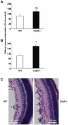
C3 deficiency results in increased angiogenesis in ROP. WT or C3−/− mice were subjected to the ROP model. (A) Retinal neovascularization was quantified on day p17 in WT and C3−/− mice as described under “Hypoxia-induced retinal vascularization, retinopathy of prematurity model.” Retinal neovascularization is presented as the number of epiretinal neovascular nuclei per section. Data are mean ± SEM (n = 12-16 pups per group) and are shown as percentage of control. Number of epiretinal nuclei in WT mice represents the 100% control. *P < .001. (B) Paraffin-embedded axial sections (6 μm) of the retina were stained with PAS and hematoxylin. Pathological neovascularization invades into the vitreous cavity anterior to the internal limiting membrane. C3−/− mice subjected to the ROP model had more neovascular regions anterior to the internal limiting membrane compared with WT mice. The arrows indicate the neovascular tufts anterior to the internal limiting membrane. Scale bars, 150 μm.
Figure 2
Figure 2
C5a receptor deficiency results in increased angiogenesis in ROP. WT, C3aR−/−, or C5aR−/− mice were subjected to the ROP model. (A) Retinal neovascularization was quantified on day p17 in WT and C3aR−/− mice. Retinal neovascularization is presented as the number of epiretinal neovascular nuclei per section. Data are mean ± SEM (n = 12-13 pups per group) and are shown as percentage of control. Number of epiretinal nuclei in WT mice represents the 100% control. There was no significant difference between both groups. (B) Retinal neovascularization was quantified on day p17 in WT and C5aR−/− mice. Data are mean ± SEM (n = 16-17 pups per group) and are shown as percentage of control. Number of epiretinal nuclei in WT mice represents the 100% control. *P < .005. (C) Paraffin-embedded axial sections (6 μm) of the retina were stained with PAS and hematoxylin, and neovascular tufts were observed anterior to the internal limiting membrane (arrows). C5aR−/− mice displayed more neovascular tufts. Scale bars, 150 μm.
Figure 3
Figure 3
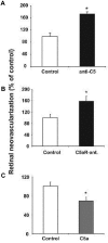
The role of C5a and C5aR in pathological angiogenesis in ROP. (A) WT mice subjected to the ROP model were treated on day p12.5 with an i.p. injection of anti-C5 or an isotype control antibody. Data are mean ± SEM (n = 6-7 pups per group) and are shown as percentage of control. Number of epiretinal nuclei in control treated mice represents the 100% value. *P < .01. (B) WT mice were subjected to the ROP model and were treated from day p12-16 with daily intraperitoneal injections of C5a receptor antagonist (C5aR-ant.) or control antagonist. Data are mean ± SEM (n = 17-19 pups per group) and are shown as percentage of control. Number of epiretinal nuclei in control treated mice represents the 100% value. *P < .05. (C) WT mice subjected to the ROP model were treated from p12-16 with daily intraperitoneal injections of C5a or vehicle control. Data are mean ± SEM (n = 17 pups per group) and are shown as percentage of control. Number of epiretinal nuclei in control treated mice represents the 100% value. *P < .05.
Figure 4
Figure 4
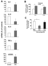
Macrophages stimulated with C5a display an antiangiogenic phenotype. (A) Isolated primary bone marrow derived mouse macrophages treated with C5a showed a polarization toward an antiangiogenic phenotype as verified by significantly increased mRNA levels of IL-6, TNF-α, and soluble VEGFR-1 and significantly reduced levels of IL-10 mRNA using quantitative real-time PCR. IL-12 mRNA levels were not significantly changed by C5a treatment. The respective mRNA was normalized against actin. Data are mean ± SEM (n = 4) and are shown as percentage of control. mRNA of macrophages treated with vehicle control represents the 100% value. *P < .05. (B) Mouse macrophages were incubated with C5a (100nM) or vehicle control for 4 or 12 hours and supernatants were analyzed for sVEGFR1 by ELISA. Data are mean ± SEM (n = 3-4) and are shown as percentage of control. Detected sVEGFR1 in supernatant of control treated macrophages represents the 100% value. *P < .05. (C) HUVEC were treated with supernatants of human monocytes that were stimulated without (vehicle control) or with C5a (100nM) or C3a (100nM) for 12 hours and analyzed in the in vitro Matrigel tube formation assay. To assess for VEGF-induced tube formation, growth factor-reduced Matrigel supplemented with VEGF was used. The supernatant of C5a-stimulated human monocytes inhibited the VEGF induced tube formation of HUVEC. Length of forming tubes was quantified using Metamorph software. Data are mean ± SEM (n = 3) and are shown as percentage of control. Tube length of endothelial cells incubated with supernatant of control–treated macrophages represents the 100% value. *P < .05.
Figure 5
Figure 5
Increased angiogenesis due to C3-deficiency is dependent on macrophages. WT or C3−/− mice were subjected to the ROP model and animals were injected with control liposomes or clodronate liposomes to deplete macrophages. Retinal neovascularization was quantified on day p17. Retinal neovascularization is presented as the number of epiretinal neovascular nuclei per section. Data are mean ± SEM (n = 12-17 pups per group) and are shown as percentage of control. Number of epiretinal nuclei in WT mice treated with control liposomes represents the 100% control. *P < .05; ns, not significant.
Figure 6
Figure 6
Matrigel plug angiogenesis in C3-deficient mice. Angiogenesis was assessed using the in vivo Matrigel plug assay as described under “In vivo Matrigel plug assay.” (A) Six hours after Matigel implantation, WT and C3−/− mice were injected with control liposomes or clodronate liposomes to deplete macrophages and after 7 days angiogenesis was assessed within the explanted Matrigels. Data are mean ± SEM (n = 14 Matrigels per group) and are shown as percentage of control. Area of neovascularization in WT mice treated with control liposomes represents the 100% control. *P < .01; ns, not significant. (B) Frozen sections of the explanted Matrigels were stained with H&E. C3−/− mice showed significantly more Matrigel plug neovascularization. Scale bars, 150 μm.
Figure 7
Figure 7
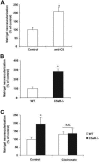
C5aR plays an inhibitory role in Matrigel plug angiogenesis. (A) Angiogenesis was assessed using the in vivo Matrigel plug assay. (A) Six hours and 3 days after Matrigel implantation, C57BL/6 WT mice were injected with control IgG or anti-C5 (clone BB5.1, 750 μg per mouse). After 7 days, angiogenesis was assessed within the explanted Matrigels. Data are mean ± SEM (n = 10 Matrigels per group) and are shown as percentage of control. Area of neovascularization in WT mice treated with IgG control represents the 100% value. *P < .005. (B) Matrigel angiogenesis was quantified in WT and C5aR−/− mice. Data are mean ± SEM (n = 5 Matrigels per group) and are shown as percentage of control. The area of neovascularization of WT mice represents the 100% control. *P < .01. (C) Six hours after Matrigel implantation, WT and C5aR−/− mice were injected with control liposomes or clodronate liposomes to deplete macrophages and after 7 days angiogenesis was assessed within the explanted Matrigels. Data are mean ± SEM (n = 8 Matrigels per group) and are shown as percentage of control. Area of neovascularization in WT mice treated with control liposomes represents the 100% control. *P < .05; ns, not significant.

Comment in

References

    1. Ricklin D, Hajishengallis G, Yang K, Lambris JD. Complement: a key system for immune surveillance and homeostasis. Nat Immunol. 2010;11(9):785–797. - PMC - PubMed
    1. Guo RF, Ward PA. Role of C5a in inflammatory responses. Annu Rev Immunol. 2005;23:821–52. - PubMed
    1. Huber-Lang M, Sarma JV, Zetoune FS, et al. Generation of C5a in the absence of C3: a new complement activation pathway. Nat Med. 2006;12(6):682–687. - PubMed
    1. Sjoberg AP, Trouw LA, Blom AM. Complement activation and inhibition: a delicate balance. Trends Immunol. 2009;30(2):83–90. - PubMed
    1. Strey CW, Markiewski M, Mastellos D, et al. The proinflammatory mediators C3a and C5a are essential for liver regeneration. J Exp Med. 2003;198(6):913–923. - PMC - PubMed

Publication types

MeSH terms