. 2010 Jul 6;5(7):e11440.
doi: 10.1371/journal.pone.0011440.

Adolescents, adults and rewards: comparing motivational neurocircuitry recruitment using fMRI

Affiliations

Adolescents, adults and rewards: comparing motivational neurocircuitry recruitment using fMRI

James M Bjork et al. PLoS One. .

Abstract

Background: Adolescent risk-taking, including behaviors resulting in injury or death, has been attributed in part to maturational differences in mesolimbic incentive-motivational neurocircuitry, including ostensible oversensitivity of the nucleus accumbens (NAcc) to rewards.

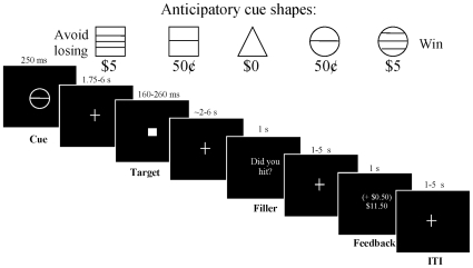
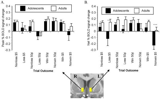
Methodology/principal findings: To test whether adolescents showed increased NAcc activation by cues for rewards, or by delivery of rewards, we scanned 24 adolescents (age 12-17) and 24 adults age (22-42) with functional magnetic resonance imaging while they performed a monetary incentive delay (MID) task. The MID task was configured to temporally disentangle potential reward or potential loss anticipation-related brain signal from reward or loss notification-related signal. Subjects saw cues signaling opportunities to win or avoid losing $0, $.50, or $5 for responding quickly to a subsequent target. Subjects then viewed feedback of their trial success after a variable interval from cue presentation of between 6 to 17 s. Adolescents showed reduced NAcc recruitment by reward-predictive cues compared to adult controls in a linear contrast with non-incentive cues, and in a volume-of-interest analysis of signal change in the NAcc. In contrast, adolescents showed little difference in striatal and frontocortical responsiveness to reward deliveries compared to adults.

Conclusions/significance: In light of divergent developmental difference findings between neuroimaging incentive paradigms (as well as at different stages within the same task), these data suggest that maturational differences in incentive-motivational neurocircuitry: 1) may be sensitive to nuances of incentive tasks or stimuli, such as behavioral or learning contingencies, and 2) may be specific to the component of the instrumental behavior (such as anticipation versus notification).


Conflict of interest statement

Competing Interests: The authors have declared that no competing interests exist.

Figures

Figure 1
Figure 1. Modified monetary incentive delay (MID) task.
Each trial began with presentation of one of five anticipatory cues. The cue signaled the opportunity to either win money (circle series), avoid losing money (square series), or win/lose no money (triangle) by recording a button press while the following white square target was presented on the screen. After target presentation, the subject then waited across a variable delay for notification (feedback) of whether he or she hit the target. During this delay, a lexical filler stimulus (“Did you hit?”) was presented. Intervals between trial stimuli were pseudorandomly varied as indicated, and trials were also separated by a 1–5 s variable intertrial interval (ITI) following each notification.
Figure 2
Figure 2. MID task behavior.
Mean reaction time (RT) to targets (A) showed significant main effects of trial incentive and time. Specifically, subjects responded more quickly over time, from runs 1 to 3 of the task, and subjects responded more quickly to incentivized, compared to non-incentivized targets. Accordingly, there was a significant main effect of incentive amount on overall task hit rates (B), with a greater proportion of incentivized versus non-incentivized targets hit. There were no main or interactive effects of age group on either RT or hit rates. ** denotes P<.05 per simple-effect t-test.
Figure 3
Figure 3. MID task affective ratings.
On a post-scan questionnaire, participants reported greater happiness (A) and excitement (B) when seeing anticipatory cues as the potential reward amounts increased. There were no significant main or interactive effects of age group on positive affect ratings. Similarly, subjects reported greater unhappiness (C) and fearfulness (D) as potential loss amounts increased. There were main effects of group (F(1,46)  = 5.338, p<.05) on unhappiness ratings across the combined non-incentive and loss-trial types, with greater self-reported unhappiness in adolescents compared to adults. There were no other significant main or interaction effects of age group negative affect ratings. * denotes P<.10 and ** denotes P<.05 per simple-effect t-test.
Figure 4
Figure 4. Activation by anticipation of responding for rewards.
In these and subsequent statistical maps: 1) all images are right-left reversed per radiological convention, 2) the underlay is a T1-weighted structural image from a representative subject, 3) the Talairach coordinate of the image plane is indicated, 4) illuminated voxels in group-wise maps feature contrast activation that survives false discovery rate (FDR) correction to P<.05, and 5) illuminated voxels in the inset group-difference t-statistic maps do not survive FDR correction, but illustrate differences in NAcc recruitment as the structure of a priori interest. Anticipation of responding for rewards contrasted with anticipation of responding for no incentive activated portions of ventral striatum (VS) insula, and posterior mesofrontal cortex in both adolescents (A,C) and in adults (B,D). In the inset uncorrected map of the direct voxel-wise age-group difference in activation by this contrast, relatively lower VS activation in adolescents is depicted in cool colors.
Figure 5
Figure 5. Activation by anticipation of responding to avoid losses.
Anticipation of responding to avoid losses contrasted with anticipation of responding for no incentive activated striatal voxels in both adolescents (A,C) and in adults (B,D). In the inset uncorrected map of the direct voxel-wise age-group difference in activation by this contrast, relatively lower VS activation in adolescents is depicted in cool colors.
Figure 6
Figure 6. Activation by notification of rewards and losses.
Notification of rewards (contrasted with notification of failure to win reward) activated the VS and mesofrontal cortex (mFC) in both adolescents (A,C) and in adults (B,D). Notification of all losses (versus notification of successful loss avoidance) did not activate any voxels above threshold in adolescents (E), but activated anterior cingulate cortex in adults (F).
Figure 7
Figure 7. Anticipatory signal change in NAcc VOI.
Time series data were extracted from a two-voxel mask in Talairach space in each of right and left NAcc (inset), for each trial type separately. Group mean peak modeled anticipatory signal changes (∼6 s post-cue) are presented as absolute signal change from baseline in parts A and B, and as a net difference from the signal change following presentation of the nonincentive cue (parts C and D). Net signal change elicited by high-reward cues correlated with age in right (E) but not left (F) NAcc. * denotes P<.10 and ** denotes P<.05 per simple-effect t-test.
Figure 8
Figure 8. Notification-elicited signal change in NAcc VOI.
Trial-outcome-averaged time series data were extracted from each of the right and left NAcc masks (inset). Group mean modeled peak outcome-elicited signal changes (∼6 s post-cue) are presented here as signal change from baseline. * denotes P<.10 and ** denotes P<.05 per simple-effect t-test.

References

    1. Johnson SB, Blum RW, Giedd JN. Adolescent maturity and the brain: the promise and pitfalls of neuroscience research in adolescent health policy. J Adolesc Health. 2009;45:216–221. - PMC - PubMed
    1. Castellanos FX, Lee PP, Sharp W, Jeffries NO, Greenstein DK, et al. Developmental trajectories of brain volume abnormalities in children and adolescents with attention-deficit/hyperactivity disorder. Jama. 2002;288:1740–1748. - PubMed
    1. Giedd JN, Blumenthal J, Jeffries NO, Castellanos FX, Liu H, et al. Brain development during childhood and adolescence: a longitudinal MRI study. Nat Neurosci. 1999;2:861–863. - PubMed
    1. Sowell ER, Thompson PM, Holmes CJ, Jernigan TL, Toga AW. In vivo evidence for post-adolescent brain maturation in frontal and striatal regions. Nat Neurosci. 1999;2:859–861. - PubMed
    1. Durston S, Hulshoff Pol HE, Casey BJ, Giedd JN, Buitelaar JK, et al. Anatomical MRI of the developing human brain: what have we learned? J Am Acad Child Adolesc Psychiatry. 2001;40:1012–1020. - PubMed

Publication types