. 2010 Aug;38(4):239-47.
doi: 10.1007/s00240-010-0296-z. Epub 2010 Jul 13.

Plaque and deposits in nine human stone diseases

Affiliations

Plaque and deposits in nine human stone diseases

Fredric L Coe et al. Urol Res. 2010 Aug.

Abstract

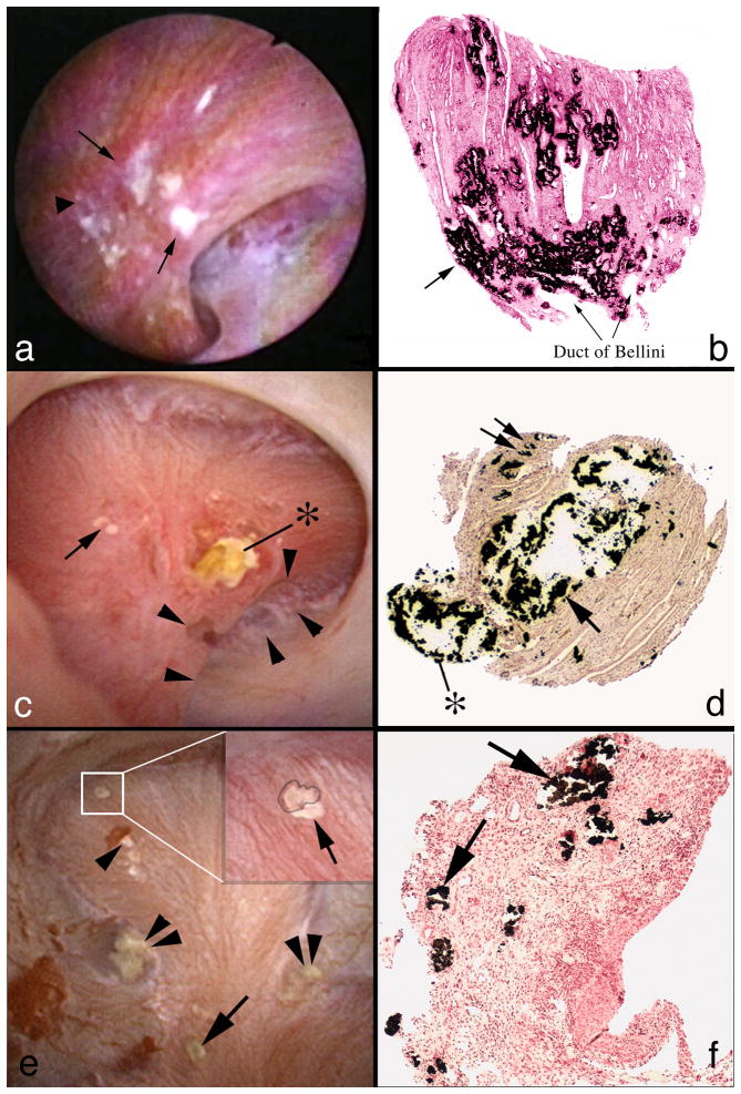
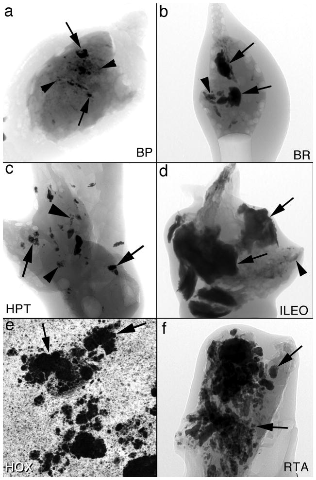
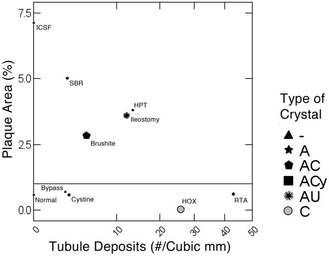
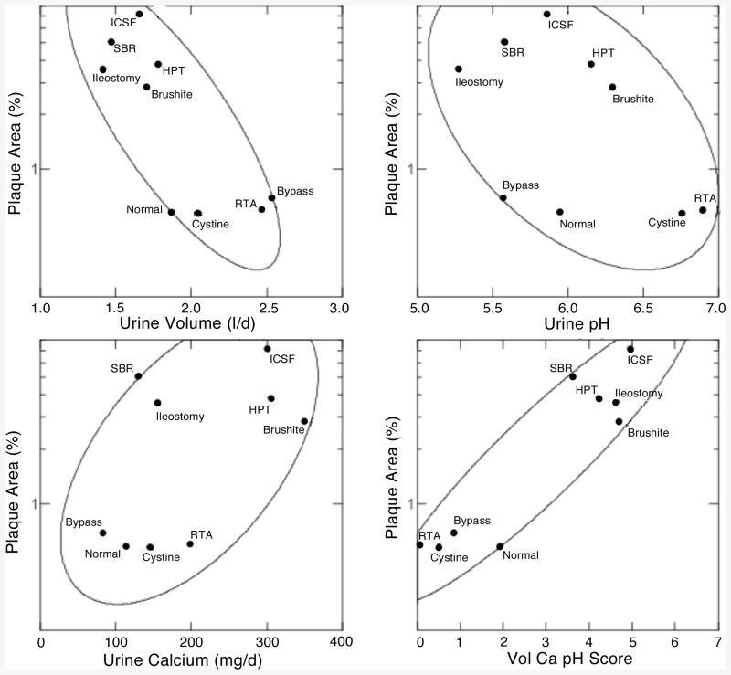
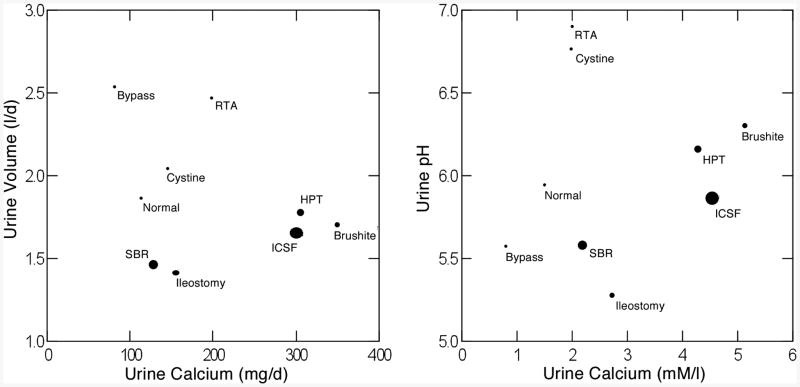
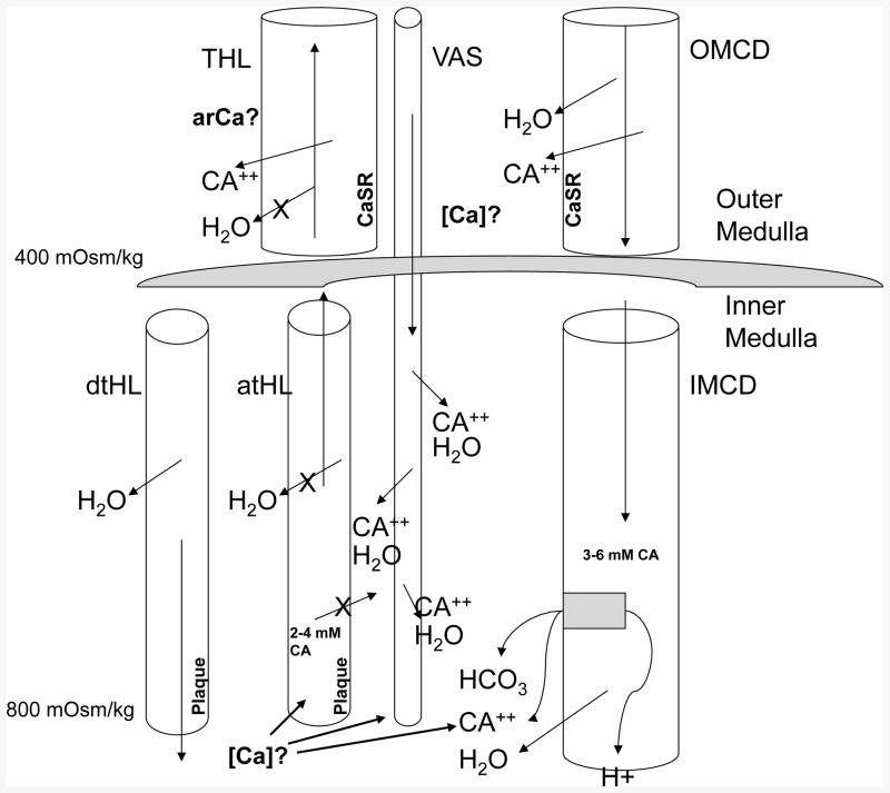
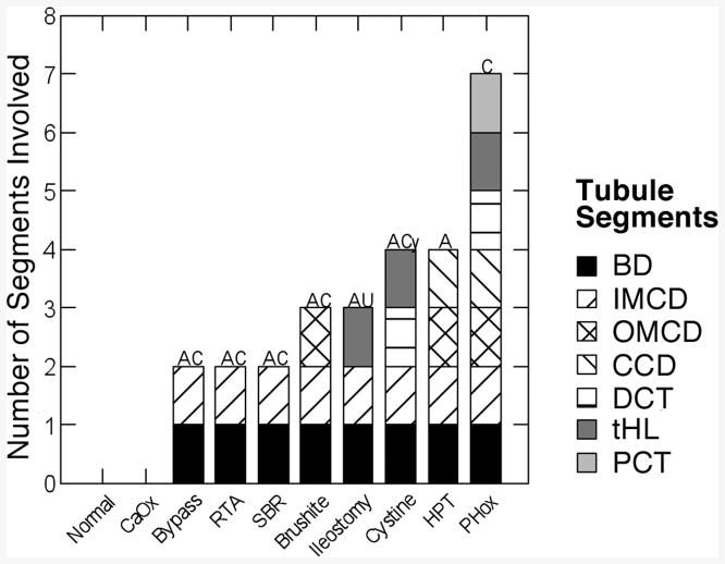
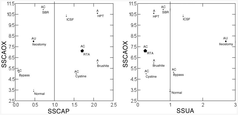
Data concerning nine forms of human stone disease, along with observations on normal people give new insights into formation of interstitial apatite plaque and intra-tubular crystal deposits. In general, across multiple disease states, one can reproduce the same relationships between plaque abundance as is seen among patients within individual disease states, so that the link between plaque and high urine calcium excretion, and low urine volume and pH seems increasingly secure. From this, one can propose a specific model of plaque formation, susceptible to experimental test. In many diseases, formation of inner medullary collecting duct and Bellini duct deposits is compatible with simple crystallization driven by urine supersaturations; this is expected in that these segments contain tubule fluid quite close in composition to final urine. But in ileostomy, small bowel disease and obesity bypass patients, crystals found in deposits are not those expected: apatite and urates in deposits, despite formation of highly acidic urine. Also, this discrepancy suggests the possibility of divergence between bulk urine pH and pH of focal collecting ducts, a new kind of possibility that is susceptible to experimental test.


Figures

Figure 1
Figure 1
Surgical anatomy and histopathology of plaque and deposits in three stone diseases. Plaque is seen as a white sub-urothelial reposition during endoscopic surgery (panel a, arrows) in an ICSF. On biopsy, plaque is stained black with Yasue stain; tubule lumens show through the interstitial deposits (panel b). In a brushite SF (panel c) a large deposit extends out of the mouth of a greatly dilated Bellini ducts (BD) (*). Arrowheads outline a deformity of the papillum representing fibrosis. Yellow plaque, the gross reflection of inner medullary collecting ducts (IMCD) deposits is at the arrow. On biopsy (panel d) the large IMCD and BD deposit(arrow) has destroyed epithelial cells and protrudes (*); interstitial fibrosis surrounds the dilated tubule. Double arrow points to interstitial plaque. In a hyperparathyroid stone former (panels e & f) a small CaOx stone is attached over white plaque (insert box and blowup, panel e). Double arrowheads point to yellow IMCD lumen deposits. Single arrow points to white plaque. The arrowhead points to white plaque where an attached stone has been removed. Biopsy shows numerous plugged IMCD (panel f, arrows) with extensive interstitial fibrosis.
Figure 2
Figure 2
Micro-CT analysis of deposits in six stone diseases. In all 6 panels, arrows point to intratubular deposits and arrowheads to interstitial plaque. From the micro-CT reconstructions we calculate density of deposits in cubic mm of tissue as well as the size of the deposit in mm. Panels A, B, C, D and F are micro-CT shadow images while panel E is a micro-CT cross sectional image. Abbreviations as in Table 1.
Figure 3
Figure 3
Plaque surface coverage (y-axis) vs. tubule deposit density (x-axis) in 9 conditions. Disease abbreviations as in Table 1. Size of symbol gauges mean size of individual deposits. Deposit density and size are from micro-CT analysis. The horizontal line represents the upper boundary of plaque abundance in normal renal tissue. A, apatite; C, CaOx; Cy, cystine; AU, sodium and ammonium urate crystals; - denotes no deposits, shown as triangles for normal and ICSF.
Figure 4
Figure 4
Relationship between plaque surface area and urine chemistry measurements. Ellipses are 68% non-parametric containment estimates. Vol Ca pH score, multivariate combined effects of urine volume, pH and calcium excretion.
Figure 5
Figure 5
Relationship between urine volume and calcium excretion (left panel) and urine pH and urine calcium molarity (right panel) in 9 conditions. Size of symbol denoted plaque surface area. Note clustering of high (large symbols) plaque states to the lower right of both panels and marked separation from low plaque states.
Figure 6
Figure 6
Schematic diagram of outer and inner medulla showing elements of proposed vas washdown hypothesis – details presented in text. THL, thick ascending limb of Henle's loop; Ca, calcium ion; [Ca}, interstitial calcium concentration; CaSR calcium sensing receptor; OMCD, outer medullary collecting duct; dtHL and atHL, descending and ascending limbs of Henle's loop; Vas, vas recta; X, blockade of transepithelial movement; estimates of osmolality and tubule fluid concentrations are for illustrative purposes.
Figure 7
Figure 7
Segments containing deposits in 10 conditions. BD, Bellini ducts; IMCD, OMCD and CCD, inner and outer medullary and cortical collecting ducts; DCT, distal convoluted tubule; tHL, thin limbs of Henle's loop; PCT, proximal tubule.
Figure 8
Figure 8
Relationship between urine CaOx and CaP (left panel) and CaOx and uric acid SS (right panel) in 9 conditions. Size of symbol reflects density of deposits in number per cubic mm of tissue assessed by micro-CT analysis. Vertical lines denote SS values of 1, the point of solubility. Type of crystal deposit is above each symbol: A, apatite; C, CaOx; Cy, cystine; U, sodium and ammonium urate; -, no deposits occur. Details presented in text.

References

    1. Evan AP, Lingeman JE, Coe FL, et al. Randall's plaque of patients with nephrolithiasis begins in basement membranes of thin loops of Henle. J Clin Invest. 2003;111:607–616. - PMC - PubMed
    1. Evan AP, Coe FL, Lingeman JE, et al. Anat Rec. Vol. 290. Hoboken: 2007. Mechanism of formation of human calcium oxalate renal stones on Randall's plaque; pp. 1315–1323. - PubMed
    1. Kuo RL, Lingeman JE, Evan AP, et al. Urine calcium and volume predict coverage of renal papilla by Randall's plaque. Kidney Int. 2003;64:2150–2154. - PubMed
    1. Evan AP, Lingeman JE, Coe FL, et al. Intra-tubular deposits, urine and stone composition are divergent in patients with ileostomy. Kidney Int. 2009;76:1081–1088. - PMC - PubMed
    1. Evan AP, Coe FL, Lingeman JE, et al. Renal crystal deposits and histopathology in patients with cystine stones. Kidney Int. 2006;69:2227–2235. - PubMed

Publication types

LinkOut - more resources