Review
. 2011 Mar 15;14(6):1049-63.
doi: 10.1089/ars.2010.3400. Epub 2010 Oct 28.

Thiol-based redox switches and gene regulation

Affiliations
Review

Thiol-based redox switches and gene regulation

Haike Antelmann et al. Antioxid Redox Signal. .

Abstract

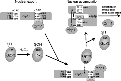
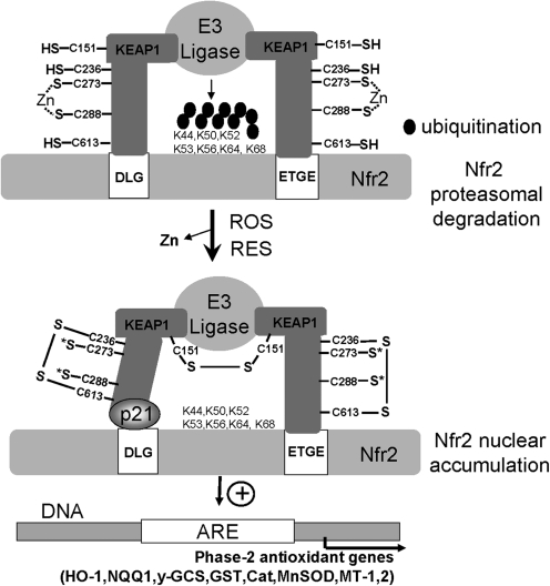
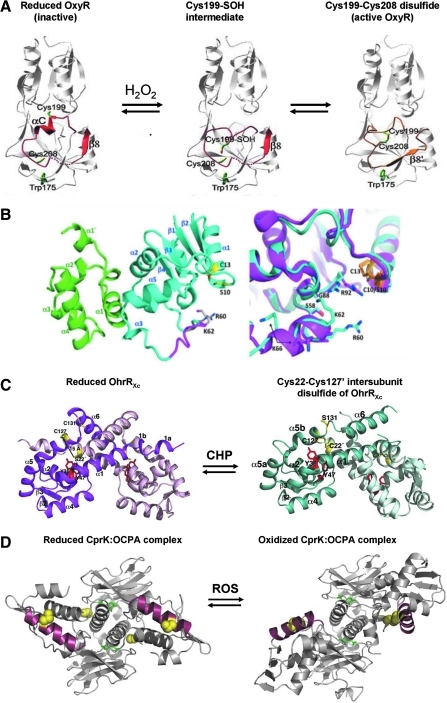
Cysteine is notable among the universal, proteinogenic amino acids for its facile redox chemistry. Cysteine thiolates are readily modified by reactive oxygen species (ROS), reactive electrophilic species (RES), and reactive nitrogen species (RNS). Although thiol switches are commonly triggered by disulfide bond formation, they can also be controlled by S-thiolation, S-alkylation, or modification by RNS. Thiol-based switches are common in both prokaryotic and eukaryotic organisms and activate functions that detoxify reactive species and restore thiol homeostasis while repressing functions that would be deleterious if expressed under oxidizing conditions. Here, we provide an overview of the best-understood examples of thiol-based redox switches that affect gene expression. Intra- or intermolecular disulfide bond formation serves as a direct regulatory switch for several bacterial transcription factors (OxyR, OhrR/2-Cys, Spx, YodB, CrtJ, and CprK) and indirectly regulates others (the RsrA anti-σ factor and RegB sensory histidine kinase). In eukaryotes, thiol-based switches control the yeast Yap1p transcription factor, the Nrf2/Keap1 electrophile and oxidative stress response, and the Chlamydomonas NAB1 translational repressor. Collectively, these regulators reveal a remarkable range of chemical modifications exploited by Cys residues to effect changes in gene expression.


Figures

FIG. 1.
FIG. 1.
Post-translational cysteine modifications involved in redox-sensing mechanisms. Cysteine can be reversibly oxidized by ROS to the unstable Cys sulfenic acid (R-SOH; a), which can then form an intramolecular disulfide (b), an intermolecular disulfide (c), an S-thiolated adduct with a LMW thiol (RSH) (d), or a cyclic sulfenamide with a polypeptide backbone amide (e). Irreversible Cys modifications include overoxidation to Cys sulfinic (R-SO2H) (f) and sulfonic acids (R-SO3H) (g). RNS, such as nitric oxide (NO) or peroxynitrite (ONOO), modify Cys by S-nitrosothiol formation (R-SNO) (h) or S-nitrothiol formation (R-SNO2) (i). RES with partial positively charged carbon centers (δ+), such as quinones or aldehydes, lead to thiol-(S)-alkylation of cysteine, resulting in quinone-S-adducts (j) by benzoquinone or S-hydroxymethylthiol modification by formaldehyde (k). This figure is modified from (71).
FIG. 2.
FIG. 2.
The disulfide-switch model for transcriptional activation of OxyR in Escherichia coli. OxyR responds to hydrogen peroxide (H2O2) in E. coli and other bacteria. The conserved C199 and C208 residues are essential for redox-sensing activity. C199 is initially oxidized to the sulfenic acid intermediate that rapidly reacts further to form an intramolecular disulfide with Cys208. Oxidized OxyR binds as a tetramer to promoter regions to activate transcription of genes encoding proteins with antioxidant functions, including GrxA, which is involved in the reduction of oxidized OxyR.
FIG. 3.
FIG. 3.
Redox regulation of the Zn-binding antisigma factor RsrA of Streptomyces coelicolor. RsrA is a zinc-binding, redox-sensitive anti-σ factor that sequesters σR under reducing conditions. Diamide treatment causes intramolecular disulfide-bond formation in RsrA, resulting in the release of Zn(II) and active σR. Free σR activates transcription of σR regulon genes, including trxBA and trxC (thioredoxin and thioredoxin reductase), mshA (a glycosyltransferase catalyzing the first step in MSH synthesis), and mca (mycothiol-S-conjugate amidase). Collectively, these thiol-reducing systems function in reduction of oxidized RsrA and restore the thiol redox balance. σR and the unstable σR′protein (with a destabilizing N-terminal extension) are positively autoregulated (see text).
FIG. 4.
FIG. 4.
Mechanisms regulating Spx levels in response to disulfide stress in Bacillus subtilis. Under nonstress conditions, Spx interacts with the Zn(II)-binding YjbH adaptor protein, which targets Spx to ClpXP proteolytic degradation. YjbH and ClpX both contain Cys-rich Zn-binding sites that are oxidized to an intramolecular disulfide, resulting in the release of Zn(II). The levels of Spx are increased by diamide because of the inactivation of YjbH and ClpX and subsequent decreased proteolysis. The spx gene is regulated by the repressors YodB and PerR, which are both inactivated by diamide stress. Spx has a redox-active CxxC motif that is oxidized by diamide and converts it to a transcriptional activator. Oxidized Spx contacts αCTD of RNAP to activate transcription of trxA and trxB and thereby maintain the thiol-redox state of the cell. (Note: the figure does not specify the precise sites of binding of the regulatory proteins to promoter DNA).
FIG. 5.
FIG. 5.
Redox-sensing mechanisms of the 1-Cys and 2-Cys type OhrR family proteins. OhrR controls the OhrA thiol-dependent peroxidase and contains one conserved reactive Cys15 residue in B. subtilis and three Cys residues (C22, C127, and C131) in X. campestris. (a) The 1-Cys OhrR protein of B. subtilis is initially oxidized by CHP to the Cys sulfenic acid, which further undergoes reversible S-thiolation with Cys and BSH in vivo and may also generate a cyclic sulfenamide when thiols are depleted. In the presence of the strong oxidant LHP, C15 is irreversibly overoxidized to the sulfinic and sulfonic acids. (b) The 2-Cys OhrR protein of X. campestris is regulated by intersubunit disulfide formation between C22 and C127’ of opposing subunits. Both Cys residues are essential for redox sensing in vivo, as shown by mutagenesis (73).
FIG. 6.
FIG. 6.
CprK regulation in Desulfitobacterium dehalogenans. CprK is present in a reduced state in the cell under anaerobic conditions. In response to oxidative stress, CprK forms a Cys11-Cys200′ intersubunit disulfide bond and a Cys105-Cys200 intramolecular disulfide, which inhibit CprK DNA-binding activity. Oxidized and reduced CprK bind the effector OCPA with similar affinity. Reduced, ligand-bound CprK binds DNA and activates transcription of the cpr gene cluster for halorespiration.
FIG. 7.
FIG. 7.
Regulation of yeast transcription factor Yap1p in response to peroxide stress. The activity of Yap1p is regulated by nuclear localization. Yap1p contains two Cys-rich domains (C-terminal and N-terminal CRD) that harbor redox-sensitive Cys residues that are reduced under nonstress conditions. The nuclear export sequence (NES) can bind to the nuclear export receptor (Crm1), and Yap1p is exported to the cytosol, preventing its accumulation in the nucleus. On peroxide stress, C36 of the glutathione peroxidase (Gpx3) is oxidized to sulfenate and interacts with C598 of Yap1p through intermolecular disulfide formation to a Yap1-Gpx3 intermediate that binds the Yap1-binding protein 1 (Ybp1). Thiol-disulfide exchange reactions lead subsequently to C303-C598 disulfide formation and other intramolecular disulfide bonds (e.g., C310-C629) in Yap1p, accompanied by conformational changes of Yap1p and the release of Gpx3. The NES export signal is not accessible for Crm1 interaction, which leads to accumulation of Yap1p in the nucleus. This in turn leads to activation of transcription of Yap1p antioxidant target genes (5).
FIG. 8.
FIG. 8.
Redox regulation of the KEAP1/Nfr2 system in response to ROS and RES. The Keap1 sensor protein contains more than 25 Cys residues, including the redox-sensitive C151 and the C273 and C288 residues that coordinate Zn. Under nonstress conditions, Keap1 binds to the DLG and ETGE sites in the Nfr2 transcription factor, positioning the lysine residues of Nfr2 optimal for ubiquitination by the E3 ligase and subsequent degradation of Nfr2. On exposure to ROS, the C151 residues form an intersubunit disulfide bond in Keap1, and intramolecular disulfide bonds between C236-C613 have also been detected. Oxidation of C273 and C288 leads to Zn release. S* indicates unknown oxoforms (75). RES leads to alkylation of many different Cys residues, including C151, C273, and C288 and others. Disulfide bonds or S-alkylation of Keap1 decreases binding to the DLG site of Nfr2. Oxidation of Keap1 also masks the NES, leading to nuclear accumulation of Nfr2, which is further stabilized by binding to p21. This leads to transcriptional activation of Nfr2 that binds to antioxidant response elements (AREs) in the promoters of phase-2 genes encoding heme oxygenase (HO-1), quinone oxidoreductase (NQQ1), γ-glutamylcysteine synthetase (γ-GCS), glutathione S-transferase (GST), catalase (Cat), superoxide dismutase (MnSod), or metallothioneins (MT-1,2) and other antioxidant-function genes. This figure is adapted from (75).
FIG. 9.
FIG. 9.
Structures of reduced and oxidized forms of the thiol-based redox sensors OxyR, Spx, OhrR (2-Cys), and CprK. (A) Structures are shown for OxyR in its reduced, Cys199 sulfenic acid intermediate and Cys199-Cys208 intramolecular disulfide conformations. In reduced OxyR, C199 and C208 are separated by 17 Å. The interaction of C199 with the side chains of T100 and R266 stabilizes the C199 thiolate anion. The residues 195 to 223, including αC and β8, are shown in red in reduced and gold in oxidized OxyR and are involved in the structural transition on oxidation. C199 is initially oxidized to the sulfenic acid intermediate that rapidly reacts further to form an intramolecular disulfide with Cys208. During OxyR oxidation, the αC helix melts, and the αC/β8 loop shifts positions. This figure is adapted from (47). (B) Structures are shown for reduced SpxC10S in complex with the αCTD. Left: Spx and αCTD are shown as teal and green ribbons, respectively. Helix α4 is colored magenta in oxidized Spx. Residues S10 and C13 are shown as sticks with carbon and sulfur atoms colored yellow and the γ-oxygen of S10, red. Right: Close-up of the region surrounding helix α4 and residues C10/S10 and C13 after the superposition of the oxidized and reduced αCTD-Spx complex structures. Reduced Spx is shown as a magenta ribbon, and oxidized Spx, as a teal ribbon. The C10-C13 disulfide bond is shown in orange sticks, and S10 and C13 from the reduced structure are shown as yellow sticks. This figure was reproduced from (62). (C) The structures of the 2-Cys OhrR of Xanthomonas campestris OhrRXc in its reduced form (left) and oxidized Cys22-Cys127′ intersubunit disulfide-linked state (right). In reduced OhrR, one subunit is colored magenta, and the other, light pink. In the reduced form, Cys22 and Cys127′ are separated by 15.5 Å. The hydrogen bonds provided by the Y36′ and Y46′ side chains (red) are involved in stabilization of the Cys22 thiolate (yellow). In the oxidized intersubunit disulfide version of the OhrR, one subunit is colored teal, and the other, light blue. On oxidation of OhrR, Cys22 is oxidized to the Cys22-SOH intermediate, disrupting the Y36′-C22-Y47′ hydrogen-bonding network and distorting α5. This allows the movement of Cys127′ by 135-degrees rotation and 8-Å translation, formation of the Cys22-Cys127′ intersubunit disulfide bond, and α6-α6′ helix-swapped reconfiguration, resulting in 28-degree rigid body rotations of the HTH motifs and dissociation from the DNA. This figure is reproduced from (64). (D) The Desulfitobacterium hafniense CprK structure is shown in ligand (OCPA) bound, reduced complex (left) and in the oxidized complex (right) containing intersubunit disulfides between C11 and C200′ (in the DNA-binding HTH motif). The C11 and C200 cysteines are indicated by yellow spheres, the bound OCPA ligand is shown in stick form (green), and the DNA-recognition helix is in red. Note that in the reduced form (left), the two DNA-binding helices are approximately parallel and appropriately spaced for interaction with their cognate DNA operator. Oxidation distorts the protein, disrupting the DNA interaction surface and preventing transcription activation. For further details of the structural changes induced in CprK by ligand binding and protein oxidation, please see (37, 54). This figure is kindly provided by Dr. David Leys, Manchester, UK.

References

    1. Anjem A. Varghese S. Imlay JA. Manganese import is a key element of the OxyR response to hydrogen peroxide in Escherichia coli. Mol Microbiol. 2009;72:844–858. - PMC - PubMed
    1. Antelmann H. Hecker M. Zuber P. Proteomic signatures uncover thiol-specific electrophile resistance mechanisms in Bacillus subtilis. Expert Rev Proteom. 2008;5:77–90. - PubMed
    1. Bae JB. Park JH. Hahn MY. Kim MS. Roe JH. Redox-dependent changes in RsrA, an anti-sigma factor in Streptomyces coelicolor: zinc release and disulfide bond formation. J Mol Biol. 2004;335:425–435. - PubMed
    1. Barford D. The role of cysteine residues as redox-sensitive regulatory switches. Curr Opin Struct Biol. 2004;14:679–686. - PubMed
    1. Brandes N. Schmitt S. Jakob U. Thiol-based redox switches in eukaryotic proteins. Antioxid Redox Signal. 2009;11:997–1014. - PMC - PubMed

Publication types

MeSH terms

LinkOut - more resources