. 2010 Jul 13:8:15.
doi: 10.1186/1477-3155-8-15.

PVP-coated silver nanoparticles block the transmission of cell-free and cell-associated HIV-1 in human cervical culture

Affiliations

PVP-coated silver nanoparticles block the transmission of cell-free and cell-associated HIV-1 in human cervical culture

Humberto H Lara et al. J Nanobiotechnology. .

Abstract

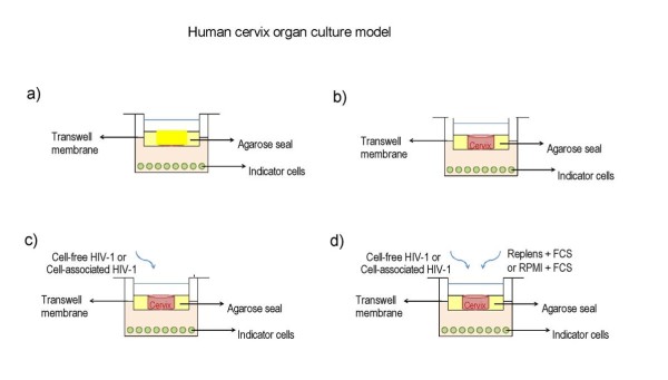
Background: Previous in vitro studies have demonstrated that polyvinylpyrrolidone coated silver nanoparticles (PVP-coated AgNPs) have antiviral activity against HIV-1 at non-cytotoxic concentrations. These particles also demonstrate broad spectrum virucidal activity by preventing the interaction of HIV-1 gp120 and cellular CD4, thereby inhibiting fusion or entry of the virus into the host cell. In this study, we evaluated the antiviral activity of PVP-coated AgNPs as a potential topical vaginal microbicide to prevent transmission of HIV-1 infection using human cervical culture, an in vitro model that simulates in vivo conditions.

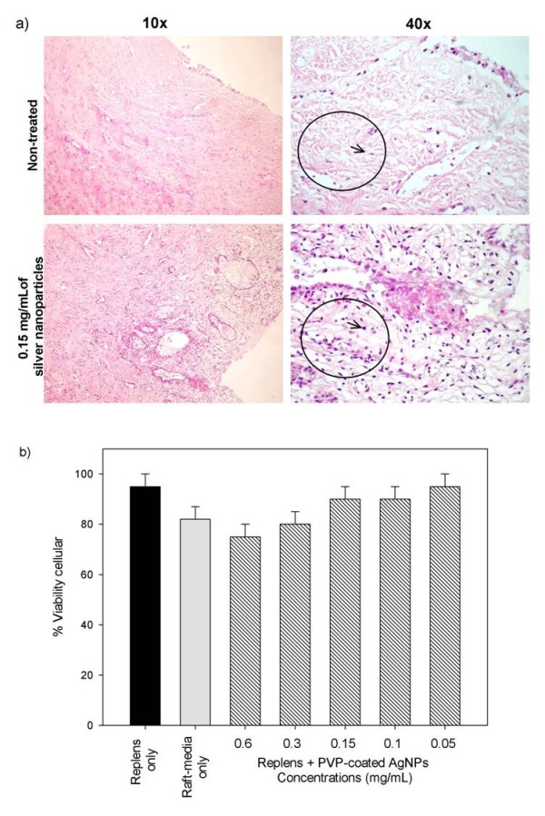
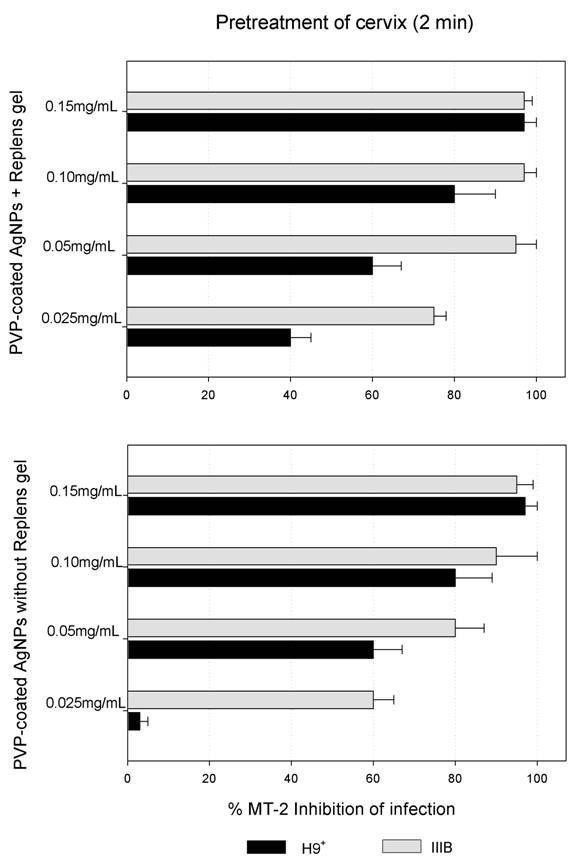
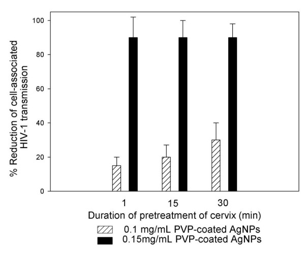
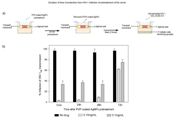
Results: When formulated into a non-spermicidal gel (Replens) at a concentration of 0.15 mg/mL, PVP-coated AgNPs prevented the transmission of cell-associated HIV-1 and cell-free HIV-1 isolates. Importantly, PVP-coated AgNPs were not toxic to the explant, even when the cervical tissues were exposed continuously to 0.15 mg/mL of PVP-coated AgNPs for 48 h. Only 1 min of PVP-coated AgNPs pretreatment to the explant was required to prevent transmission of HIV-1. Pre-treatment of the cervical explant with 0.15 mg/mL PVP-coated AgNPs for 20 min followed by extensive washing prevented the transmission of HIV-1 in this model for 48 h.

Conclusions: A formulation of PVP-coated AgNPs homogenized in Replens gel acts rapidly to inhibit HIV-1 transmission after 1 min and offers long-lasting protection of the cervical tissue from infection for 48 h, with no evidence of cytotoxicity observed in the explants.Based on this data, PVP-coated AgNPs are a promising microbicidal candidate for use in topical vaginal/cervical agents to prevent HIV-1 transmission, and further research is warranted.


Figures

Figure 1
Figure 1
Human cervical culture model. a) To rule out possible leaks in the agarose seal, Dextran blue was added to the upper chamber on day 6 of culture, and its presence in the lower chamber was determined 20 h later to all Transwells used in the experiments and negative control well with agarose only, b) other negative control with tissue alone without treatment and without challenge with virus and c) positive control well with tissue alone infected with only with HIV-1 virus. d) Inhibition of HIV-1 transmission, Cervical tissue is treated with PVP-coated AgNPs at different concentrations in a Replens gel or RPMI + 10% FCS media, which was then infected with HIVIIIB. HIV transmission or inhibition of transmission across the mucosa was determined in the lower chamber by formation of syncytia using indicator cells (MT-2).
Figure 2
Figure 2
Toxicity of the PVP-coated AgNPs to the cervical tissue. a) Normal squamous epithelium and the stroma of ecto-cervical tissues were exposed to Replens gel mixed with 0.15 mg/mL PVP-coated AgNPs for 48 h. Ecto-cervical explants (5 mm) were exposed to either Replens gel alone, which served as a control, or to PVP-coated AgNPs. After 48 h of incubation in a 37°C humidified incubator, the tissues were washed, embedded in paraffin, and stained with hematoxylin and eosin. b) Cervical explants in the upper Transwell chambers were exposed to Replens alone as a control, Raft-medium, or Replens gel containing different concentrations of PVP-coated AgNPs (0.6, 0.3, 0.15, 0.1, 0.05 and 0 mg/mL). After 24 h, the medium containing PVP-coated AgNPs was removed and washed three times with culture media. Cell viability was measured by the CellTiter-Glo® assay. Graphs show values of the means ± standard deviations from three separate experiments. Graphs were created using the SigmaPlot 10.0 software.
Figure 3
Figure 3
Inhibition of HIV-1 transmission with and without Replens gel using the cervical culture model. The upper chamber of the Transwell with the cervical explant was exposed for 2 min to different concentrations of PVP-coated AgNPs (0.025, 0.05, 0.1 and 0.15 mg/mL), either alone or mixed with the Replens gel. After thoroughly washing extracellular PVP-coated AgNPs from the cervical explant, cell-free (HIV-1IIIB) [(5 × 10⁵ TCID50)], or cell-associated virus (H9+) (5 × 10⁵ cells) were added. To evaluate inhibition of the HIV-1 infection, indicator cells (MT-2) in the lower chamber were cultured and formation of syncytia was monitored for ten days. Graphs show values of the means ± standard deviations from three separate experiments. Graphs were created using the SigmaPlot 10.0 software.
Figure 4
Figure 4
Time needed for PVP-coated AgNPs to confer protection from the transmission of HIV-1 through the cervical. Cervical explants were pretreated for 1, 15 and 30 minutes with 0.1 or 0.15 mg/mL PVP-coated AgNPs. After thoroughly washing extracellular PVP-coated AgNPs from the cervical tissue, cell-associated virus (H9+) (5 × 10⁵ cells) was added to the upper chamber of the Transwell. Indicator cells (MT-2) were cultured in the lower chamber to evaluate the inhibition of HIVIIIB infection. Graphs show values of the means ± standard deviations from three separate experiments. Graphs were created using the SigmaPlot 10.0 software.
Figure 5
Figure 5
Protection from HIV-1 infection following pre-treatment of the cervical explant with PVP-coated AgNPs. a) Cervical explants were exposed to 0.1 or 0.15 mg/mL PVP-coated AgNPs in RPMI + 10% FCS media for 20 minutes. After thoroughly washing extracellular PVP-coated AgNPs from the cervical explant and after 1 minute, 24 h, 48 h and 72 h, cell-free virus (HIV-1IIIB) [(5 × 10⁵ TCID50)] was added to the upper chamber. To verify the neutralization of HIV-1 transmission, we cultured the indicator cells (MT-2) in the lower chamber and evaluated inhibition of the HIV-1 infection. b) Cervical explants were exposed to HIV-1 in the absence of PVP-coated AgNPs as a control and to 0.1 or 0.15 mg/mL of PVP-coated AgNPs as pretreatment. Graphs show values of the means ± standard deviations from three separate experiments. Graphs were created using the SigmaPlot 10.0 software.

References

    1. Ray N. Maraviroc in the treatment of HIV infection. Drug Des Devel Ther. 2009;2:151–161. - PMC - PubMed
    1. Walker PR, Worobey M, Rambaut A, Holmes EC, Pybus OG. Epidemiology: Sexual transmission of HIV in Africa. Nature. 2003;422:679. doi: 10.1038/422679a. - DOI - PubMed
    1. Elias C, Coggins C. Acceptability research on female-controlled barrier methods to prevent heterosexual transmission of HIV: Where have we been? Where are we going? J Womens Health Gend Based Med. 2001;10:163–173. doi: 10.1089/152460901300039502. - DOI - PubMed
    1. Kalichman SC, Williams EA, Cherry C, Belcher L, Nachimson D. Sexual coercion, domestic violence, and negotiating condom use among low-income African American women. J Womens Health. 1998;7:371–378. doi: 10.1089/jwh.1998.7.371. - DOI - PubMed
    1. van der Straten A, King R, Grinstead O, Serufilira A, Allen S. Couple communication, sexual coercion and HIV risk reduction in Kigali, Rwanda. AIDS. 1995;9:935–944. doi: 10.1097/00002030-199508000-00016. - DOI - PubMed