. 2010 Jun 15;18(6):950-60.
doi: 10.1016/j.devcel.2010.02.019.

The tripartite motif protein MADD-2 functions with the receptor UNC-40 (DCC) in Netrin-mediated axon attraction and branching

Affiliations

The tripartite motif protein MADD-2 functions with the receptor UNC-40 (DCC) in Netrin-mediated axon attraction and branching

Joe C Hao et al. Dev Cell. .

Abstract

Neurons innervate multiple targets by sprouting axon branches from a primary axon shaft. We show here that the ventral guidance factor unc-6 (Netrin), its receptor unc-40 (DCC), and the gene madd-2 stimulate ventral axon branching in C. elegans chemosensory and mechanosensory neurons. madd-2 also promotes attractive axon guidance to UNC-6 and assists unc-6- and unc-40-dependent ventral recruitment of the actin regulator MIG-10 in nascent axons. MADD-2 is a tripartite motif protein related to MID-1, the causative gene for the human developmental disorder Opitz syndrome. MADD-2 and UNC-40 proteins preferentially localize to a ventral axon branch that requires their function; genetic results indicate that MADD-2 potentiates UNC-40 activity. Our results identify MADD-2 as an UNC-40 cofactor in axon attraction and branching, paralleling the role of UNC-5 in repulsion, and provide evidence that targeting of a guidance factor to specific axonal branches can confer differential responsiveness to guidance cues.


Figures

Figure 1
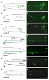
Figure 1. Axon Branching and Guidance Are Disrupted in madd-2 Mutants
(A–F) ADL neurons visualized with srh-220::gfp transgene and schematics. (A and B) The wild-type ADL cell body projects an axon laterally into the nerve ring, where it branches into a dorsal (arrow) and a ventral (arrowhead) process. (C and D) madd-2(ky592) ADL; normal dorsal branch (arrow) and no ventral branch. (E and F) madd-2(ky592) ADL; defective guidance of the ADL primary axon (arrow) into the nerve ring. (G–J) PLM neurons visualized with mec-4::gfp transgene and schematics. (G and H) The wild-type PLM neuron extends a primary axon anteriorly and projects a ventrally directed axon branch (arrow) near the vulva. (I and J) madd-2(ky592) PLM defective in ventral branch (arrow). (K–N) AVM neurons visualized with mec-4::gfp transgene and schematics. (K and L) The wild-type AVM axon projects ventrally (arrow) and then extends anteriorly. (M and N) madd-2(ky592) AVM axon growing anteriorly (arrow) in a lateral position instead of ventrally. Anterior is to the left and dorsal is at the top in all panels. Scale bars, 2 μm.
Figure 2
Figure 2. madd-2 Acts in the unc-6/unc-40 Pathway for AVM Ventral Guidance
(A) AVM ventral guidance. The expression of both SAX-3 (Robo) and UNC-40 (DCC) receptors in AVM (green) allows its axon to extend toward the ventral UNC-6 (Netrin) attractive cue (blue) made by neurons and away from the dorsal SLT-1 repulsive cue made by muscles (red). Ventral muscles are shown in gray. (B and C) AVM ventral guidance defects in single and double mutants, scored using a mec-4::gfp transgene. (B) madd-2; unc-6 and unc-40; madd-2 double mutants exhibit no enhancement in AVM defects compared to single mutants (n = 73–297). (C) madd-2; slt-1 double mutants exhibit enhanced defects compared to single mutants (n = 45–367). Error bars represent the standard error of proportion. Asterisks indicate different from single mutants by χ2 test at p < 0.01. (D) Mutations in madd-2 do not suppress the outgrowth phenotypes associated with MYR:: UNC-40 expression. The percentage of excess AVM outgrowth, labeled by a mec-4::gfp transgene, was determined for animals carrying the MYR::UNC-40, MYR::UNC-40ΔP1, or MYR:: UNC-40ΔP2 transgenes (Gitai et al., 2003) alone or as double mutants with madd-2(ky592) (n = 58–153). Error bars represent the standard error of proportion.
Figure 3
Figure 3. Molecular Analysis of madd-2
(A) Genetic map position of madd-2, deficiencies used for mapping, and clones used for rescue experiments, showing the genomic organization of the madd-2 coding region with sites of mutations. Exons are indicated by black boxes and the 5′ SL1 trans-splice leader sequence by an open box. (B) madd-2 genomic and cDNA subclones generated for rescue experiments and GFP expression studies. (C) Predicted protein domains in MADD-2. The percent identities between MADD-2 and either CG31721 or mouse TRIM9 are shown for each domain. The sites of madd-2 mutations are indicated. (D) Amino acid sequence alignment for MADD-2, CG31721, and mouse TRIM9. The bars highlight the conserved domains and correspond to colors used for the domains shown in (C).
Figure 4
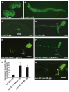
Figure 4. madd-2::gfp Expression and Preferential Localization of MADD-2 and UNC-40 to the ADL Ventral Branch
(A) Comma stage (bottom) and 1.5-fold stage (top) embryos. The anterior cells that express madd-2::gfp include developing neurons of the nerve ring. Expression is also detected in the ventral and posterior embryo, including motor neurons and hypodermal cells. (B) L1 stage larva. Expression is high in head neurons that project into the nerve ring (arrow). Muscles of the head also express the transgene (arrowhead). (C) L3 stage larva. The HSN motor neuron expresses the madd-2::gfp transgene. (D) An srh-220::gfp transgene uniformly labels both dorsal (black arrowhead) and ventral (arrow) branches of the ADL neuron. (E and F) In both larval (E) and adult (F) animals, the srh-220::madd-2::gfp transgene preferentially labels the ADL ventral branch (arrow). (G) Quantitation of the fluorescence intensity ratio between the ventral and dorsal branches in adult animals expressing GFP, MADD-2::GFP, or UNC-40::GFP in ADL. Asterisks indicate a significant enrichment in ventral branch fluorescence as compared to GFP alone (p < 0.005, t test). Error bars represent the standard error of the mean. (H) An L1 larva expressing srh-220::unc-40::gfp displays preferential expression of UNC-40::GFP in the ADL ventral branch (arrow). For (D)–(F) and (H), the ADL branch point is indicated by a white arrowhead. Anterior is to the left and dorsal is at the top in all panels. Scale bars equal 5 μm.
Figure 5
Figure 5. MADD-2 Overexpression Causes UNC-40-Dependent Ventral Outgrowth and Ectopic Branching of the ALM Neuron
(A and B) The ALM neuron projects an axon anteriorly to the head in a lateral position (arrow) in wild-type animals, as diagrammed or labeled by a mec-4::gfp transgene. (C–H) mec-7::madd-2(kyEx638) transgenic animals: (C and D) the ALM axon projects to the ventral midline before reaching the nerve ring (arrow); (E and F) the ALM cell body (arrow) is ventrally displaced compared to wild-type; (G and H) the ALM axon exhibits an ectopic branch (arrow). Anterior is to the left and dorsal is at the top in all panels. Scale bar equals 5 μm. (I) ALM phenotypes in madd-2-overexpressing strains, with or without unc-40 and unc-6 mutations (n = 136–184). The same transgene, mec-7::madd-2(kyEx638), rescues AVM and causes gain-of-function phenotypes in ALM. Error bars represent the standard error of proportion.
Figure 6
Figure 6. MADD-2 Is Required for Ventral MIG-10 Localization in HSN
(A) Diagram of molecules affecting HSN polarization and ventral guidance. (B and C) HSN neurons labeled with a membrane-bound GFP in L3 larval stage. (B) Wild-type animal with ventral HSN growth cones (arrowheads). (C) madd-2 mutant with unpolarized HSN. (D and E) HSNs expressing functional, ventrally localized UNC-40::GFP protein in L3 larval stage wild-type (D) and madd-2 mutant (E) animals. (F–H) HSNs expressing functional MIG-10::GFP protein in L3 larval stage wild-type (F), madd-2 mutant (G), and unc-40 mutant (H) animals. (I) Quantification of HSN defects at different developmental stages; polarized ventral growth in the L2 stage is followed by ventral neurite outgrowth in the L3 stage (Adler et al., 2006). Scale bars equal 5 μm.

References

    1. Acebes A, Ferrús A. Cellular and molecular features of axon collaterals and dendrites. Trends Neurosci. 2000;23:557–565. - PubMed
    1. Adler CE, Fetter RD, Bargmann CI. UNC-6/Netrin induces neuronal asymmetry and defines the site of axon formation. Nat. Neurosci. 2006;9:511–518. - PMC - PubMed
    1. Alexander M, Chan KK, Byrne AB, Selman G, Lee T, Ono J, Wong E, Puckrin R, Dixon SJ, Roy PJ. An UNC-40 pathway directs postsynaptic membrane extension in Caenorhabditis elegans. Development. 2009;136:911–922. - PubMed
    1. Bagnard D, Lohrum M, Uziel D, Puschel AW, Bolz J. Semaphorins act as attractive and repulsive guidance signals during the development of cortical projections. Development. 1998;125:5043–5053. - PubMed
    1. Bagri A, Cheng HJ, Yaron A, Pleasure SJ, Tessier-Lavigne M. Stereotyped pruning of long hippocampal axon branches triggered by retraction inducers of the semaphorin family. Cell. 2003;113:285–299. - PubMed

Publication types

MeSH terms

LinkOut - more resources