. 2010 Jun 15;18(6):1022-9.
doi: 10.1016/j.devcel.2010.05.015.

Nkx6 transcription factors and Ptf1a function as antagonistic lineage determinants in multipotent pancreatic progenitors

Affiliations

Nkx6 transcription factors and Ptf1a function as antagonistic lineage determinants in multipotent pancreatic progenitors

Ashleigh E Schaffer et al. Dev Cell. .

Abstract

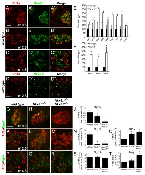
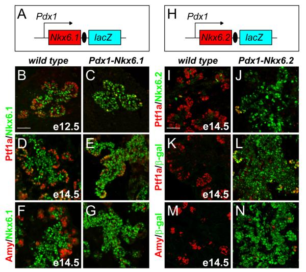
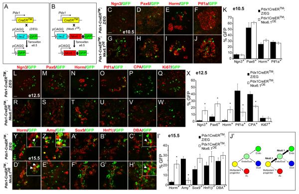
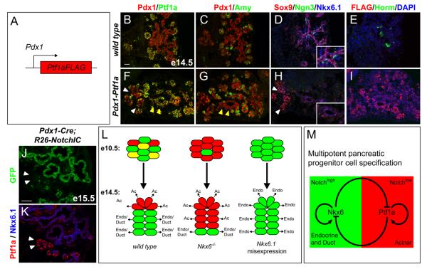
The molecular mechanisms that underlie cell lineage diversification of multipotent progenitors in the pancreas are virtually unknown. Here we show that the early fate choice of pancreatic progenitors between the endocrine and acinar cell lineage is restricted by cross-repressive interactions between the transcription factors Nkx6.1/Nkx6.2 (Nkx6) and Ptf1a. Using genetic loss- and gain-of-function approaches, we demonstrate that Nkx6 factors and Ptf1a are required and sufficient to repress the alternative lineage program and to specify progenitors toward an endocrine or acinar fate, respectively. The Nkx6/Ptf1a switch only operates during a critical competence window when progenitors are still multipotent and can be uncoupled from cell differentiation. Thus, cross-antagonism between Nkx6 and Ptf1a in multipotent progenitors governs the equilibrium between endocrine and acinar cell neogenesis required for normal pancreas development.


Figures

Figure 1
Figure 1. Endocrine to acinar fate conversion in Nkx6-deficient embryos
(A-D) Immunofluorescence for Ptf1a and Nkx6.1 or Nkx6.2 in the embryonic mouse pancreas. At e12.5, Nkx6.1 and Nkx6.2 become excluded from the Ptf1a+ tip domain. (E-F) Quantification of mRNA levels for acinar markers by microarray (E) and qRT-PCR (F) in e15.5 pancreas. Nkx6.1-deficiency results in increased expression of acinar-specific genes. (G-I,K-M,P-R) Immunofluorescence staining reveals expansion of the Ptf1a+ domain into the trunk region in Nkx6.1−/− and Nkx6.1−/−;Nkx6.2−/− pancreata at e12.5. Concomitantly, Ngn3+ cells are reduced. At e14.5, Nkx6-deficient embryos show increased numbers of acinar cells. (J,N,O,S,T) Quantification of marker+ cell numbers relative to total pancreatic area. Scale bar=50 μm; Try, trypsin; Klk, kallikrein; Ela, elastase; Clps, co-lipase; Cel, carboxylester-lipase; Amy, amylase.
Figure 2
Figure 2. Nkx6 proteins suppress acinar cell differentiation
(A,H) Schematic of the Pdx1-Nkx6.1 and Pdx1-Nkx6.2 transgenes. Immunofluorescence staining of pancreata from Pdx1-Nkx6.1 embryos at e12.5 and e14.5 (B-G) and Pdx1-Nkx6.2 embryos at e14.5 (I-N) shows that ectopic expression of Nkx6.1 or Nkx6.2 blocks expression of Ptf1a and amylase.
Figure 3
Figure 3. Nkx6.1 functions as an endocrine fate determinant
(A,B) A tamoxifen-inducible form of Cre recombinase (CreER™) expressed under the control of the Pdx1 promoter removes a floxed Bgeo (lacZ) cassette to permanently activate expression of green fluorescent protein (GFP) from the Z/EG transgene (A) or Nkx6.1 and GFP from the Nkx6.1OE transgene (B). Immunofluorescence staining of pancreas sections from e10.5 (C-J), e12.5 (L-W) and e15.5 (Y-H′) Pdx1-CreER;Z/EG and Pdx1-CreER;Nkx6.1OE embryos injected with one dose of tamoxifen at e8.5. Quantification of GFP+/marker+ cells relative to the total number of GFP+ cells at e10.5 (K), e12.5 (X), or e15.5 (I′). At e10.5, similar numbers of GFP+ cells have initiated expression of the endocrine markers Ngn3, Pax6, and hormones in Pdx1-CreER;Nkx6.1OE and Pdx1-CreER;Z/EG control embryos (K). Similar analysis at e12.5 reveals a preference of GFP+ cells in Pdx1-CreER;Nkx6.1OE embryos to express Ngn3, Pax6, and hormones, while showing a decreased propensity to express Ptf1a and CPA (X). Expression of the proliferation marker Ki67 does not show a difference between the two groups (X). At e15.5, GFP+ cells in Pdx1-CreER;Z/EG embryos express endocrine hormones (Y) as well as the acinar marker amylase (Z) (arrowheads in inset, Y,Z). By contrast, in Pdx1-CreER;Nkx6.1OE embryos, GFP+/amylase+ cells are rarely found (E′), while GFP+/hormone+ cells are abundantly detected (arrowheads in inset, D′). GFP+ cells from Pdx1-CreER;Nkx6.1OE and Pdx1-CreER;Z/EG embryos have a similar potential to differentiate into DBA+ ductal cells (arrowheads in inset show GFP+/DBA+ cells, C′,H′). (J′) Model of cell fate shifts induced by heritable Nkx6.1 misexpression in multipotent pancreatic progenitors. Scale bar=50 μm; triangles represent loxP sites; filled ovals, IRES; Amy, amylase; Horm, hormone (insulin, glucagon, somatostatin, pancreatic polypeptide); Endo, endocrine; Ac, acinar.
Figure 4
Figure 4. Ptf1a is a repressor of endocrine fate
(A) Schematic of the Pdx1-Ptf1a transgene. (B-I) Immunofluorescence staining of adjacent pancreas sections from e14.5 embryos shows ectopic expression of Ptf1a (F) and FLAG (I) in the trunk region of Pdx1-Ptf1a embryos. Ptf1a misexpression does not uniformly induce amylase expression (yellow arrow heads, F,G). Ectopic Ptf1a represses Nkx6.1, Sox9 and Ngn3 (H), and prevents endocrine cell differentiation (I). White arrowheads in (F,H) point to a region that does not misexpress Ptf1a and therefore expresses Sox9 and Nkx6.1 (H). Insets in (D,H) show higher magnifications. (J) Summary of the observed cell fate changes in Nkx6 loss- and gain-of-function models. (K,L) Immunofluorescence staining of adjacent pancreatic sections from Pdx1-Cre(mosaic);R26NotchIC embryos at e15.5 shows expression of Nkx6.1 in all cells that have recombined the R26NotchIC allele and therefore activated NotchIC/GFP expression. Ptf1a is excluded from the GFP+ domain, but is expressed in areas that have not recombined the R26NotchIC allele (arrowheads, K,L). (M) Our data support a model whereby a regulatory circuit comprised of the counter-antagonistic repressive activities of Nkx6 and Ptf1a restricts multipotent pancreatic progenitors to ductal/endocrine and acinar fates, respectively. Positive autoregulatory feedback of Nkx6 and Ptf1a creates bistability of the fate choice. High Notch activity appears to favor the Nkx6.1+ state. Note, for simplicity the redundant factors Nkx6.1 and Nkx6.2 are referred to as Nkx6. Scale bar=50 μm; Amy, amylase; Horm, hormone (insulin, glucagon, somatostatin, pancreatic polypeptide), Endo, endocrine; Ac, acinar.

References

    1. Apelqvist A, Li H, Sommer L, Beatus P, Anderson DJ, Honjo T, Hrabe de Angelis M, Lendahl U, Edlund H. Notch signalling controls pancreatic cell differentiation. Nature. 1999;400:877–881. - PubMed
    1. Briscoe J, Pierani A, Jessell TM, Ericson J. A homeodomain protein code specifies progenitor cell identity and neuronal fate in the ventral neural tube. Cell. 2000;101:435–445. - PubMed
    1. Burlison JS, Long Q, Fujitani Y, Wright CV, Magnuson MA. Pdx-1 and Ptf1a concurrently determine fate specification of pancreatic multipotent progenitor cells. Dev Biol. 2008;316:74–86. - PMC - PubMed
    1. Cras-Meneur C, Li L, Kopan R, Permutt MA. Presenilins, Notch dose control the fate of pancreatic endocrine progenitors during a narrow developmental window. Genes Dev. 2009;23:2088–2101. - PMC - PubMed
    1. Dong PD, Provost E, Leach SD, Stainier DY. Graded levels of Ptf1a differentially regulate endocrine and exocrine fates in the developing pancreas. Genes Dev. 2008;22:1445–1450. - PMC - PubMed

Publication types

MeSH terms