. 2010 Aug 6;398(4):752-8.
doi: 10.1016/j.bbrc.2010.07.017. Epub 2010 Jul 11.

Antiviral immune responses in H5N1-infected human lung tissue and possible mechanisms underlying the hyperproduction of interferon-inducible protein IP-10

Affiliations

Antiviral immune responses in H5N1-infected human lung tissue and possible mechanisms underlying the hyperproduction of interferon-inducible protein IP-10

Arunee Thitithanyanont et al. Biochem Biophys Res Commun. .

Abstract

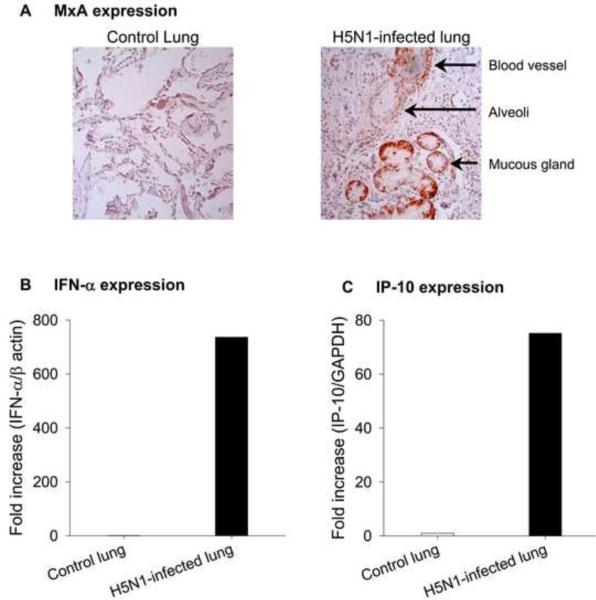
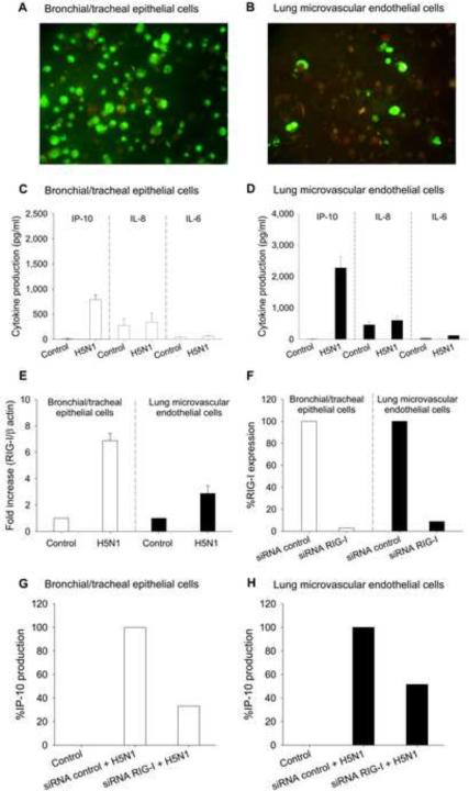
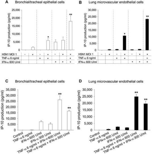
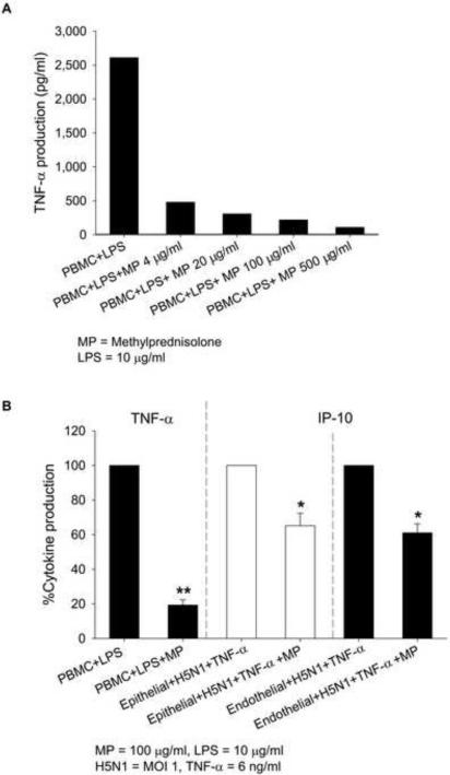
Information on the immune response against H5N1 within the lung is lacking. Here we describe the sustained antiviral immune responses, as indicated by the expression of MxA protein and IFN-alpha mRNA, in autopsy lung tissue from an H5N1-infected patient. H5N1 infection of primary bronchial/tracheal epithelial cells and lung microvascular endothelial cells induced IP-10, and also up-regulated the retinoic acid-inducible gene-I (RIG-I). Down-regulation of RIG-I gene expression decreased IP-10 response. Co-culturing of H5N1-infected pulmonary cells with TNF-alpha led to synergistically enhanced production of IP-10. In the absence of viral infection, TNF-alpha and IFN-alpha also synergistically enhanced IP-10 response. Methylprednisolone showed only a partial inhibitory effect on this chemokine response. Our findings strongly suggest that both the H5N1 virus and the locally produced antiviral cytokines; IFN-alpha and TNF-alpha may have an important role in inducing IP-10 hyperresponse, leading to inflammatory damage in infected lung.


Figures

Fig. 1
Fig. 1
Expression of MxA (Panel A), IFN-α (Panel B), and IP-10 (Panel C) in autopsy lung tissue from human subjects with and without influenza H5N1 infection.
Fig. 2
Fig. 2
In vitro infectivity by influenza H5N1 virus and induction of IP-10, IL-8, and IL-6 in bronchial/tracheal epithelial cells (Panels A and C) and lung microvascular endothelial cells (Panels B and D). H5N1 up-regulated RIG-I expression in bronchial/tracheal epithelial cells and lung microvascular endothelial cells (Panel E). RIG-I expression (%) in epithelial and endothelial cells transfected with control siRNA or siRNA RIG-I after overnight culture (Panel F). IP-10 expression (%) in H5N1 infected epithelial cells (Panel G) and endothelial cells (Panel H), transfected with control siRNA or siRNA RIG-I. Data in Panels F, G and H are representative results from one of two experiments. Data in Panels C, D and E are shown as mean values ± SEM of 4 independent experiments using cells derived from two individual donors.
Fig. 3
Fig. 3
IP-10 induction with treatment by H5N1, TNF-α, IFN-α, or different combinations in bronchial/tracheal epithelial cells (Panel A) and lung microvascular endothelial cells (Panel B). Panels C and D show IP-10 production with treatment of TNF-α and IFN-α in the absence of viral infection. Data are shown as mean values ± SEM of 4 independent experiments using cells derived from two individual donors. P < 0.05 compared with the sum of the individual effects. **P < 0.01 compared with the sum of the individual effects.
Fig. 4
Fig. 4
TNF-α expression in LPS-treated PBMC in the presence of different concentrations of methylprednisolone (MP) (Panel A). Cytokine expression in stimulated PBMC (TNF-α), stimulated bronchial/tracheal epithelial cells (IP-10) and stimulated lung microvascular endothelial cells (IP-10) with and without MP treatment (Panel B). Data in panel A are representative results from one of two experiments. Cytokine expression in treated cells (panel B) is shown as mean percentage of TNF-α and IP-10 expression ± SEM of 4 independent experiments using cells derived from two individual donors. *P < 0.05 compared with the same cell type without MP treatment. **P < 0.01 compared with the same cell type without MP treatment.

References

    1. Chan MC, Cheung CY, Chui WH, Tsao SW, Nicholls JM, Chan YO, Chan RW, Long HT, Poon LL, Guan Y, Peiris JS. Proinflammatory cytokine responses induced by influenza A (H5N1) viruses in primary human alveolar and bronchial epithelial cells. Respir Res. 2005;6:135. - PMC - PubMed
    1. Thitithanyanont A, Engering A, Ekchariyawat P, Wiboon-ut S, Limsalakpetch A, Yongvanitchit K, Kum-Arb U, Kanchongkittiphon W, Utaisincharoen P, Sirisinha S, Puthavathana P, Fukuda MM, Pichyangkul S. High susceptibility of human dendritic cells to avian influenza H5N1 virus infection and protection by IFN-alpha and TLR ligands. J Immunol. 2007;179:5220–5227. - PubMed
    1. de Jong MD, Simmons CP, Thanh TT, Hien VM, Smith GJ, Chau TN, Hoang DM, Chau NV, Khanh TH, Dong VC, Qui PT, Cam BV, Ha do Q, Guan Y, Peiris JS, Chinh NT, Hien TT, Farrar J. Fatal outcome of human influenza A (H5N1) is associated with high viral load and hypercytokinemia. Nat Med. 2006;12:1203–1207. - PMC - PubMed
    1. Peiris JS, Yu WC, Leung CW, Cheung CY, Ng WF, Nicholls JM, Ng TK, Chan KH, Lai ST, Lim WL, Yuen KY, Guan Y. Re-emergence of fatal human influenza A subtype H5N1 disease. Lancet. 2004;363:617–619. - PMC - PubMed
    1. To KF, Chan PK, Chan KF, Lee WK, Lam WY, Wong KF, Tang NL, Tsang DN, Sung RY, Buckley TA, Tam JS, Cheng AF. Pathology of fatal human infection associated with avian influenza A H5N1 virus. J Med Virol. 2001;63:242–246. - PubMed

Publication types

MeSH terms