. 2011 Feb;10(2):M110.001479.
doi: 10.1074/mcp.M110.001479. Epub 2010 Jul 12.

Pressurized pepsin digestion in proteomics: an automatable alternative to trypsin for integrated top-down bottom-up proteomics

Affiliations

Pressurized pepsin digestion in proteomics: an automatable alternative to trypsin for integrated top-down bottom-up proteomics

Daniel López-Ferrer et al. Mol Cell Proteomics. 2011 Feb.

Abstract

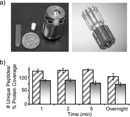
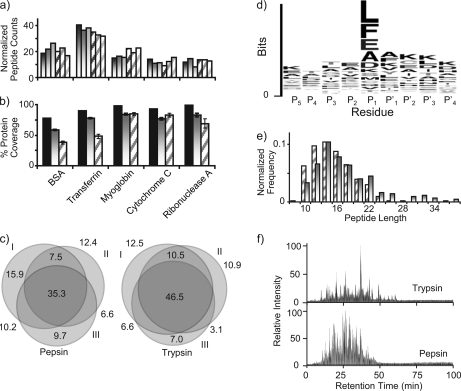
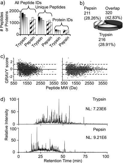
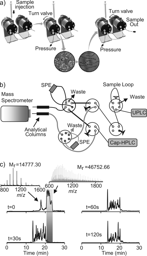
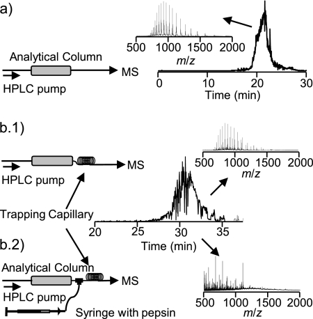
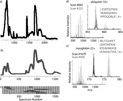
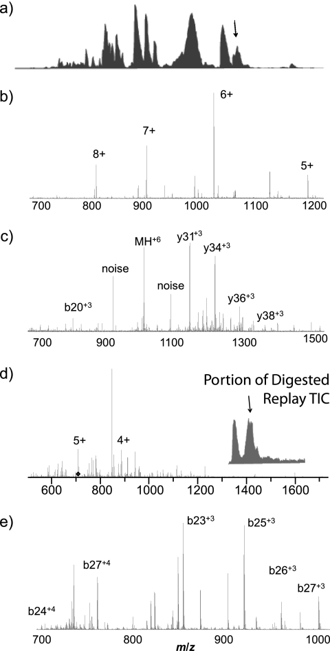
Integrated top-down bottom-up proteomics combined with on-line digestion has great potential to improve the characterization of protein isoforms in biological systems and is amendable to high throughput proteomics experiments. Bottom-up proteomics ultimately provides the peptide sequences derived from the tandem MS analyses of peptides after the proteome has been digested. Top-down proteomics conversely entails the MS analyses of intact proteins for more effective characterization of genetic variations and/or post-translational modifications. Herein, we describe recent efforts toward efficient integration of bottom-up and top-down LC-MS-based proteomics strategies. Since most proteomics separations utilize acidic conditions, we exploited the compatibility of pepsin (where the optimal digestion conditions are at low pH) for integration into bottom-up and top-down proteomics work flows. Pressure-enhanced pepsin digestions were successfully performed and characterized with several standard proteins in either an off-line mode using a Barocycler or an on-line mode using a modified high pressure LC system referred to as a fast on-line digestion system (FOLDS). FOLDS was tested using pepsin and a whole microbial proteome, and the results were compared against traditional trypsin digestions on the same platform. Additionally, FOLDS was integrated with a RePlay configuration to demonstrate an ultrarapid integrated bottom-up top-down proteomics strategy using a standard mixture of proteins and a monkey pox virus proteome.


Figures

Fig. 1.
Fig. 1.
a, the left panel shows a photograph of the MicroTube insert, and MicroTube holder used for off-line pressure digestion experiments with a dime for size comparison. The right panel shows how MicroTubes are inserted into the holder. b, the histogram depicts the average number of unique identified peptides and average protein coverage obtained from two duplicate LC-MS/MS runs of bovine serum albumin digested with pepsin for three different lengths of time at 25,000 p.s.i. and an overnight digestion at 37 °C and ambient pressure. The percentage of protein coverage is depicted as a solid bar, and uniquely identified peptides are depicted as diagonal lines. Error bars represent one standard deviation.
Fig. 2.
Fig. 2.
Comparison of enzyme performance for pepsin and trypsin pressure-assisted digestions using a five-standard protein mixture consisting of BSA, transferrin, myoglobin, cytochrome c, and ribonuclease A. a and b, histograms comparing normalized peptide counts and percentage of protein coverage for three technical replicates of pressurized digestions using either trypsin (shaded gradation bars) or pepsin (diagonal striped bars) for 1 min. Solid black bars indicate the total unique protein coverage when data from both digestions were combined. c, Venn diagrams showing the peptide overlap between three technical digestion replicates (I–III), including the percentage of identified peptides in each region. d, sequence logo for the amino acid sequence motif of pepsin-generated peptides identified by MS/MS analysis. e, histogram depicting the frequency of peptide length identified from the peptides identified either by trypsin (shaded gradation bars) or pepsin (diagonal striped bars). f, representative chromatograms corresponding to the LC-MS/MS of a pressure-assisted digestion for trypsin (top) and pepsin (bottom).
Fig. 3.
Fig. 3.
Comparison of enzyme performance and peptide properties for pepsin and trypsin pressure-assisted digestions using S. oneidensis protein extract as model proteome. a, histograms comparing the performance of 1-min pressure-assisted digestion with trypsin (shaded gradation bars) and pepsin (diagonal striped bars) across three technical replicates. b, pie chart representing the overlap of enzyme exclusive peptide identifications. c, GRAVY values plotted against the calculated molecular masses for the identified peptic (left) and tryptic (right) peptides. d, representative chromatograms corresponding to the LC-MS/MS of pressure-assisted digestion for trypsin (top) and pepsin (bottom) after a 2-min digestion.
Fig. 4.
Fig. 4.
Setup and validation of FOLDS for pepsin digestions. a, illustration showing the FOLDS platform. b, schematic diagram of the two-column FOLDS. The components include three six-port injection valves and four four-port valves. The system is coupled to a two-column RPLC system, and an LTQ mass spectrometer was used for MS/MS analysis. c, comparison of the chromatograms (and representative spectra) obtained from the two-column FOLDS pepsin digestion of enolase as a function of the digestion time. UPLC, ultraperformance LC; Cap-HPLC, capillary HPLC.
Fig. 5.
Fig. 5.
LC-MS setup schematics (left) and total ion chromatogram (right) for on-line digestion of myoglobin. Insets show mass spectra extracted at the indicated LC elution times. a, control experiment (no digestion). b, on-line digestion. b.1 and b.2, effect of the insertion of the delay/trapping capillary without (b.1) and with (b.2) pepsin infusion.
Fig. 6.
Fig. 6.
LC-MS and RePlay pepsin digestion analysis of protein standard mixture containing carbonic anhydrase, lactoglobulins A and B, cytochrome c, ubiquitin, and myoglobin. a, a total ion chromatogram of the initial intact protein RPLC analysis. b, the corresponding total ion chromatogram for the RePlay pepsin digestion analysis. c, a plot of the normalized number of digested peptides corresponding to a particular protein as a function of elution time. The darker boxes indicate more peptides were observed. Each row contains data corresponding to a different protein. From top to bottom, these proteins are cytochrome c, ubiquitin, lactoglobulin A, lactoglobulin B, myoglobin, and carbonic anhydrase. d and e, mass spectra of both the initial intact RPLC separation plotted in black and the RePlay pepsin digestion analysis plotted in grey. A significant number of intact ubiquitin (d) and myoglobin (e) peaks were only detected in the initial RPLC run, and digestion products were only observed in the RePlay run as illustrated in the zoomed-in regions.
Fig. 7.
Fig. 7.
LC-MS/MS of isolated monkey pox viral proteins with RePlay pepsin digestion MS/MS. a, the total ion chromatogram of the initial intact protein RPLC separation. b, a typical MS scan. The arrow in a indicates when in the RPLC this spectrum was acquired. c, the 6+ peak near 1,021 m/z in b was selected for isolation and MS/MS. De novo MS/MS analysis using in-house tools identified a fragment from the monkey pox structural protein VP8 based on the observed fragment peptides. d, representative mass spectrum from the RePlay pepsin digestion corresponding to a. e, tandem mass spectrum acquired for the 5+ peak in d, which corresponds to a pepsin digestion product of the original larger protein fragment based on de novo analysis of the observed fragmentation pattern.

References

    1. Kelleher N. L., Taylor S. V., Grannis D., Kinsland C., Chiu H. J., Begley T. P., McLafferty F. W. (1998) Efficient sequence analysis of the six gene products (7–74 kDa) from the Escherichia coli thiamin biosynthetic operon by tandem high-resolution mass spectrometry. Protein Sci. 7, 1796–1801 - PMC - PubMed
    1. Zubarev R. A., Horn D. M., Fridriksson E. K., Kelleher N. L., Kruger N. A., Lewis M. A., Carpenter B. K., McLafferty F. W. (2000) Electron capture dissociation for structural characterization of multiply charged protein cations. Anal. Chem. 72, 563–573 - PubMed
    1. Washburn M. P., Wolters D., Yates J. R., 3rd (2001) Large-scale analysis of the yeast proteome by multidimensional protein identification technology. Nat. Biotechnol. 19, 242–247 - PubMed
    1. Gevaert K., Goethals M., Martens L., Van Damme J., Staes A., Thomas G. R., Vandekerckhove J. (2003) Exploring proteomes and analyzing protein processing by mass spectrometric identification of sorted N-terminal peptides. Nat. Biotechnol. 21, 566–569 - PubMed
    1. Parks B. A., Jiang L., Thomas P. M., Wenger C. D., Roth M. J., Boyne M. T., 2nd, Burke P. V., Kwast K. E., Kelleher N. L. (2007) Top-down proteomics on a chromatographic time scale using linear ion trap Fourier transform hybrid mass spectrometers. Anal. Chem. 79, 7984–7991 - PMC - PubMed

Publication types

LinkOut - more resources