. 2010 Oct;335(1):32-41.
doi: 10.1124/jpet.110.170225. Epub 2010 Jul 13.

Therapeutic role of rifaximin in inflammatory bowel disease: clinical implication of human pregnane X receptor activation

Affiliations

Therapeutic role of rifaximin in inflammatory bowel disease: clinical implication of human pregnane X receptor activation

Jie Cheng et al. J Pharmacol Exp Ther. 2010 Oct.

Abstract

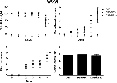
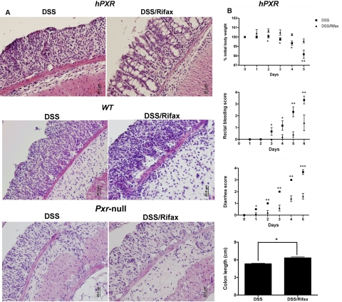
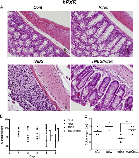
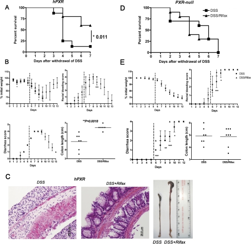
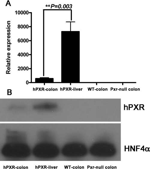
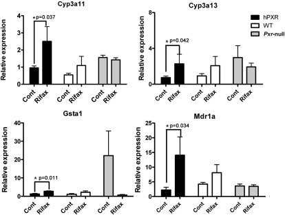
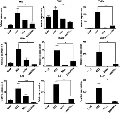
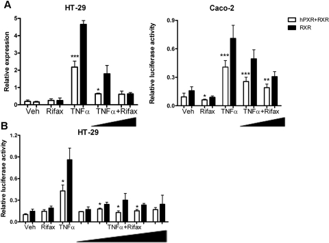
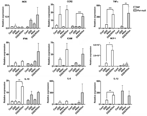
Human pregnane X receptor (PXR) has been implicated in the pathogenesis of inflammatory bowel disease (IBD). Rifaximin, a human PXR activator, is in clinical trials for treatment of IBD and has demonstrated efficacy in Crohn's disease and active ulcerative colitis. In the current study, the protective and therapeutic role of rifaximin in IBD and its respective mechanism were investigated. PXR-humanized (hPXR), wild-type, and Pxr-null mice were treated with rifaximin in the dextran sulfate sodium (DSS)-induced and trinitrobenzene sulfonic acid (TNBS)-induced IBD models to determine the protective function of human PXR activation in IBD. The therapeutic role of rifaximin was further evaluated in DSS-treated hPXR and Pxr-null mice. Results demonstrated that preadministration of rifaximin ameliorated the clinical hallmarks of colitis in DSS- and TNBS-treated hPXR mice as determined by body weight loss and assessment of diarrhea, rectal bleeding, colon length, and histology. In addition, higher survival rates and recovery from colitis symptoms were observed in hPXR mice, but not in Pxr-null mice, when rifaximin was administered after the onset of symptoms. Nuclear factor κB (NF-κB) target genes were markedly down-regulated in hPXR mice by rifaximin treatment. In vitro NF-κB reporter assays demonstrated inhibition of NF-κB activity after rifaximin treatment in colon-derived cell lines expressing hPXR. These findings demonstrated the preventive and therapeutic role of rifaximin on IBD through human PXR-mediated inhibition of the NF-κB signaling cascade, thus suggesting that human PXR may be an effective target for the treatment of IBD.


Figures

Fig. 1.
Fig. 1.
Colitis assessment of hPXR mice treated with 2.5% DSS after rifampicin (RIF) pretreatment. hPXR male mice were placed into six groups (n ≥ 6 per group): control group (Cont), DSS treatment alone (DSS), RIF (3 mg/kg/day) treatment alone (RIF3), RIF (10 mg/kg/day) treatment alone (RIF10), DSS treatment after RIF (3 mg/kg/day) pretreatment (DSS/RIF3), and DSS treatment after RIF (10 mg/kg/day) pretreatment (DSS/RIF10). Each bar represents the mean ± S.D. n ≥ 6. *, p < 0.05; **, p < 0.01.
Fig. 2.
Fig. 2.
Rifaximin protects hPXR mice in a DSS-induced colitis model. A, representative H&E-stained colon sections of hPXR, WT, and Pxr-null treated with DSS alone (DSS) or DSS/rifaximin (Rifax). Magnification: 200×. B, colitis assessment of hPXR mice treated with 2.5% DSS (DSS) or 2.5% DSS after rifaximin pretreatment (DSS/Rifax). Each bar represents the mean ± S.D. n ≥ 6. *, p < 0.05; **, p < 0.01; ***, p < 0.001.
Fig. 3.
Fig. 3.
Rifaximin protects hPXR mice in a TNBS-induced colitis model. A, representative H&E-stained colon sections of hPXR treated with control (left top), rifaximin (right top), TNBS (left bottom), and TNBS/rifaximin (right bottom). Magnification: 200×. B, body weight loss of hPXR mice treated with control (Cont), rifaximin (Rifax), TNBS, and TNBS/rifaximin (TNBS/Rifax). C, colon length of hPXR mice treated with control (Cont), rifaximin (Rifax), TNBS, and TNBS/rifaximin (TNBS/Rifax). Each bar represents the mean ± S.D. n ≥ 6. *, p < 0.05; **, p < 0.01; ***, p < 0.001.
Fig. 4.
Fig. 4.
Therapeutic role of rifaximin on DSS-induced IBD in hPXR mice. A, survival curve of hPXR mice comparing rifaximin after treatment to no rifaximin treatment. B, colitis assessment of hPXR mice comparing rifaximin after treatment to no rifaximin treatment. C, representative H&E-stained colon sections of hPXR treated with DSS (left) or rifaximin after DSS-induced colitis (center) (magnification: 200×) and macroscopic observation of colon length (left: DSS; right: DSS/Rifax). D, survival curves of Pxr-null mice comparing rifaximin after treatment to no rifaximin treatment. E, colitis assessment of Pxr-null mice comparing rifaximin after treatment to no rifaximin treatment. Each bar represents the mean ± standard deviations. n ≥ 10. *, p < 0.05; **, p < 0.01; ***, p < 0.001.
Fig. 5.
Fig. 5.
Basal expression of human PXR in the colon of hPXR mice. A, qPCR analysis of human PXR expression in colon and liver of hPXR mice and in colon of WT and Pxr-null mice. Mouse β-actin mRNA served as an internal control. B, Western blot analysis of human PXR expression in colon and liver of hPXR mice and in colon of WT and Pxr-null mice. Pooled nuclear protein (30 μg) was loaded for each sample (n = 3 per group). The monoclonal antibody against human PXR (H5017) specifically recognizes human PXR but not mouse PXR or other liver proteins. HNF4α was used as a loading control.
Fig. 6.
Fig. 6.
mRNA analysis of hPXR target genes in colon tissue. Expression of mRNAs encoding Cyp3a11, Cyp3a13, Gsta1, and Mdr1a were determined by qPCR from colon epithelial cells isolated from hPXR, WT, and Pxr-null mice treated with control (Cont) and rifaximin (Rifax). Data were normalized to β-actin. Each bar represents the mean ± standard deviations. n ≥ 6. *, p < 0.05.
Fig. 7.
Fig. 7.
mRNA analysis of proinflammatory mediators in colon tissue from hPXR mice. Colon RNA was isolated from hPXR mice treated with control (Cont), DSS, rifaximin (Rifax), and DSS/rifaximin (DSS/Rifax). Expression of mRNAs encoding iNOS, CCR2, TNFα, IFNα, ICAM-1, MCP-1, IL-10, IL-6, and IL-1β was determined by qPCR. Data were normalized to β-actin. Each bar represents the mean ± standard deviations. n ≥ 6. *, p < 0.05; **, p < 0.01; ***, p < 0.001.
Fig. 8.
Fig. 8.
Rifaximin inhibited TNFα-activated NF-κB luciferase reporter through human PXR. A, HT-29 cells and Caco-2 cells (5 × 10⁴ cells/well) were cotransfected with NF-κB luciferase reporter (0.2 mg/well) and mouse RXR (0.04 μg/well) or cotransfected with NF-κB luciferase reporter (0.2 μg/ml/well), mouse RXR (0.04 μg/well), and hPXR (0.04 μg/well). Twenty-four hours after transfection cells were incubated with vehicle (Veh, DMSO), Rifax (1 or 100 μM, dissolved in DMSO), or TNFα (10 ng/ml) or coincubated with TNFα and rifaximin for 24 h (TNFα+ Rifax). B, HT-29 cells were cotransfected with NF-κB luciferase reporter (0.2 μg/ml/well) and mouse RXR (0.04 μg/well) or cotransfected with NF-κB luciferase reporter (0.2 μg/ml/well), mouse RXR (0.04 μg/well), and hPXR (0.04 μg/well). Twenty-four hours after transfection cells were incubated with vehicle (Veh), Rifax (100 μM), and TNFα (10 ng/ml) or coincubated with rifaximin (0.001, 0.01, 0.1, 1, or 100 μM) and TNFα (10 ng/ml) for 24 h (TNFα+ Rifax). Standard dual luciferase assays were performed on cell extracts. Each bar represents the mean ± standard deviation. n ≥ 6. *, p < 0.05; **, p < 0.01; ***, p < 0.001.
Fig. 9.
Fig. 9.
mRNA analysis of proinflammatory factors in colon tissue from WT and Pxr-null mice. Colon RNA was isolated from WT and Pxr-null mice treated with control (Cont), DSS, Rifax, and DSS/Rifax. Expression of mRNAs encoding iNOS, CCR2, TNFα, IFNα, ICAM-1, MCP-1, IL-10, IL-6, and IL-1β was determined by qPCR. Data were normalized to β-actin. Each bar represents the mean ± standard deviations. n ≥ 6. *, p < 0.05; **, p < 0.01; ***, p < 0.001.

References

    1. Akbar DH, Al-Shehri HZ, Al-Huzali AM, Falatah HI. (2003) A case of rifampicin induced pseudomembraneous colitis. Saudi Med J 24:1391–1393 - PubMed
    1. Bertilsson G, Heidrich J, Svensson K, Asman M, Jendeberg L, Sydow-Bäckman M, Ohlsson R, Postlind H, Blomquist P, Berkenstam A. (1998) Identification of a human nuclear receptor defines a new signaling pathway for CYP3A induction. Proc Natl Acad Sci USA 95:12208–12213 - PMC - PubMed
    1. Chen C, Shah YM, Morimura K, Krausz KW, Miyazaki M, Richardson TA, Morgan ET, Ntambi JM, Idle JR, Gonzalez FJ. (2008) Metabolomics reveals that hepatic stearoyl-CoA desaturase 1 down-regulation exacerbates inflammation and acute colitis. Cell Metab 7:135–147 - PMC - PubMed
    1. Chen TC, Lu PL, Lin WR, Lin CY, Wu JY, Chen YH. (2009) Rifampin-associated pseudomembranous colitis. Am J Med Sci 338:156–158 - PubMed
    1. Cheng J, Ma X, Krausz KW, Idle JR, Gonzalez FJ. (2009) Rifampicin-activated human pregnane X receptor and CYP3A4 induction enhance acetaminophen-induced toxicity. Drug Metab Dispos 37:1611–1621 - PMC - PubMed

Publication types

MeSH terms

LinkOut - more resources