. 2010 Dec 1;88(3):520-9.
doi: 10.1093/cvr/cvq230. Epub 2010 Jul 13.

TGFBR2 mutations alter smooth muscle cell phenotype and predispose to thoracic aortic aneurysms and dissections

Affiliations

TGFBR2 mutations alter smooth muscle cell phenotype and predispose to thoracic aortic aneurysms and dissections

Sakiko Inamoto et al. Cardiovasc Res. .

Abstract

Aims: Transforming growth factor-β (TGF-β) signaling is critical for the differentiation of smooth muscle cells (SMCs) into quiescent cells expressing a full repertoire of contractile proteins. Heterozygous mutations in TGF-β receptor type II (TGFBR2) disrupt TGF-β signaling and lead to genetic conditions that predispose to thoracic aortic aneurysms and dissections (TAADs). The aim of this study is to determine the molecular mechanism by which TGFBR2 mutations cause TAADs.

Methods and results: Using aortic SMCs explanted from patients with TGFBR2 mutations, we show decreased expression of SMC contractile proteins compared with controls. Exposure to TGF-β1 fails to increase expression of contractile genes in mutant SMCs, whereas control cells further increase expression of these genes. Analysis of fixed and frozen aortas from patients with TGFBR2 mutations confirms decreased in vivo expression of contractile proteins relative to unaffected aortas. Fibroblasts explanted from patients with TGFBR2 mutations fail to transform into mature myofibroblasts with TGF-β1 stimulation as assessed by expression of contractile proteins.

Conclusions: These data support the conclusion that heterozygous TGFBR2 mutations lead to decreased expression of SMC contractile protein in both SMCs and myofibroblasts. The failure of TGFBR2-mutant SMCs to fully express SMC contractile proteins predicts defective contractile function in these cells and aligns with a hypothesis that defective SMC contractile function contributes to the pathogenesis of TAAD.


Figures
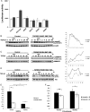
Figure 1
Figure 1
Assessment of TGF-β signaling in cells from individuals with TGFBR2 mutations. (A) In vitro analysis shows impaired activity of receptors harbouring TGFBR2 mutations. TGFBR2 constructs were co-transfected with a TGF-β responsive luciferase reporter plasmid (p3TP-Lux) into TGFBR2-deficient DR-26 cells, standardized to a β-galactosidase expression plasmid. Wild -type TGFBR2 transfected cells showed an increase in the luciferase activity in response to TGF-β stimulation (black bar) when compared with basal activity in the absence of TGF-β (white bars), while those transfected with the vector alone, a dominant negative TGFBR2 lacking the entire cytoplasmic domain (DNIIR), a missense mutation previously reported in an MFS patient (R537C), or the two missense mutations found in familial TAAD patients (R460C and R460H) did not show an increase in luciferase activity, suggesting that these mutations impair TGFBR2 function. Error bars represent ± standard deviation. (B) Assessment of Smad2 phosphorylation in aortic SMCs from an affected patient and age-matched control after TGF-β1 for various times up to 48 h (figure represents similar data collected from one of the three patients’ SMC cell strains assessed). (C) Western blot analysis of p38MAPK and phosphorylated- p38MAPK protein in SMCs on stimulation with TGF-β shows reduced phosphorylation of p38 in TGFBR2-mutant SMCs (R528H) compared with control SMCs. (D) Western blot analysis of Akt and phosphorylated- Akt protein in SMCs on stimulation with TGF-β shows reduced phosphorylation of Akt in TGFBR2-mutant SMCs (R528H) compared with control SMCs. (E) SMCs with TGFBR2 mutations proliferate significantly less rapidly than control SMCs at baseline and after 72 h of stimulation with TGF-β. *P< 0.0001 (F) Fibroblasts with TGFBR2 mutations proliferate significantly less rapidly than control fibroblasts at baseline and after 72 h of TGF-β stimulation. *P= 0.015, **P= 0.00015.
Figure 2
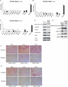
Figure 2
Decreased contractile protein expression and protein levels in patients’ explanted aortic cells and tissue. (AC) Q-PCR analysis of mRNA isolated from serum-starved SMCs from patients with TGFBR2 mutations (black bars) and matched controls (white bars). The affected patients’ SMCs consistently have reduced expression of the SMC contractile complex genes, no change in expression of cytoskeletal genes (ACTB, ACTG1) and increased expression of S100A4. Gene expression level was standardized to GAPDH. *P< 0.01, **P< 0.001. (D) Immunoblot analysis of SMC lysates from patients’ SMCs versus matched control showed reduced SMC contractile protein levels in patients’ SMCs compared with control. (E) Immunoblot analysis of proteins isolated from frozen aortic tissue from a patient showed reduced SMC contractile protein levels compared with control tissue. (F to G) Immunohistochemistry staining of aortic tissue from patients shows decreased intensity of staining of the SMC contractile proteins calponin (F) and β-myosin (G) compared with controls. Patient images are shown below their age-matched control images. Magnification is 200x, and scale bars represent 100 µm.
Figure 3
Figure 3
TGFBR2-mutant SMCs fail to demonstrate significantly increased expression of contractile proteins and fail to assemble contractile fibres with exposure to TGF-β. (AC) Q-PCR analysis of mRNA isolated from TGFBR2 R528H (A), R460C (B) and R460H SMCs (C) harvested 72 h after exposure to TGF-β fails to show increased expression of contractile protein genes ACTA2, CNN1 and MYH11 but showed significantly decreased expression of S100A4 (*P< 0.05). Control SMCs demonstrate significant increases in the expression of these same genes (*P< 0.05). For this experiment, each sample's gene expression was normalized to its own 0h timepoint to assess the changes that occurred with TGF-β stimulation (except S100A4). (D to E) Immunofluorescence analysis of α-actin (D) and calponin (E) and stress fibres in cultured SMCs from patients and controls. In the control cells, there is extensive co-localization of either α-actin (D, green) or calponin (E, green) and polymerized actin (stained red with phalloidin). The stress fibres containing α-actin tend to cross the cell body, whereas the calponin tends to be incorporated into stress fibres near the nucleus of the cell, as previously reported. In contrast, the intensity of α-actin and calponin stain is diminished in all four of the patients’ SMCs (shown below their age-matched control) with minimal staining at the periphery of the cells. SMC nuclei were counterstained with DAPI (blue). Scale bars 40µm.
Figure 4
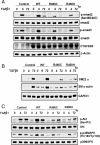
Figure 4
Disruption of TGF-β signaling by over-expressed TGFBR2 mutants R460C and R460H in 10T1/2 cells. Western blot analysis shows decreased phosphorylation levels of Smad2/3 (A) and Akt and p38 (C) in cells transfected with mutant receptors. Contractile protein expression (B) was also decreased in cells transfected with mutant compared with wild-type receptors.
Figure 5
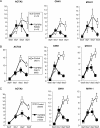
Figure 5
Defective transformation of quiescent fibroblasts to myofibroblasts with TGF-β in patient and control fibroblasts. (A) Q-PCR analysis of mRNA expression of SMC contractile protein genes in fibroblasts various times after exposure to TGF-β1 showed reduced expression in TGFBR2-mutant cells (R460C, n = 6) compared with matched controls (n = 6). (B to C) Similarly expression analysis of mRNA or SMC contractile protein genes in fibroblasts explanted from two unrelated patients with TGFBR2 mutations (C461Y, n = 1) in panel B, and R528H, (n = 1) in panel (C) also showed reduced expression of these genes.

References

    1. Owens GK, Kumar MS, Wamhoff BR. Molecular regulation of vascular smooth muscle cell differentiation in development and disease. Physiol Rev. 2004;84:767–801. doi:10.1152/physrev.00041.2003. - DOI - PubMed
    1. Sinha S, Hoofnagle MH, Kingston PA, McCanna ME, Owens GK. Transforming growth factor-beta1 signaling contributes to development of smooth muscle cells from embryonic stem cells. Am J Physiol Cell Physiol. 2004;287:C1560–C1568. doi:10.1152/ajpcell.00221.2004. - DOI - PubMed
    1. Mizuguchi T, Collod-Beroud G, Akiyama T, Abifadel M, Harada N, Morisaki T, et al. Heterozygous TGFBR2 mutations in Marfan syndrome. Nat Genet. 2004;36:855–860. doi:10.1038/ng1392. - DOI - PMC - PubMed
    1. Pannu H, Fadulu VT, Chang J, Lafont A, Hasham SN, Sparks E, et al. Mutations in transforming growth factor-beta receptor type II cause familial thoracic aortic aneurysms and dissections. Circulation. 2005;112:513–520. doi:10.1161/CIRCULATIONAHA.105.537340. - DOI - PubMed
    1. Loeys BL, Chen J, Neptune ER, Judge DP, Podowski M, Holm T, et al. A syndrome of altered cardiovascular, craniofacial, neurocognitive and skeletal development caused by mutations in TGFBR1 or TGFBR2. Nat Genet. 2005;37:275–281. doi:10.1038/ng1511. - DOI - PubMed

Publication types

MeSH terms

Substances