. 2010 Aug;120(8):2967-78.
doi: 10.1172/JCI39222. Epub 2010 Jul 12.

Low doses of killed parasite in CpG elicit vigorous CD4+ T cell responses against blood-stage malaria in mice

Affiliations

Low doses of killed parasite in CpG elicit vigorous CD4+ T cell responses against blood-stage malaria in mice

Alberto Pinzon-Charry et al. J Clin Invest. 2010 Aug.

Abstract

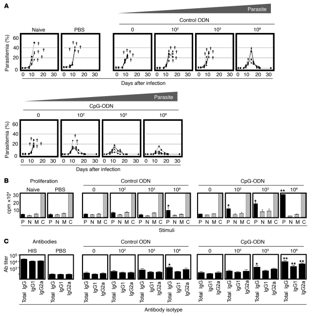
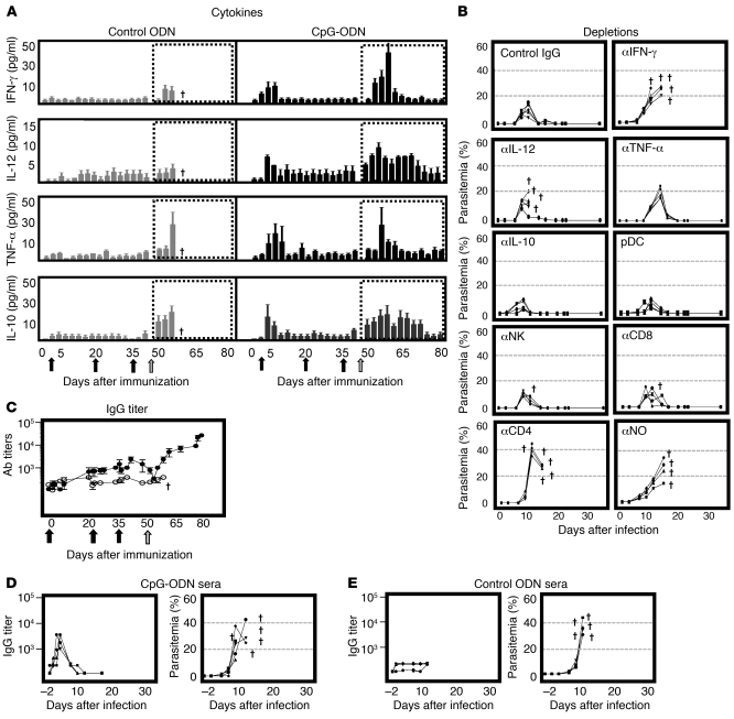
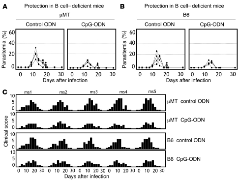
Development of a vaccine that targets blood-stage malaria parasites is imperative if we are to sustainably reduce the morbidity and mortality caused by this infection. Such a vaccine should elicit long-lasting immune responses against conserved determinants in the parasite population. Most blood-stage vaccines, however, induce protective antibodies against surface antigens, which tend to be polymorphic. Cell-mediated responses, on the other hand, offer the theoretical advantage of targeting internal antigens that are more likely to be conserved. Nonetheless, few of the current blood-stage vaccine candidates are able to harness vigorous T cell immunity. Here, we present what we believe to be a novel blood-stage whole-organism vaccine that, by combining low doses of killed parasite with CpG-oligodeoxynucleotide (CpG-ODN) adjuvant, was able to elicit strong and cross-reactive T cell responses in mice. Our data demonstrate that immunization of mice with 1,000 killed parasites in CpG-ODN engendered durable and cross-strain protection by inducing a vigorous response that was dependent on CD4+ T cells, IFN-gamma, and nitric oxide. If applicable to humans, this approach should facilitate the generation of robust, cross-reactive T cell responses against malaria as well as antigen availability for vaccine manufacture.


Figures

Figure 1
Figure 1. Definition of a low-dose whole-parasite vaccine.
(A) To define the low-dose vaccine, A/J mice were immunized with decreasing doses of P. c. chabaudi AS (AS) antigen equivalent to 108, 103, 102, or 0 (adjuvant alone) prbc in CpG-ODN or control ODN. 2 weeks after immunization, mice were challenged i.v. with 105 homologous (AS) prbc and the outcome of infection monitored as parasitemia. Disease severity is also described in Supplemental Figure 1. Data for individual mice are shown. Animals that succumbed to infection. (B) To assess cellular immune responses induced by vaccination, antigen-specific proliferation of splenic lymphocytes was assessed at the time of challenge infection. Spleen cells were cultured for 96 hours in the presence of fresh (AS) parasitized rbc (P), normal rbc (N), medium (M), or ConA (C) and proliferation assessed by thymidine uptake. Results show mean ± SEM. (C) To assess vaccine-induced antibody responses, parasite-specific IgG titers against homologous (AS) antigen were assessed by ELISA at the time of challenge. Reciprocal median total IgG titers and interquartile ranges are shown. All results are representative of 5 mice per group of 3 independent experiments performed. Significant differences compared with adjuvant alone (control ODN or CpG-ODN) are shown. *P < 0.05; **P < 0.01. HIS, hyperimmune sera.
Figure 2
Figure 2. Mechanisms of protection.
(A) To determine the profile of cytokine secretion, levels of IFN-γ (first panel), IL-12 (second panel), TNF-α (third panel), and IL-10 (fourth panel) were assessed in sera collected from A/J mice during immunization with 103 (AS) prbc in control ODN (gray bars) or CpG-ODN (black bars) as well as following challenge (dotted boxes). Immunizations (1 priming and 2 boost injections) are indicated as black arrows and challenge as a gray arrow on the x axis (days after immunization). Results represent mean ± SEM. Animals that succumbed to infection. (B) To identify cytokines and cell types essential for protection, A/J mice were immunized with 103 prbc in CpG-ODN and subsequently depleted of IFN-γ, IL-12, TNF-α, IL-10, pDC, NK, CD8, CD4, or NO as described in Methods. Mice were then challenged i.v. with 105 homologous (AS) prbc and the outcome of infection monitored. Data for individual mice are shown. (C) To determine the kinetics of antibody responses during immunization and challenge, A/J mice were immunized with 103 (AS) prbc in CpG-ODN (black circles) or control ODN (white circles) and titers of parasite-specific IgG against homologous antigen assessed. Reciprocal median total IgG titers and interquartile ranges are shown. (D, E) To ascertain a role for antibodies in protection, naive A/J mice were adoptively transferred on days –1, 0, and 1 with 200 ml i.p. of serum collected from immune (103 [AS] prbc in CpG-ODN) or control (103 [AS] prbc in control ODN) donors. Recipients were challenged with homologous 105 (AS) prbc on day 0. Parasitemia and parasite-specific IgG titers against homologous (AS) were monitored. Reciprocal median total IgG titers and interquartile ranges are shown. All data sets are representative of 5 mice per group of 2 independent experiments performed.
Figure 3
Figure 3. Protection in B cell–deficient mice.
To define the role of B cells in protection following immunization, B cell–deficient (μMT) or B cell–sufficient (B6) mice were immunized with 103 (AS) prbc in CpG-ODN or control ODN. 2 weeks later, mice were challenged i.v. with 105 homologous (AS) prbc and (A) parasitemia or (B) disease severity monitored. Disease severity was assessed by determining clinical scores (C). Data for individual mice are shown and are representative of 3 independent experiments. Note that mice of B6 background (B6 or μMT) are naturally resistant to P. c. chabaudi AS and thus, protection was determined by reduced parasitemia and disease severity. ms1, mouse 1.
Figure 4
Figure 4. Cellular responses and cross-reactivity.
(A) To determine CD4 response, A/J mice were immunized with 103 (AS) prbc in CpG-ODN (black bars) or control ODN (gray bars). Spleen cells collected 2 weeks after immunization were cultured with homologous (AS) or heterologous (P. c. chabaudi AJ [AJ]; P. yoelii YM [YM]) parasites, normal rbc (nrbc), or ConA. After 96 hours, supernatants were assayed for TNF-α, IFN-γ, IL-2 (Th1 cytokines), and IL-4 or IL-5 (Th2 cytokines) assessed. Results are mean ± SEM. (B) For proliferation, spleen cells from A/J mice immunized as above were purified at 2, 8, or 12 weeks and incubated for 96 hours with AS, AJ or YM prbc, nrbc, medium, or ConA. Numbers represent percentage of CFSEdim CD4 T cells. (C) For cytokine secretion, spleen cells from A/J mice immunized as above and purified at 2, 8, or 12 weeks were incubated for 96 hours with AS, AJ, or YM prbc, nrbc, or medium and stained for intracellular IFN-γ. Numbers represent percentage of IFN-γ+ CD4 T cells. (D) For cross-reactive antibodies, sera from A/J mice immunized with 103 (AS) prbc in CpG-ODN (black bars), control ODN (gray bars), or hyperimmune sera (HIS, white bars) were tested for IgG titers against homologous (AS) or heterologous (AJ and YM) parasites. Results represent reciprocal median total IgG titers and interquartile ranges. All data sets are representative of 5 mice per group of 3 independent experiments performed. Significant differences compared with controls are shown. *P < 0.05.
Figure 5
Figure 5. Heterologous immunity.
To assess for cross-protection, different strains of mice including A/J [H-2a], B6 [H-2b] and BALB/c [H-2d] mice were immunized with 103 (AS) prbc and challenged i.v. with homologous (AS) or heterologous (AJ or YM) parasites at 2, 8, or 12 weeks after vaccination. Rechallenge was performed at 12 weeks after parasite clearance. The outcome of infection was monitored by assessing parasitemia. Data for individual mice are shown. nd, not determined. Animals that succumbed to infection.
Figure 6
Figure 6. Homologous immunity.
To assess for homologous protection against P. yoelii YM, different strains of mice including A/J, B6 and BALB/c mice were immunized with 103 (YM) prbc in CpG-ODN and challenged i.v. with homologous parasites at 2 weeks after vaccination. The outcome of infection was monitored by assessing parasitemia. In all data sets, results for individual mice are shown and are representative of 3 independent experiments performed. Animals that succumbed to infection.
Figure 7
Figure 7. Induction of CD4 T cell memory.
(A) To assess the role of CD4 T cells in protracted protection, A/J mice immunized with 103 (AS) prbc in control ODN or CpG-ODN were depleted of CD4 T cells 12 weeks after immunization. Mice were challenged i.v. with 105 homologous (AS) prbc immediately after depletion (left panels, effector) or 21 days after depletion to allow for naive/helper CD4 T cells to replenish (right panels, helper). Parasitemias were monitored. Results are representative of 3 separate experiments and data for individual mice are shown. Animals that succumbed to infection. (B) To assess for cytokine secretion in memory CD4 T cells, spleen cells from A/J mice immunized with 103 (AS) prbc in CpG-ODN were purified 12 weeks after immunization, incubated for 96 hours in the presence of homologous (AS) prbc, and cells stained for intracellular IFN-γ and IL-2. Numbers represent percentages of cytokine+ CD4 T cells. (C) To identify the specificity of CD4 T cell memory subsets, IFN-γ secretion and expression of lymph node homing receptor (CD62L) were assessed by harvesting spleen cells from A/J mice immunized with 103 (AS) prbc in CpG-ODN at 12 weeks after immunization. Cells were incubated for 96 hours in the presence of homologous (AS) prbc and stained. Numbers represent percentages of IFN-γ+ CD62LloCD4 T cells. (D) To determine the existence of memory populations, spleen cells from A/J mice immunized with 103 (AS) prbc in CpG-ODN were FACS-sorted into CM (CD44hiCD62Lhi) and EM (CD44hiCD62Llo) populations directly ex vivo. CM and EM cells were then incubated for 96 hours in the presence of homologous (AS) prbc and stained for intracellular cytokine (IFN-γ) and proliferation (BrDU). Numbers represent percentages of CD4 T cells in corresponding quadrants. Results are representative of 3 independent experiments performed.

References

    1. Good MF. Vaccine-induced immunity to malaria parasites and the need for novel strategies. Trends Parasitol. 2005;21(1):29–34. doi: 10.1016/j.pt.2004.10.006. - DOI - PubMed
    1. Fox JP. Modes of action of poliovirus vaccines and relation to resulting immunity. Rev Infect Dis. 1984;6 suppl 2:S352–S355. - PubMed
    1. Mielcarek N, et al. Live attenuated B. pertussis as a single-dose nasal vaccine against whooping cough. PLoS Pathog. 2006;2(7):e65. doi: 10.1371/journal.ppat.0020065. - DOI - PMC - PubMed
    1. McGregor IA. Mechanisms of acquired immunity and epidemiological patterns of antibody responses in malaria in man. Bull World Health Organ. 1974;50(3–4):259–266. - PMC - PubMed
    1. Hoffman SL, et al. Naturally acquired antibodies to sporozoites do not prevent malaria: vaccine development implications. Science. 1987;237(4815):639–642. doi: 10.1126/science.3299709. - DOI - PubMed

Publication types