. 2010 Oct;18(10):1796-802.
doi: 10.1038/mt.2010.152. Epub 2010 Jul 13.

Duration of expression and activity of Sleeping Beauty transposase in mouse liver following hydrodynamic DNA delivery

Affiliations

Duration of expression and activity of Sleeping Beauty transposase in mouse liver following hydrodynamic DNA delivery

Jason B Bell et al. Mol Ther. 2010 Oct.

Abstract

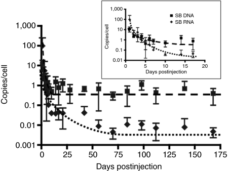
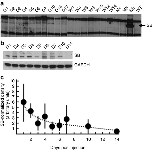
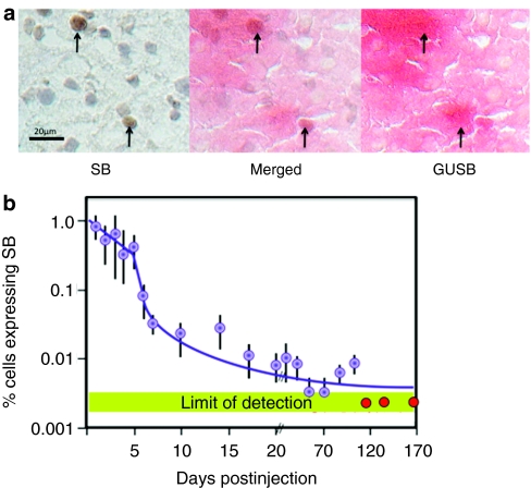
The Sleeping Beauty (SB) transposon system can direct integration of DNA sequences into mammalian genomes. The SB system comprises a transposon and transposase that "cuts" the transposon from a plasmid and "pastes" it into a recipient genome. The transposase gene may integrate very rarely and randomly into genomes, which has led to concerns that continued expression might support continued remobilization of transposons and genomic instability. Consequently, we measured the duration of SB11 transposase expression needed for remobilization to determine whether continued expression might be a problem. The SB11 gene was expressed from the plasmid pT2/mCAGGS-Luc//UbC-SB11 that contained a luciferase expression cassette in a hyperactive SB transposon. Mice were imaged and killed at periodic intervals out to 24 weeks. Over the first 2 weeks, the number of plasmids with SB11 genes and SB11 mRNA dropped about 90 and 99.9%, respectively. Expression of the luciferase reporter gene in the transposon declined about 99% and stabilized for 5 months at nearly 1,000-fold above background. In stark contrast, transposition-supporting levels of SB11 mRNA lasted only about 4 days postinfusion. Thus, within the limits of current technology, we show that SB transposons appear to be as stably integrated as their viral counterparts.


Figures

Figure 1
Figure 1
Delivery of the Sleeping Beauty (SB) transposon system by hydrodynamic injection. (a) Transposon and transposase-encoding plasmid used in this study. pT2/mCAGGS-Luc//UbC-SB11 contains a firefly luciferase transposon behind a mCAGGS promoter. Outside of the transposon, a 340-bp PCR-generated human ubiquitin C (UbC) promoter regulating expression of the SB11 transposase gene. (b) Twenty-four hour bioluminescent values from C57BL/6 mice injected with 25 µg pT2/mCAGGS-Luc//UbC-SB11. The inset shows a representative mouse after bioluminescent imaging. Of the injected mice 85% had photon fluxes above 109 photons/second/cm2.
Figure 2
Figure 2
Sustained expression of luciferase reporter. (a) Persistence of the Sleeping Beauty (SB) gene depends on the co-delivered transgene. Top: the expression cassette for hyperactive SB11 transposase is delivered on the same plasmid with the transposon that harbors an expression cassette for either of the three reporter genes, two encoding lysosomal enzymes (hIDUA and hGUSB) that escape the cells or one encoding the firefly luciferase (Luc) protein that is cytoplasm-restricted. The three reporter genes in a T2 transposon were driven by an mCAGGS promoter; the SB11 transposase gene was driven by a short PCR-generated UbC promoter. Bottom: comparison of SB11 gene copy numbers following co-delivery with three different reporter genes. C57BL/6 mice were injected with either of the three transposon plasmids shown on top. Mice were killed on selected days and DNA for real-time PCR was isolated from nitrogen-pulverized whole livers. The lines represent best-fit curves. (b) Time course of Luc activity from C57BL/6 mice injected with 25 µg pT2/mCAGGS-Luc//UbC-SB11. Circles represent the mean ± SD of bioluminescence values (n = 5 mice/time point). hGUSB, human β-glucuronidase; hIDUA, human α-ℓ-iduronidase.
Figure 3
Figure 3
Expression of SB11 transposase mRNA relative to Sleeping Beauty (SB) gene copy number. Over the first 2 weeks the SB11 gene number declined by 90% and then stabilized, whereas the mRNA copy number declined ~99.9%. SB genes were quantified by quantitative PCR and SB mRNA by quantitative reverse transcription-PCR. The gene copy number assumes 2 copies of the glyceraldehyde-3-phosphate dehydrogenase as an internal standard. The inset shows in greater detail the loss of gene and mRNA copy numbers for the first 2.5 weeks. Mice were hydrodynamically injected with pT2/mCAGGS-Luc//UbC-SB11 and killed at selected time points, 5 mice/group. Data are presented as mean ± SD.
Figure 4
Figure 4
Expression of SB11 protein as a function of time. (a) Western blot demonstrating expression of Sleeping Beauty (SB) transposase protein from the hydrodynamic injection of the pT2/mCAGGS-Luc//UbC-SB11 vector. Liver lysates from five mice for each collection time were pooled for each sample and separated by polyacrylamide gel electrophoresis for western blotting. Samples D1-D17, W3-W14, and M4-M5 represent days, weeks, and months postinjection, respectively. SB lane shows SB expression from the Rosa26-SB11 transgenic mouse (positive control) and wild-type (WT) lane shows the proteins from liver of sham-treated mice injected with empty plasmid (negative control). The samples were probed with a monoclonal antibody against SB transposase and reprobed with an anti-GAPDH antibody. The heavy bands above and below the SB band (38 kd) are due to the presence of the heavy and light immunoglobulin chains that are found at high levels in hydrodynamically injected mice; these bands are detected using only the secondary antibody when the primary anti-SB monoclonal antibody is not included in the blotting procedure (data not shown). (b) Western blots of SB and Gapdh, protein used for a loading control, over the first 2 weeks. GAPDH, glyceraldehyde-3-phosphate dehydrogenase. (c) Normalized expression of SB protein from analysis of four sets of western blots.
Figure 5
Figure 5
Immunohistochemical analysis of Sleeping Beauty (SB) gene expression in liver tissue. (a) Colocalization of expression of SB transposase and GUSB genes 2 days following injection of pT2/mCAGGS-GUSB//CMV-SB11. Serial 4-µm sections prepared from optimal cutting temperature–embedded frozen liver samples were postfixed and exposed alternately to either anti-SB monoclonal antibody for analysis of SB expression (left panel) or to a histochemical stain for GUSB (right panel). The middle panel shows superimposed SB-immunohistochemistry (IHC) and GUSB-stained sections. The arrows indicate areas where the SB-positive cells matched GUSB-positive cells. Bar = 20 µm. (b) Percentage of liver cells from mice injected with pT2/mCAGGS-Luc//UbC-SB11 showing positive IHC activity for SB protein. The green bar indicates the limit of detection of this assay. Each point represents mean ± SD from five mice, one slide/mouse. Filled purple circles: measurements above the limits of detection; red circles: measurements at background levels. Each slide had two sections; stained cells from each section were counted in three independent fields.
Figure 6
Figure 6
Transposition as a function of time following injection of pT2/mCAGGS-Luc//UbC-SB11. SB11 transposase activity was evaluated by the production of excision products (EPs) as a function of time. The level of Sleeping Beauty (SB) mRNA is reproduced from Figure 3. The inset shows in greater detail EP and mRNA time courses over the first 2.5 weeks. Data are presented as mean ± SD, n = 5 mice/group.

References

    1. Aiuti A., and, Roncarolo MG. Ten years of gene therapy for primary immune deficiencies. Hematology Am Soc Hematol Educ Program. 2009. pp. 682–689. - PubMed
    1. Ott MG, Schmidt M, Schwarzwaelder K, Stein S, Siler U, Koehl U, et al. Correction of X-linked chronic granulomatous disease by gene therapy, augmented by insertional activation of MDS1-EVI1, PRDM16 or SETBP1. Nat Med. 2006;12:401–409. - PubMed
    1. Aiuti A, Cattaneo F, Galimberti S, Benninghoff U, Cassani B, Callegaro L, et al. Gene therapy for immunodeficiency due to adenosine deaminase deficiency. N Engl J Med. 2009;360:447–458. - PubMed
    1. Cartier N, Hacein-Bey-Abina S, Bartholomae CC, Veres G, Schmidt M, Kutschera I, et al. Hematopoietic stem cell gene therapy with a lentiviral vector in X-linked adrenoleukodystrophy. Science. 2009;326:818–823. - PubMed
    1. Manno CS, Pierce GF, Arruda VR, Glader B, Ragni M, Rasko JJ, et al. Successful transduction of liver in hemophilia by AAV-Factor IX and limitations imposed by the host immune response. Nat Med. 2006;12:342–347. - PubMed

Publication types

LinkOut - more resources