. 2010 Jul 8;6(7):e1000991.
doi: 10.1371/journal.ppat.1000991.

Plus- and minus-end directed microtubule motors bind simultaneously to herpes simplex virus capsids using different inner tegument structures

Affiliations

Plus- and minus-end directed microtubule motors bind simultaneously to herpes simplex virus capsids using different inner tegument structures

Kerstin Radtke et al. PLoS Pathog. .

Abstract

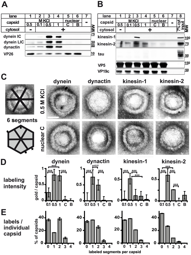
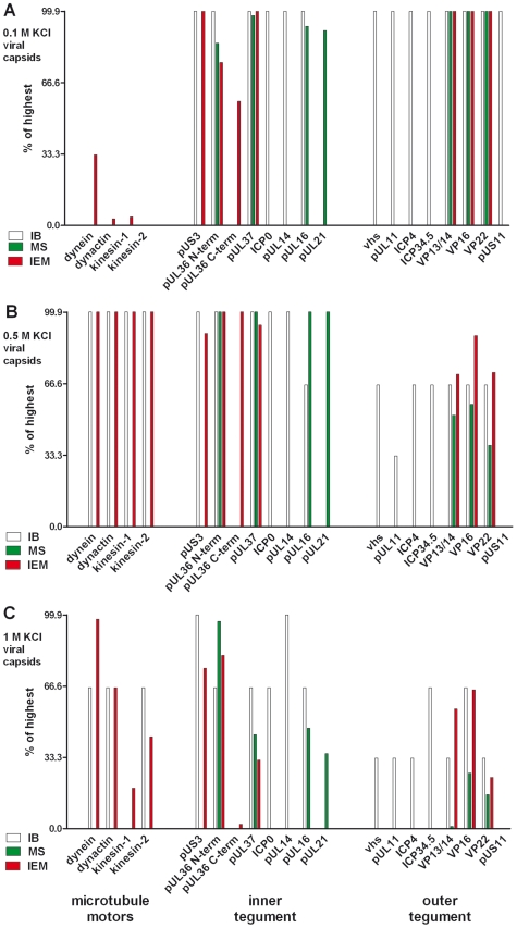
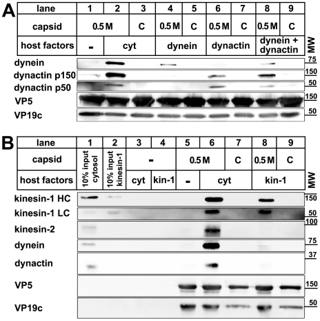
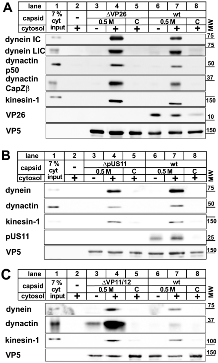
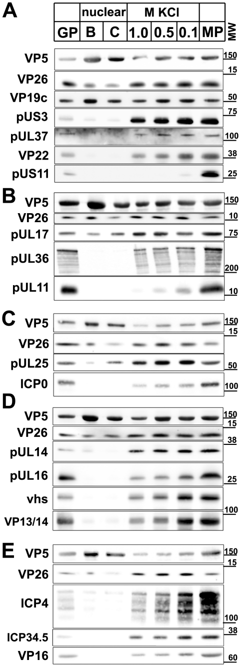
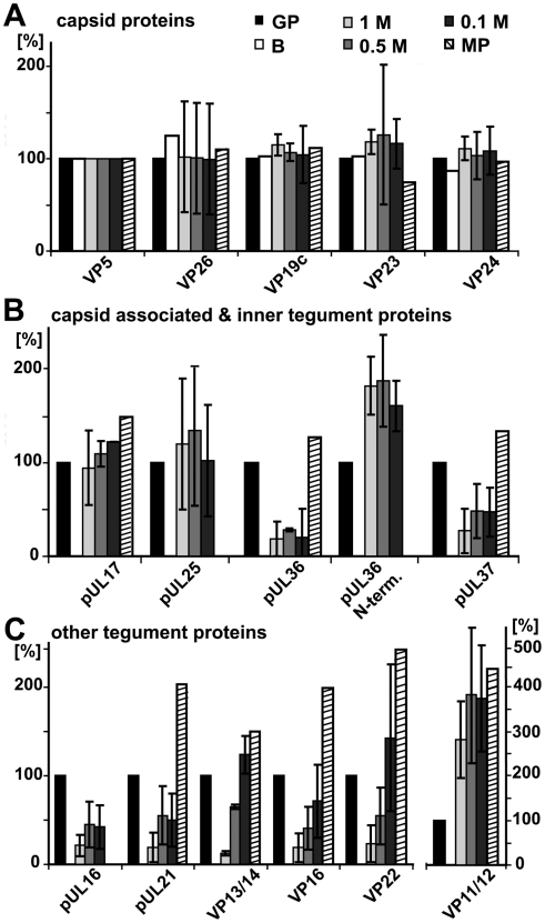
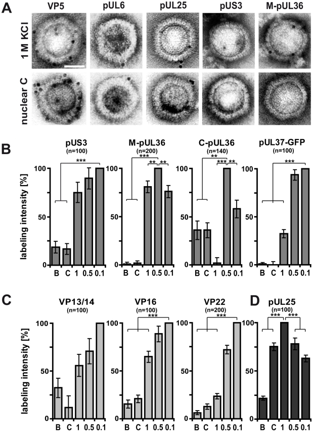
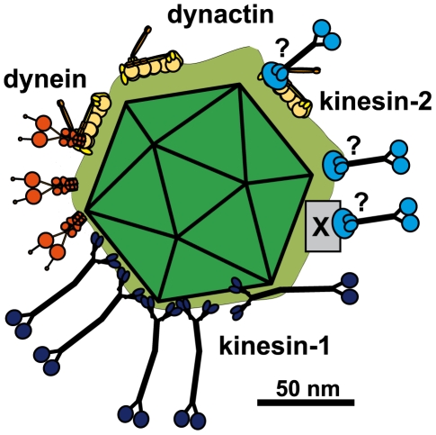
Many viruses depend on host microtubule motors to reach their destined intracellular location. Viral particles of neurotropic alphaherpesviruses such as herpes simplex virus 1 (HSV1) show bidirectional transport towards the cell center as well as the periphery, indicating that they utilize microtubule motors of opposing directionality. To understand the mechanisms of specific motor recruitment, it is necessary to characterize the molecular composition of such motile viral structures. We have generated HSV1 capsids with different surface features without impairing their overall architecture, and show that in a mammalian cell-free system the microtubule motors dynein and kinesin-1 and the dynein cofactor dynactin could interact directly with capsids independent of other host factors. The capsid composition and surface was analyzed with respect to 23 structural proteins that are potentially exposed to the cytosol during virus assembly or cell entry. Many of these proteins belong to the tegument, the hallmark of all herpesviruses located between the capsid and the viral envelope. Using immunoblots, quantitative mass spectrometry and quantitative immunoelectron microscopy, we show that capsids exposing inner tegument proteins such as pUS3, pUL36, pUL37, ICP0, pUL14, pUL16, and pUL21 recruited dynein, dynactin, kinesin-1 and kinesin-2. In contrast, neither untegumented capsids exposing VP5, VP26, pUL17 and pUL25 nor capsids covered by outer tegument proteins such as vhs, pUL11, ICP4, ICP34.5, VP11/12, VP13/14, VP16, VP22 or pUS11 bound microtubule motors. Our data suggest that HSV1 uses different structural features of the inner tegument to recruit dynein or kinesin-1. Individual capsids simultaneously accommodated motors of opposing directionality as well as several copies of the same motor. Thus, these associated motors either engage in a tug-of-war or their activities are coordinately regulated to achieve net transport either to the nucleus during cell entry or to cytoplasmic membranes for envelopment during assembly.


Conflict of interest statement

The authors have declared that no competing interests exist.

Figures

Figure 1
Figure 1. Analysis of host factor recruitment to HSV1 capsids.
Partially tegumented viral capsids were generated from mature extracellular particles released from HSV1 infected cells by detergent lysis to remove the viral envelope in the presence of 0.1 M, 0.5 M or 1 M KCl to extract different amounts of tegument (light green), and purified through sucrose cushions. Untegumented nuclear B and C capsids (dark green) were isolated from the nuclei of HSV1 infected cells by gradient sedimentation. The 5 different capsid types were resuspended in BRB80 buffer using tip sonification, treated with DNase and RNase, repelleted, and then incubated with cytosolic extracts or purified MAPs (colored circles). Capsids were analyzed for bound host factors either after sedimentation through a sucrose cushion by immunoblot or directly by electron microscopy after immunolabeling and negative contrasting.
Figure 2
Figure 2. Plus- and minus-end directed MT motors bind to HSV1 capsids.
A and B: Immunoblot analysis of MAP binding to capsids. HSV1(F) viral (0.5 M KCl, lanes 1 and 3; 0.1 M KCl, lanes 2; 1 M KCl, lanes 4) or nuclear capsids (C capsids, lanes 5; B capsids, lanes 6) or mock samples lacking capsids (lanes 7) were incubated in 0.25 mg/ml (A, lanes 2 to 7) or 0.75 (B; lanes 2 to 7) mg/ml pig brain cytosol, and sedimented through sucrose cushions. The host proteins were analyzed by immunoblot using antibodies against dynein (A: MAB1618 against intermediate chain; pAb anti-LIC2 against light intermediate chain), and dynactin (A: mAb anti-p50), or kinesin-1 (B: MAB1613 against heavy chain), kinesin-2 (B: mAb against KAP3A) and tau (B: pAb). The amount of capsids in each sample was estimated by labeling the major capsid proteins VP26 (A: pAb anti-VP26), VP5 (B: pAb NC-1), or VP19c (B: pAb NC-2). As a loading control, 7% of the amount of the input cytosol was also directly analyzed (B; lane 8). C: Immunoelectron microscopy images of HSV1(F) capsids incubated with 0.75 mg/ml pig brain cytosol and labeled with mouse monoclonal antibodies against dynein (MAB1618 against intermediate chain), dynactin (mAb anti-p50), kinesin-1 (MAB1613 against heavy chain) or kinesin-2 (mAb K2.4 against KIF3A) followed by rabbit-anti-mouse antibodies, protein-A gold and negative staining. Scale bar: 50 nm. D: Quantification of the labeling intensity of different capsid types. After immunogold-labeling with antibodies against MAPs, negative staining and electron microscopy, the number of gold particles per capsid was counted for 100 to 170 capsids. Labeling of capsids incubated with buffer instead of cytosol was considered background and subtracted. Error bars: SEM. Three asterisks denote P<0.0001 as determined in a two-sided Student's t-test. E: Labeling frequency for different MAPs on individual viral capsid treated with 0.5 M KCl. In a two-dimensional projection, the icosahedral capsid appears as a hexagon that can be divided into 6 segments (c.f. C; schematic capsid). The number of capsids that had labeling on 0, 1, 2, or more of such segments was counted. Gold particles were counted as multiple labeling events when they were more than 40 nm apart (C; circles on schematic capsid). Labels in closer proximity were scored as only one label (C; stars on schematic capsid). Error bars: SEM. The quantifications (D, E) were compiled from three experiments analyzing dynein, dynactin and kinesin-1 and two experiments for kinesin-2.
Figure 3
Figure 3. Characterization of tegumented, viral HSV1 capsids.
The MAP binding (Fig. 2; left 4 data sets) to the three viral capsid types treated with 0.1, 0.5 or 1 M KCl (panels A, B and C, respectively) and their inner (middle 8 data sets) and outer tegument (right 8 data sets) organization (Figs. 6,7,8) have been analyzed by immunoblot (IB), quantitative mass spectrometry (MS), and quantitative immunoelectron microscopy (IEM). While IB and MS indicate the amount of different tegument proteins on the capsids, the IEM determines to what extent such tegument proteins were accessible on the capsid surfaces to antibodies or host factors. Please note that nuclear capsids did not bind to MAPs and contained very little tegument (c.f. Figs. 2, 6, 7, 8; data not shown in this figure). For further comparative analysis, we normalized the results of the IB, MS and IEM for the viral capsids such that the amount of a given MAP or a tegument protein on the capsids with the highest amount was set to 100%, and recalculated accordingly for the other capsid types (% of highest). These results are also listed in supplementary Table S1. Please note that in contrast to MS and IEM, the IB data were not quantitative. Instead, we only estimated the amount of the respective proteins based on the band intensities into 4 classes: absent (0%), minor (33%), major (66%) or highest (100%) amounts.
Figure 4
Figure 4. Dynein, dynactin, and kinesin-1 bind directly to tegumented HSV1 capsids.
HSV1 (F) capsids binding do dynein and or dynactin (A) or kinesin-1 (B) was analyzed as follows: HSV1(F) capsids generated by detergent lysis of extracellular virions in the presence of 0.5 M KCl, or nuclear C capsids were incubated with buffer (A: lane 1), 0.25 mg/ml pig brain cytosol (A: lanes 2 and 3), 15 µg/ml purified, native dynein (A: lanes 4 and 5), 15 µg/ml purified, native dynactin (A: lanes 6 and 7) or native dynein and dynactin (A: lanes 8 and 9) or with buffer (B: lane 5), 0.75 mg/ml pig brain cytosol (B: lanes 6 and 7) or 33 µg/ml purified, native kinesin-1 (B: lanes 8 and 9), and sedimented through a sucrose cushion. As controls, cytosol (B: lanes 1 and 3) or purified kinesin-1 (B: lanes 2 and 4) were also directly analyzed (B: lanes 1 and 2) or sedimented in the absence of capsids (B: lanes 3 and 4). Host proteins were detected by immunoblotting with antibodies against dynein (A & B: MAB1618 against intermediate chain), dynactin (A: mAb anti-p150, mAb anti-p50, B: anti-CapZβ mAb3F2.3), kinesin-1 (B: MAB1613 against heavy chain, MAB1616 against light chain), kinesin-2 (B: mAb against KAP3A). As loading controls, the samples were probed with antibodies against the capsid proteins VP5 (A and B: pAb NC-1) or VP19c (A and B: pAb NC-2). These blots show one of three independent experiments yielding similar results.
Figure 5
Figure 5. MAPs bind to HSV1 capsids lacking VP26, pUS11, or VP11/12.
Capsids of HSV1(KOS)-ΔVP26 (A: lanes 3 to 5), HSV1(F)-ΔUS11 (B: lanes 3 to 5), or HSV1(F)-ΔVP11/12 (C: lanes 3 to 5) were isolated by detergent lysis of trypsin-treated extracellular virions in the presence of 0.5 M KCl or from nuclei of infected cells (C capsids). HSV1(F) wild-type was used as control (A, B, C: wt, lanes 6 to 8). The capsids were incubated in 0.75 mg/ml pig brain cytosol, and after sedimentation analyzed by immunoblot with antibodies against dynein (A, B, C: MAB1618 against intermediate chain; A: pAb anti-LIC2 against light intermediate chain), dynactin (A, B: mAb anti-p50; A, C: mAb3F2.3 against CapZβ), or kinesin-1 (A, B, C: MAB1613 against heavy chain). Labeling with antibodies against VP26 (A: pAb anti-VP26) or pUS11 (B: mAb #28) confirmed the lack of these proteins on mutant capsids. As loading control, the samples were probed with antibodies against the capsid protein VP5 (A, B, C: pAB NC-1). As controls, cytosol alone was directly analyzed (A, B, C: lanes 1) or sedimented in the absence of capsids (A, B, C: lanes 2). These blots show one of two or more experiments yielding similar results.
Figure 6
Figure 6. Immunoblot characterization of nuclear and viral HSV1 capsids.
The protein composition of HSV1 capsids (nuclear B capsids, nuclear C capsids, viral capsids treated with 1.0, 0.5 or 0.1 M KCl), virions sedimented from cell culture supernatants (MP, medium pellet) and gradient purified virions (GP) were analyzed by immunoblot using antibodies directed against the capsid proteins VP5 (A–E: pAb NC-1), VP26 (A–E: pAb anti-VP26), VP19c (A: pAb NC-2), pUL17 (B: mAb #203), pUL25 (C: pAb ID1), pUL16 (D: anti-pUL16), or the tegument proteins pUS3 (A: pAb anti-pUS3), pUL37 (A: pAb 780), VP22 (A: pAb AGV30), pUS11 (A: mAb #28), pUL36 (B: pAb #147; anti-middle-pUL36), pUL11 (B: pAb anti-pUL11), ICPO (C: mAb 11060), pUL14 (D: anti-pUL14), vhs (D: pAb 11.388), VP13/14 (D: pAb R220), ICP4 (E: mAb 58S), ICP34.5 (E: pAb anti-ICP34.5) and VP16 (E: pAb SW7). Each blot shows one of three indendent experiments. The panels A, B, C, D, and E represent separate membranes. These data were after a further normalization also integrated into Fig. 3 (white columns).
Figure 7
Figure 7. Mass spectrometric characterization of nuclear and viral HSV1 capsids.
The protein composition of HSV1 capsids (B: nuclear B capsids, viral capsids treated with 1.0, 0.5 or 0.1 M KCl), virions sedimented from cell culture supernatants (MP, medium pellet) and gradient purified virions (GP) were analyzed by quantitative mass spectrometry. A: the capsid proteins VP5, VP26, VP19c, VP23, VP24, B: the capsid-associated proteins pUL17 and pUL25 and the inner tegument proteins pUL36, an N-terminal fragment of pUL36, pUL37, pUL16, pUL21 and C: the other tegument proteins pUL16, pUL21, VP13/14, VP16, VP22 and VP11/12 were analyzed. The relative protein amount in a capsid or a virus preparation is given in comparison to the gradient purified virions set as 100%. Values lower than 100% indicate removal of a protein in the respective capsid or virus preparation, whereas values higher than 100% indicate a higher amount of this protein in capsid samples than in gradient purified virions. Values are normalized to the amount of the capsid protein VP5 to account for varying amounts of capsids in each sample. Mean values of three independent experiments are given for viral capsids treated with 1, 0.5 and 0.1 M KCl, and one experiment for medium pellet and nuclear B capsids. Error bars: SD. These data were after a further normalization also integrated into Fig. 3 (green columns).
Figure 8
Figure 8. Generation of different HSV1 capsid surfaces during isolation.
HSV1(F) capsids were isolated from infected nuclei (B or C) or prepared from extracellular virions by detergent lysis in the presence of different KCL concentrations (1, 0.5 or 0.1 M KCl), and labeled with antibodies against the capsid proteins VP5 (A: pAb NC-1), pUL25 (A, D: pAb ID1) or pUL6 (A: mAb 1C), against the inner tegument proteins pUS3, (A: pAb anti-US3), pUL36 (A, B: pAb #147, anti-middle-pUL36; B: pAb anti-Cterminal-pUL36), pUL37-GFP (B: mAb anti-GFP JL-8; here capsids from strain HSV1-pUL37GFP), or the outer tegument proteins VP13/14 (C: pAb R220), VP16 (D: pAb SW7), or VP22 (D: pAb AGV30). A: Immunoelectron microscopy images of capsids after immunolabeling followed by protein-A gold and negative staining. Scale bar: 50 nm. B to D: The labeling intensity for inner tegument proteins (B), outer tegument proteins (C) or the capsid associated protein pUL25 (D) was quantified by counting the number of gold particles per capsid. After subtraction of the background without the primary antibodies, the number on the capsid type with the highest labeling was set to 100%, and recalculated accordingly for the other capsid types. These data sets were also directly integrated into Fig. 3 (red columns). Error bars: SEM. n: number of capsids (same for each capsid type with a given antibody) summarized from three experiments. Two asterisks denote p<0.001 and three asterisks indicate P<0.0001 as determined in two-sided Student's t-tests.
Figure 9
Figure 9. Scale drawing of a HSV1 viral capsids exposing inner tegument proteins and interacting with MAPs.
Tegumented (light green) HSV1 capsid (dark green) with three bound dynein (orange), three dynactin (yellow), five kinesin-1 (dark blue) and 3 kinesin-2 (light blue). The scheme has been drawn to scale and assuming that MT binding domains of the MAPs point away from the capsid to enable microtubule binding. Dynein, dynactin and kinesin-1 can interact directly and independently of each other with the capsids. Individual capsids could recruit several copies of a MAP, and different MAPs at once. Dynein and Kinesin-2 may either bind directly to tegument proteins or indirectly via dynactin. Furthermore, kinesin-2 may also utilize another, unknown host factor (X). Scale bar: 50 nm.

References

    1. Sodeik B. Mechanisms of viral transport in the cytoplasm. Trends Microbiol. 2000;8:465–472. - PubMed
    1. Smith GA, Enquist LW. BREAK INS AND BREAK OUTS: Viral Interactions with the Cytoskeleton of Mammalian Cells. Annu Rev Cell Dev Biol. 2002;18:135–161. - PubMed
    1. Greber UF, Way M. A Superhighway to Virus Infection. Cell. 2006;124:741–756. - PubMed
    1. Radtke K, Döhner K, Sodeik B. Viral interactions with the cytoskeleton: A hitchhiker's guide to the cell. Cellular Microbiology. 2006;8:387–400. - PubMed
    1. Brandenburg B, Zhuang X. Virus trafficking - learning from single-virus tracking. Nat Rev Microbiol. 2007;5:197–208. - PMC - PubMed

Publication types