. 2010 Jul 8;6(7):e1001021.
doi: 10.1371/journal.pgen.1001021.

Partial loss of ataxin-1 function contributes to transcriptional dysregulation in spinocerebellar ataxia type 1 pathogenesis

Affiliations

Partial loss of ataxin-1 function contributes to transcriptional dysregulation in spinocerebellar ataxia type 1 pathogenesis

Juan Crespo-Barreto et al. PLoS Genet. .

Abstract

Spinocerebellar ataxia type 1 (SCA1) is a dominantly inherited neurodegenerative disease caused by expansion of a CAG repeat that encodes a polyglutamine tract in ATAXIN1 (ATXN1). Molecular and genetic data indicate that SCA1 is mainly caused by a gain-of-function mechanism. However, deletion of wild-type ATXN1 enhances SCA1 pathogenesis, whereas increased levels of an evolutionarily conserved paralog of ATXN1, Ataxin 1-Like, ameliorate it. These data suggest that a partial loss of ATXN1 function contributes to SCA1. To address this possibility, we set out to determine if the SCA1 disease model (Atxn1(154Q/+) mice) and the loss of Atxn1 function model (Atxn1-/- mice) share molecular changes that could potentially contribute to SCA1 pathogenesis. To identify transcriptional changes that might result from loss of function of ATXN1 in SCA1, we performed gene expression microarray studies on cerebellar RNA from Atxn1-/- and Atxn1(154Q/+) cerebella and uncovered shared gene expression changes. We further show that mild overexpression of Ataxin-1-Like rescues several of the molecular and behavioral defects in Atxn1-/- mice. These results support a model in which Ataxin 1-Like overexpression represses SCA1 pathogenesis by compensating for a partial loss of function of Atxn1. Altogether, these data provide evidence that partial loss of Atxn1 function contributes to SCA1 pathogenesis and raise the possibility that loss-of-function mechanisms contribute to other dominantly inherited neurodegenerative diseases.


Conflict of interest statement

The authors have declared that no competing interests exist.

Figures
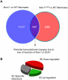
Figure 1
Figure 1. Comparison of transcriptional profiles of Atxn1154Q/+ and Atxn1−/− cerebella.
(A) Venn diagram reporting the number of significant gene expression changes in Atxn1−/− and Atxn1154Q/+cerebella. A total of 197 transcripts were significantly altered in both Atxn1−/− and Atxn1154Q/+mice, using a p-value of less than 0.01, and a minimal fold change of |±0.1| log2. (B) The majority of shared changes (68% of genes or 135 out of 197) went in the same direction.
Figure 2
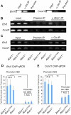
Figure 2. Chromatin immunoprecipitation (ChIP) reveals co-occupancy at the promoters of Cic target genes by Atxn1 and Cic.
(A) ChIP using Cic antisera confirmed Cic binding at the promoter of two direct targets of Capicua, Ccnd1 and Etv5, that were up-regulated in the Atxn1−/− and Atxn1154Q/+ cerebella. (B) ChIP using Atxn1 anti-sera in cerebellar extracts from Atxn1 +/−, Atxn1154Q/−, and Atxn1 −/− mice reveals a signal in mice expressing only wild-type (Atxn1 +/−) but not polyQ-Atxn1 (Atxn1154Q/−) compared to negative controls (pre-immune sera, and Atxn1 −/−) (C) ChIP as in (B), this time using Cic antibody. In contrast to (B), Cic is present at comparable levels at the target promoters in Atxn1 +/−, Atxn1154Q/−, and Atxn −/− mice. All ChIP assays were repeated three times on independent samples, representative results shown. (D) ChIP followed by quantitative PCR (ChIP-qPCR) on the promoter of Etv5 confirms that the proportion of immunoprecipitated DNA by Cic antibody is comparable in all three genotypes (as seen in C). A region containing two Capicua binding sites (promoter-CBS) of Etv5 is more enriched by Cic antibody than a region lacking CBSs (promoter-no CBS) compared to preimmune sera. (E) ChIP-qPCR on the promoter of Ccnd1 also shows similar enrichment of immunoprecipitated DNA by Cic antibody in all three genotypes (as seen in C). ChIP-qPCR using primers designed for a region within 100 bps of the CBS in the Ccnd1 promoter (which is highly conserved across species) show more Cic binding than primers designed for a poorly conserved region further downstream (∼400 bps) of the CBS at the promoter of Ccnd1, compared to preimmune sera. ChIP-qPCR experiments in (D) and (E) were performed in triplicate on three independent sets of samples (3 cerebella per genotype) using SYBR Green Dye. N.S. = not significant, ** p<0.01, *** p<0.005.
Figure 3
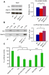
Figure 3. Atxn1Ldp stabilizes Cic protein levels in Atxn1−/− mice by enhancing Atxn1L-Cic complex formation.
(A) Western blot analysis shows that overexpression of Atxn1L rescues the reduced Cic levels in Atxn1−/− mice (* p<0.05). (B) Co-immunoprecipitation of Atxn1L using Cic antibodies show that, despite the reduced levels of Cic protein in Atxn1−/− cerebella, the relative fraction of Atxn1L co-immunoprecipitated with Cic in Atxn1−/− protein extracts was greater than in wild-type cerebella. Atxn1L overexpression further increased the levels of Atxn1L-Cic co-immunoprecipitation in Atxn1−/−; Atxn1Ldp/+ mice. Images show representative blots and the quantification of three independent experiments. Error bars in graphs represent +/−SEM. * p<0.05. (C) To determine if Atxn1L-Cic complexes are functional, we measured the transcriptional effect of Cic, Atxn1 and Atxn1L on the expression of a luciferase reporter construct containing a tandem array of Cic binding sites (CBS). Luciferase activity is expressed as fraction of the activity when the reporter is expressed alone (100%). Co-transfection of Cic and Atxn1[2Q]) results in synergistic co-repression of this luciferase reporter (Cic+Atxn1[2Q]). Interestingly, co-transfection of constructs expressing Cic and Atxn1L (Cic+Atxn1[2Q]) results in synergistic repression of the reporter similar to co-transfection of Cic and wild-type Atxn1. These results suggest that Atxn1L-Cic complexes are functional in Capicua-dependent repression. Assays were performed in duplicate in 5 independent experiments. Error bars in graph represent +/− SEM, *p<0.05,***p<0.005.
Figure 4
Figure 4. Mild overexpression of Atxn1L restores expression levels of some genes altered in Atxn1−/− cerebella.
Real-time quantitative RT-PCR on RNA extracted from wild-type (n = 8), Atxn1 −/− (n = 10) and Atxn1−/−; Atxn1Ldp/+ cerebella (n = 6) for (A) Ccnd1, (B) Igfbp5, (C) Apba2bp, (D) Robo1, and (E) Grid2 reveals that these transcripts were restored close to wild-type levels in Atxn1−/−; Atxn1Ldp/+ cerebellum. Real-time qRT-PCR experiments were performed in triplicate for each sample. Error bars represent +/−SEM, *p<0.05.
Figure 5
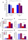
Figure 5. Atxn1Ldp suppresses the behavioral deficits in Atxn1−/− mice.
We assessed the effects of mild overexpression of Atxn1L on Atxn1−/− phenotypes (A) Mice of the following genotypes: Atxn1+/+ (n = 5), Atxn1 +/− (n = 12), Atxn1 −/− (n = 9), Atxn1 +/+; Atxn1Ldp/ +(n = 10), Atxn1 +/−; Atxn1Ldp/ +(n = 17), and Atxn1 −/−; Atxn1Ldp/ +(n = 12), were tested at 8 weeks for contextual conditioned fear. Atxn1−/− mice exhibited significantly reduced freezing behavior in the contextual fear-conditioning test compared to wild-type and Atxn1+/− littermates. However, Atxn1−/− mice carrying the Atxn1L duplication performed significantly better than Atxn1−/− littermates in this task (p<0.05). Wild-type and Atxn1+/− mice expressing the Atxn1Ldp allele performed similarly to wild-type and Atxn1+/− mice without the Atxn1L duplication. (B–E) An independent cohort of mice was generated to test the effects of Atxn1L duplication on the motor deficits of Atxn1−/− mice. Atxn1 +/− (n = 16), Atxn1 +/−; Atxn1Ldp/ +(n = 23), Atxn1 −/− (n = 14), and Atxn1 −/−; Atxn1Ldp/ + mice (n = 17) were tested on the dowel and wire hang paradigms at 8 weeks. The latency of Atxn1−/− mice to reach the side for the first time was increased compared to Atxn1+/− and Atxn1+/−; Atxn1dp/+ littermates on the rod (B); they also walked off the rod fewer times in the 120 s interval (C). In contrast, Atxn1−/−; Atxn1Ldp/+ mice took less time to walk off the dowel (B), and they also crossed the dowel more times in 120 s than Atxn1−/− littermates (C). In the wire hang test, Atxn1−/− mice showed increased latency to reach the sides of the wire compared Atxn1+/− and Atxn1+/−; Atxn1Ldp/+controls (D). Additionally, Atxn1−/− mice reached the sides fewer times than control littermates (E). In contrast, Atxn1−/− mice overexpressing Atxn1L exhibited marked reduction in the time for the first touch (D), and increased number of side touches in the 120 s interval, when compared to Atxn1−/− mice (E). Error bars in graphs represent +/− SEM, *p<0.05.
Figure 6
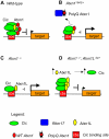
Figure 6. Model of Atxn1L rescue through Cic stabilization in Atxn1−/− mice.
(A) In wild-type mice, Atxn1-Cic and Atxn1L-Cic complexes bind the promoters of target genes and repress them effectively. (B) In mice expressing polyglutamine-expanded Atxn1, mutant Atxn1 associates preferentially with Rbm17, while the decrease in Atxn1-Cic complexes destabilizes Cic and reduces its levels at the promoters, thus leading to de-repression of its target genes. (C) A similar Cic destabilization occurs in the absence of wild-type Atxn1 in Atxn1−/− mice, also resulting in increased expression of target genes. (D) When Atxn1L is moderately overexpressed in Atxn1−/− mice, it stabilizes Cic levels by forming functional Atxn1L-Cic complexes that can substitute for Atxn1-Cic at the promoters, thus rescuing target gene repression. We propose that this mechanism might also act to rescue loss-of-function of Atxn1 in Atxn1154Q/+; Atxn1Ldp/+ mice.

References

    1. Orr HT, Zoghbi HY. Trinucleotide repeat disorders. Annu Rev Neurosci. 2007;30:575–621. - PubMed
    1. Gatchel JR, Zoghbi HY. Diseases of unstable repeat expansion: mechanisms and common principles. Nat Rev Genet. 2005;6:743–755. - PubMed
    1. Zuhlke C, Riess O, Bockel B, Lange H, Thies U. Mitotic stability and meiotic variability of the (CAG)n repeat in the Huntington disease gene. Hum Mol Genet. 1993;2:2063–2067. - PubMed
    1. Keckarevic D, Culjkovic B, Savic D, Stojkovic O, Kostic V, et al. The status of SCA1, MJD/SCA3, FRDA, DRPLA and MD triplet containing genes in patients with Huntington disease and healthy controls. J Neurogenet. 2000;14:257–263. - PubMed
    1. Lorenzetti D, Bohlega S, Zoghbi HY. The expansion of the CAG repeat in ataxin-2 is a frequent cause of autosomal dominant spinocerebellar ataxia. Neurology. 1997;49:1009–1013. - PubMed

Publication types