Review
. 2010 Jul 8;6(7):e1000942.
doi: 10.1371/journal.ppat.1000942.

Inositol hexakisphosphate-induced autoprocessing of large bacterial protein toxins

Affiliations
Review

Inositol hexakisphosphate-induced autoprocessing of large bacterial protein toxins

Martina Egerer et al. PLoS Pathog. .

Abstract

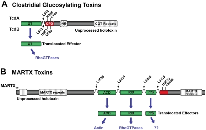
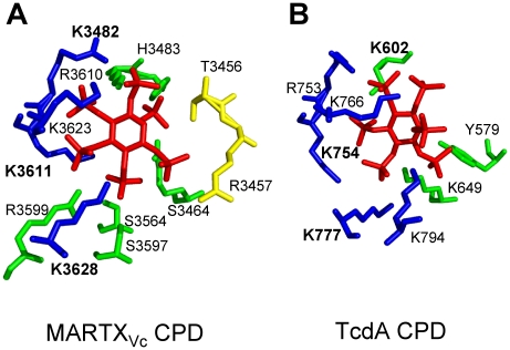
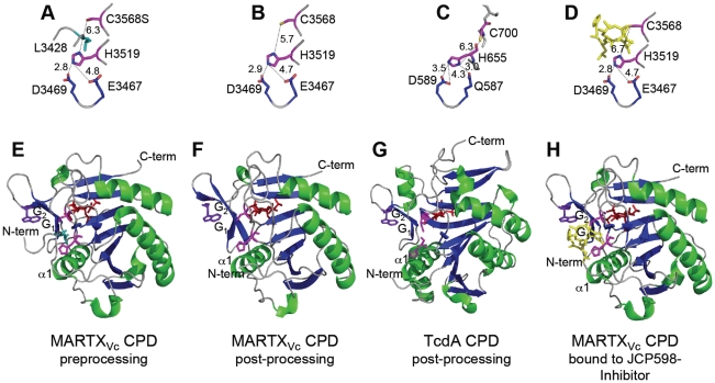
Large bacterial protein toxins autotranslocate functional effector domains to the eukaryotic cell cytosol, resulting in alterations to cellular functions that ultimately benefit the infecting pathogen. Among these toxins, the clostridial glucosylating toxins (CGTs) produced by Gram-positive bacteria and the multifunctional-autoprocessing RTX (MARTX) toxins of Gram-negative bacteria have distinct mechanisms for effector translocation, but a shared mechanism of post-translocation autoprocessing that releases these functional domains from the large holotoxins. These toxins carry an embedded cysteine protease domain (CPD) that is activated for autoprocessing by binding inositol hexakisphosphate (InsP(6)), a molecule found exclusively in eukaryotic cells. Thus, InsP(6)-induced autoprocessing represents a unique mechanism for toxin effector delivery specifically within the target cell. This review summarizes recent studies of the structural and molecular events for activation of autoprocessing for both CGT and MARTX toxins, demonstrating both similar and potentially distinct aspects of autoprocessing among the toxins that utilize this method of activation and effector delivery.


Conflict of interest statement

KJFS has a pending patent application that describes use of the CPD for biotechnological applications.

Figures

Figure 1
Figure 1. Schematic diagrams representing CPD-dependent autoprocessing sites within CGTs and MARTX toxins.
Diagrams are shown for (A) CGTs represented by TcdA and TcdB or (B) MARTX toxins represented by MARTXVc. In (A), the CGT holotoxins contain C-terminal repeats required for receptor interactions and a hydrophobic region (HR) postulated to function in translocation of the GT across the membrane of the endosome. Upon autoprocessing, the catalytically active glucosyltransferase effector (GT) is delivered to cells where it targets RhoGTPases. In (B), the MARTX holotoxin contains both N- and C-terminal repeats that likely function in translocation. Upon autoprocessing, MARTXVc delivers three effectors that have distinct cellular targets as indicated. For both diagrams, the CPD catalytic Cys and His are marked, as are processing site Leu residues (see Table 1) found in unstructured segments between effectors (indicated by arrows). For CGTs, sequence numbering above the diagram represents TcdA while numbering below the diagram represents TcdB. For MARTXVc, sequence numbering is based on the original annotation of the rtxA gene by Lin et al. and may be different than that found in cited references.
Figure 2
Figure 2. Side chain residues from CPD that contact InsP6 in the structural models derived from crystal structures of MARTXVc and TcdA CPD.
All key residues that contact InsP6 in the CPD of (A) MARTXVc and (B) TcdA are shown labeled with a single letter code, with the three Lys residues determined to be most critical for InsP6 binding shown in bold text. Interestingly, despite strong conservation of the critical Lys residues in the primary amino acid sequence, contacts with InsP6 and the orientation of InsP6 differ in the two structures. Diagram is colored to represent residues originating from the N-terminal strand (yellow), the core structure (green), and β-strands G1-G5 (blue), a structure also known as β8-β12 or the β-flap. Structural models were based on PDB (A) 3FZY and (B) 3HO6 , and figures were prepared with MacPyMol software (DeLano Scientific).
Figure 3
Figure 3. Crystal structures of MARTXVc and TcdA CPDs.
Crystal structures of the (A–D) CPD catalytic sites with distances between residues designated in angstroms and (E–G) the CPD proteins are shown at various stages of processing. (A, E) Pre-processing and (B, F) post-processing structures of MARTXVc CPD bound to InsP6 (PDB 3FZY and PDB 3EEB , respectively). (C, G) Post-processing structures of TcdA CPD bound to InsP6 (PDB 3HO6 [57]). (D, H) Post-processing structure of MARTXVc bound to z-Leu-Leu-azaLeu-epoxide inhibitor JCP598 as a surrogate substrate representing the structure of CPD after reactivation (PDB 3GCD [49]). Structures are identically oriented at a conserved Trp (purple) in the G1/G2 β-hairpin that is critical to InsP6 induction of autoprocessing . The catalytic Cys and His residues are shown in pink with InsP6 present at the backside of each structure in red. The P1 Leu (turquoise) is found only in the unprocessed structure (A) with the scissile bond oriented between the catalytic residues. Figures were prepared with MacPyMol software (DeLano Scientific).
Figure 4
Figure 4. Proposed model for cooperative activation and reactivation of MARTXVc CPD by InsP6.
I. Apo-CPD without InsP6 is an unstable protein susceptible to thermal denaturation at physiological temperature. The core structure (green) is folded but the β-flap (blue) is susceptible to proteolysis, indicating it may be only partially structured. II. Upon binding InsP6, the structure rearranges such that the N-terminus (yellow) becomes locked within the active site between the catalytic Cys (C) and His (H) in a rigid alignment amenable to substrate-activated autoprocessing. III. After autoprocessing, the MARTXVc CPD enters a transitional state that has distinct biochemical properties, including a 500-fold reduced affinity for InsP6. IV. After first binding a new substrate (grey) and then a new molecule of InsP6, the enzyme–substrate complex structure of the MARTXVc CPD is restored for additional processing events. Figure is based on multisite processing model for MARTXVc proposed by Prochazkova et al. . Current evidence from NMR studies supports the idea that stage I and II also occur for TcdA . However, binding studies with TcdB suggest CGTs likely do not undergo stage III deactivation or stage IV reactivation .

References

    1. Wooldridge K, editor. Norfolk, UK: Caister Academic Press; 2009. Bacterial Secreted Proteins: Secretory Mechanisms and Role in Pathogenesis.511
    1. Ma AT, McAuley S, Pukatzki S, Mekalanos JJ. Translocation of a Vibrio cholerae type VI secretion effector requires bacterial endocytosis by host cells. Cell Host Microbe. 2009;5:234–243. - PMC - PubMed
    1. Sanchez J, Holmgren J. Cholera toxin structure, gene regulation and pathophysiological and immunological aspects. Cell Mol Life Sci. 2008;65:1347–1360. - PMC - PubMed
    1. Young JA, Collier RJ. Anthrax toxin: receptor binding, internalization, pore formation, and translocation. Annu Rev Biochem. 2007;76:243–265. - PubMed
    1. Baldwin MR, Barbieri JT. Association of botulinum neurotoxins with synaptic vesicle protein complexes. Toxicon. 2009;54:570–574. - PMC - PubMed

Publication types

LinkOut - more resources